
Make detailed yet simple to learn format with must know facts for quiz on paediatric leukaemia and lymphoma including causes, pathogenesis, clinical features and management
acute lymphoblastic leukemia ALL bone marrow blast cells microscopy
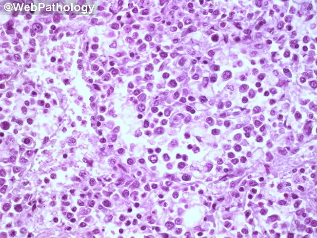
Imaging modality: light microscopy of a hematopathology slide from a bone marrow aspirate smear. Stain: Hematoxylin and Eosin (H&E). Magnification: high-power field (~400x total; 40x objective with 10x ocular). Anatomical location: bone marrow within the medullary cavity (hematopoietic tissue). Visual features: sheets of immature blasts with high nuclear-to-cytoplasmic ratio, round to oval nuclei, fine chromatin, prominent nucleoli, and scant basophilic cytoplasm; increased cellularity with near-complete effacement of normal hematopoiesis; occasional mitotic figures; minimal cytoplasmic granularity; sparse residual neutrophils, erythroid precursors, and megakaryocytes. Pathologic interpretation: diffuse marrow infiltration by blasts, consistent with an acute leukemia until immunophenotyping and genetic studies delineate subtype. Diagnostic significance: morphologic hallmark of an acute hematologic malignancy; requires ancillary tests (flow cytometry, immunohistochemistry, cytogenetics/molecular studies) for lineage classification (myeloid vs lymphoid), prognosis, and treatment planning. Differential considerations: ALL (acute lymphoblastic leukemia), AML (acute myeloid leukemia), lymphoblastic lymphoma with marrow involvement, myelodysplastic syndrome with excess blasts, or nonhematopoietic marrow infiltration. Clinical correlation: commonly presents with cytopenias, fatigue, infections, and bleeding; pediatric ALL vs adult AML guidance; urgent hematology-oncology workup is indicated. Educational use: foundational blast morphology recognition, marrow infiltration patterns, and the need for confirmatory immunophenotyping. This image is valuable for training in blast morphology, differential diagnosis, and education in hematopathology.
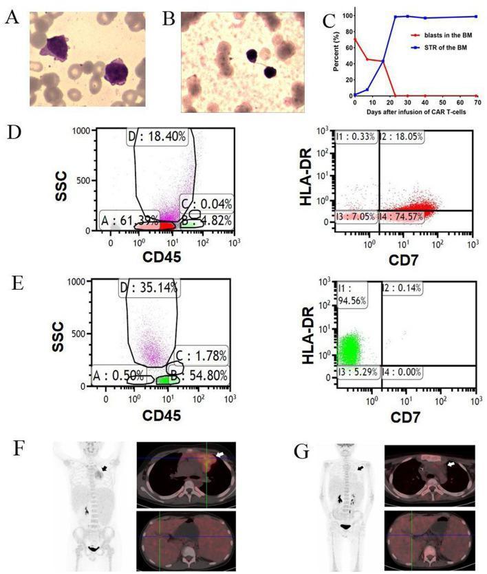
A multi-panel figure illustrating the therapeutic response of early T-cell precursor acute lymphoblastic leukemia/lymphoma (ETP-ALL/LBL) to CD7 CAR T-cell infusion. Panels A and B show bone marrow (BM) morphology, transitioning from hypercellular with prominent blasts (A) to hypocellular without visible blasts (B). Panel C is a line graph tracking the sharp decline of BM blast percentage and the simultaneous rise of donor chimerism (STR) to nearly 100% within 30 days. Panels D and E represent flow cytometry analysis of BM cells before and after treatment. D shows a large population of CD45-dim blasts (Region A: 61.39%) that are CD7-positive. E demonstrates the elimination of these blasts (Region A: 0.50%) and the emergence of CD7-negative populations (Region B: 54.80%) and CD3-positive/CD7-negative T-cells. Panels F and G display FDG PET-CT scans. The 'before' scan (F) reveals extensive hypermetabolic extramedullary disease, including a large mediastinal mass and widespread lymphadenopathy. The 'after' scan (G) shows a complete metabolic response with the disappearance of the mediastinal mass and resolution of hypermetabolic lesions across cervical, mediastinal, and abdominal regions.
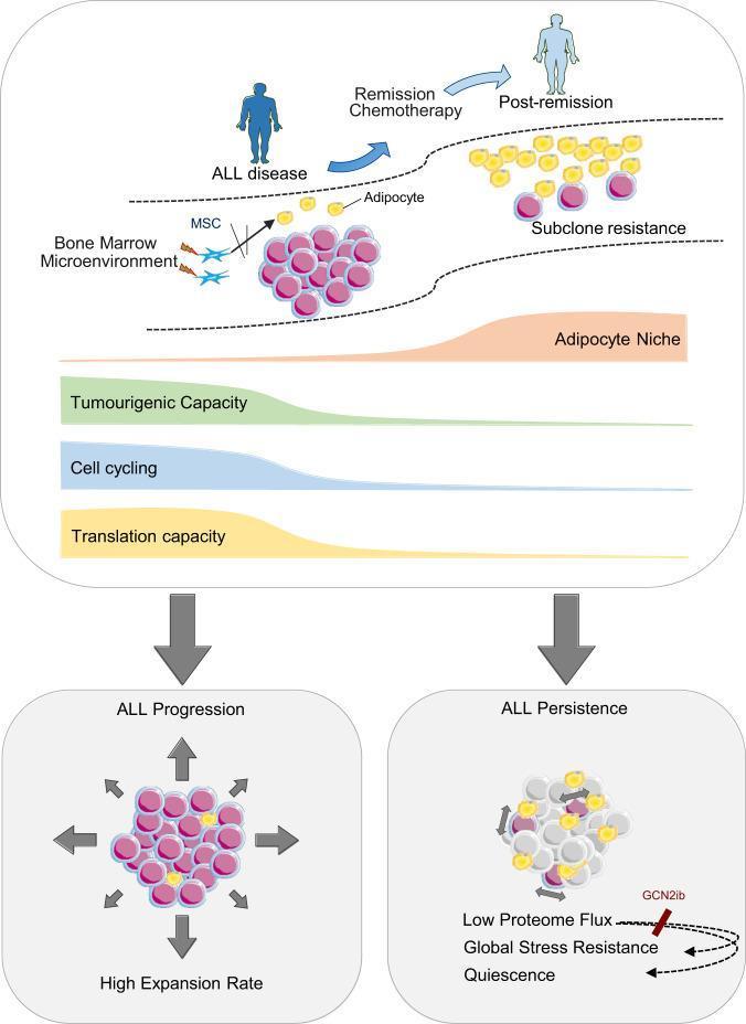
This pathophysiology diagram illustrates the remodeling of the bone marrow microenvironment (BMM) during the transition from Acute Lymphoblastic Leukemia (ALL) disease to remission. The upper panel depicts two states: 'ALL disease' and 'Post-remission'. In the disease state, the BMM is crowded with leukemia blasts (pink clusters), showing profound depletion of adipocytes (yellow spheres) and the presence of Mesenchymal Stem Cells (MSCs). Following 'Remission Chemotherapy', the post-remission niche shows adipocyte reconstitution and 'Subclone resistance' with fewer, persistent leukemia cells. Quantitative area charts indicate that as the 'Adipocyte Niche' expands, there is a reciprocal decline in 'Tumourigenic Capacity', 'Cell cycling', and 'Translation capacity'. The lower panel compares 'ALL Progression' with 'ALL Persistence'. 'ALL Progression' is characterized by a high expansion rate and active cell proliferation (indicated by outward arrows). In contrast, 'ALL Persistence' describes a dormant state defined by 'Low Proteome Flux', 'Global Stress Resistance', and 'Quiescence'. The diagram highlights how the adipocyte-rich remission niche promotes leukemia cell dormancy and chemoprotection, potentially mediated by GCN2-related pathways.
Hodgkin lymphoma Reed-Sternberg cells histology
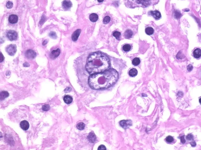
Histopathology: Light microscopy of an H&E-stained lymph node biopsy. This image highlights classic Reed-Sternberg (RS) cells within a mixed inflammatory background, a hallmark of classic Hodgkin lymphoma (cHL). The RS cells are large (up to ~100 microns) with abundant pale eosinophilic cytoplasm and prominent, bilobed to multinucleate nuclei. The nuclei show vesicular chromatin and thick nuclear membranes, with large central nucleoli creating an owl-eye appearance when two nuclei are juxtaposed. Variants, including giant RS cells and lacunar RS cells, may be seen in different HL subtypes. The surrounding milieu contains eosinophils, neutrophils, mature lymphocytes, plasma cells, and histiocytes, reflecting a characteristic reactive infiltrate. The nodal architecture is variably effaced, sometimes with formation of a rosette-like collar of T lymphocytes around RS cells. Immunophenotyping in practice typically demonstrates CD30 and CD15 positivity with weak PAX5 expression, supporting the HL diagnosis, and helps distinguish RS cells from RS-like cells in non-Hodgkin lymphomas or mimics such as anaplastic carcinoma. This histology provides essential diagnostic confirmatory evidence in suspected HL and informs prognosis and treatment planning (ABVD regimen, radiotherapy, or combined modality therapy). In some cases, classic RS cells may be absent, requiring ancillary testing and correlation with clinical findings, correlating with patient outcome.
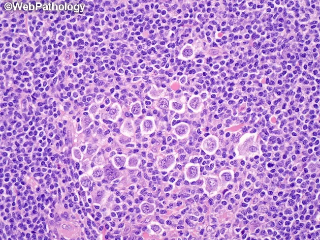
This histology image depicts lymph node tissue from a classic Hodgkin lymphoma with nodular sclerosis. The predominant cells are lacunar variants of Reed-Sternberg (RS) cells, characterized by multilobulated nuclei with coarse chromatin and small nucleoli, and a distinctive perinuclear cytoplasmic condensation that creates clear lacunar spaces in fixed tissues. The lacunae are artifacts of formalin fixation and are not present in B5-fixed specimens. Background comprises a mixed inflammatory milieu with small lymphocytes and scattered eosinophils embedded in fibrous nodules; the nodular architecture is evident with fibrous bands partitioning the lymph node into nodules, a hallmark of nodular sclerosis. RS cells may be solitary and scattered or form compact clusters and, in higher-grade nodular sclerosis (grade II), diffuse sheets. Clinical significance: The presence of lacunar RS cells in a fibrous nodal milieu supports a diagnosis of nodular sclerosis classic Hodgkin lymphoma. Differential considerations include other lymphadenopathies with RS-like cells (reactive conditions, other HL subtypes, or ALCL), but the combination of lacunar morphology, nodular sclerosis, and immunophenotype (CD30+, CD15+, PAX5 weak) is characteristic. This image is relevant for educational demonstrations of HL subtypes, fixation artifacts, and morphology-guided diagnosis, as well as educational references in pathology training and diagnostic review. Educational reference material.
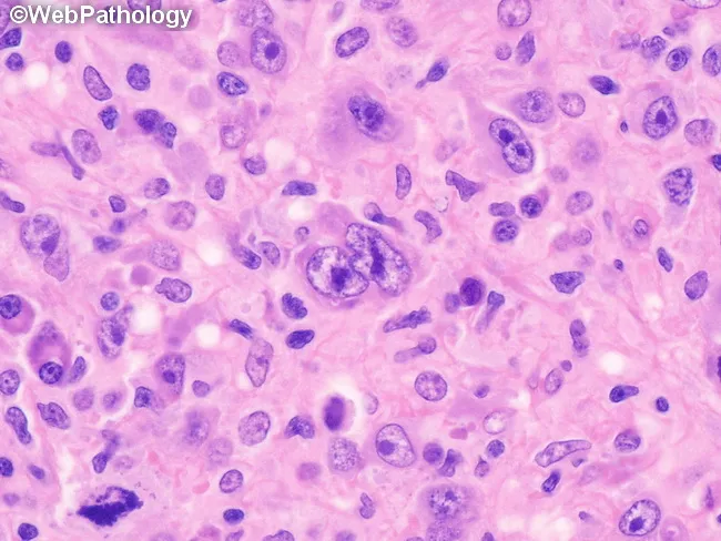
This histopathology image depicts a lymph node biopsy showing features diagnostic of lymphocyte-depleted classic Hodgkin lymphoma, reticular subtype. Using light microscopy on an H&E-stained paraffin section, the lymphoid architecture is markedly effaced by a cellular sheet of large, atypical mononuclear cells with prominent nucleoli. Occasional Reed-Sternberg cells are visible within the expansive background, including cells with multilobed or bilobed nuclei and prominent eosinophilic nucleoli, characteristic of Hodgkin lymphoma. The RS cells are scattered among numerous atypical mononuclear variants rather than forming a cohesive nodular structure. There is little-to-no fibrosis and only sparse non-neoplastic inflammatory cells, which is typical for the lymphocyte-depleted pattern. The background may show rimming by small lymphocytes and histiocytes in a reticular network, but overall cellularity is high. Immunophenotypic markers are not shown here, but in clinical practice RS cells typically express CD30 and CD15 with weaker PAX5 or B-cell markers. The diagnostic significance lies in recognizing the lymphocyte-depleted cHL morphology, which carries distinct clinical implications, often presenting with advanced stage disease and systemic symptoms. This image is useful for educational purposes, differential diagnosis conversation, and correlating histology with treatment planning (ABVD/BEACOPP regimens). Correlation with immunohistochemistry (CD30, CD15, PAX5) and EBV status further supports diagnosis in practice.
🩸 Paediatric Leukaemia & Lymphoma — Master Quiz Guide
Format key: ⚡ = High-yield quiz fact | 🔴 = Classic exam trap | 🟢 = Must memorise | 📌 = Key concept
PART 1: PAEDIATRIC LEUKAEMIA
📚 Overview & Epidemiology
| Feature | Fact |
|---|---|
| Most common childhood malignancy | Leukaemia (30-35% of all childhood cancers) |
| Most common type | ALL (Acute Lymphoblastic Leukaemia) — 75-80% |
| AML in children | ~15-20% of childhood leukaemia |
| Peak age of ALL | 2-5 years (pre-B cell type) |
| Sex | Males > Females |
🔬 Classification of Acute Leukaemia
Leukaemia
├── ALL (Acute Lymphoblastic)
│   ├── B-ALL (~85%) ← most common
│   └── T-ALL (~15%)
└── AML (Acute Myeloid)
    ├── M0–M7 (FAB classification)
    └── Acute promyelocytic (M3) — treat with ATRA
⚙️ Causes & Risk Factors
Genetic / Constitutional Factors
| Risk Factor | Associated Leukaemia | Relative Risk |
|---|---|---|
| Down syndrome (Trisomy 21) | ALL and AML | 20x increased |
| Fanconi anaemia | AML | High |
| Bloom syndrome | ALL, AML | High |
| Ataxia-telangiectasia | ALL (T-cell) | High |
| Neurofibromatosis type 1 | JMML (juvenile myelomonocytic) | High |
| Identical twin with ALL | ALL | 25% concordance |
Environmental Factors
- Ionising radiation (prenatal X-rays, post-Hiroshima)
- Benzene exposure (AML)
- Prior chemotherapy (alkylating agents → AML; topoisomerase II inhibitors → AML with 11q23 rearrangement)
- High birth weight (ALL risk)
🧬 Pathogenesis
- First hit: Prenatal somatic mutation (e.g., TEL-AML1 fusion) — clonal pre-leukaemic clone created in utero
- Second hit: Postnatal immune stimulation / infection triggers final transformation
| Cytogenetic Finding | Significance |
|---|---|
| t(12;21) TEL-AML1 [ETV6-RUNX1] | Most common (~25%), favourable prognosis |
| Hyperdiploidy (>50 chromosomes) | Favourable prognosis |
| t(9;22) BCR-ABL "Philadelphia chromosome" | Poor prognosis; treat with TKI (imatinib) |
| t(4;11) MLL-AF4 [KMT2A] | Infant ALL, very poor prognosis |
| t(1;19) E2A-PBX1 | Intermediate prognosis |
| Hypodiploidy (<44 chromosomes) | Poor prognosis |
| t(8;21), inv(16), t(15;17) | Favourable AML subtypes |
| t(15;17) PML-RARA | Acute Promyelocytic Leukaemia (M3) |
🩺 Clinical Features of ALL
Symptoms from Bone Marrow Failure (crowding out normal cells):
| Feature | Mechanism |
|---|---|
| Pallor, fatigue, anaemia | ↓ Erythropoiesis |
| Fever, infections | ↓ Normal WBCs (neutropenia) |
| Bleeding, petechiae, bruising | Thrombocytopenia |
| Bone pain, limp | Marrow expansion — classic in children, often misdiagnosed as arthritis |
Symptoms from Leukaemic Infiltration:
| Feature | Note |
|---|---|
| Lymphadenopathy | Generalised, painless |
| Hepatosplenomegaly | Very common |
| Mediastinal mass | T-ALL — can cause SVC syndrome, respiratory distress |
| CNS involvement | Headache, vomiting, cranial nerve palsies (CN VI, VII most common) |
| Testicular enlargement | Boys — painless, hard swelling (sanctuary site) |
| Orbital proptosis | Particularly in AML (chloroma/granulocytic sarcoma) |
🔬 Investigations
- Anaemia (normocytic normochromic)
- Thrombocytopenia
- WBC: variable (can be low, normal, or very high)
- Blast cells seen on peripheral smear
- ⚡ Diagnosis confirmed: ≥20% blasts in bone marrow (WHO 2016 criterion)
- Morphology, immunophenotyping (flow cytometry), cytogenetics, molecular studies
| Test | Purpose |
|---|---|
| LP (lumbar puncture) | CNS involvement |
| CXR / CT chest | Mediastinal mass (T-ALL) |
| Testicular USS | Boys with T-ALL or relapse |
| Uric acid, LDH, creatinine | Tumour lysis syndrome risk |
| TdT (Terminal deoxynucleotidyl transferase) | Positive in ALL (not AML) |
💊 Management of ALL
Phase Structure (3 Phases):
Induction (4-6 weeks)
    ↓
Consolidation/Intensification (several months)
    ↓
Maintenance (2-3 years total)
Induction Therapy (aim: achieve remission — <5% blasts)
| Drug | Role |
|---|---|
| Prednisolone/Dexamethasone | Cornerstone; kills lymphoblasts |
| Vincristine | Vinca alkaloid, disrupts mitotic spindle |
| L-Asparaginase | Depletes asparagine (tumour cells can't synthesise it) |
| Daunorubicin | Anthracycline (for high-risk disease) |
CNS Prophylaxis
- Intrathecal methotrexate (± cytarabine, hydrocortisone)
- Replaced cranial irradiation (now avoided due to neurocognitive toxicity)
Maintenance (2-3 years)
- Daily 6-mercaptopurine (6-MP)
- Weekly methotrexate (oral)
- Monthly vincristine + steroids
Risk Stratification:
| Group | Features |
|---|---|
| Standard risk | Age 1-9, WBC <50,000, good cytogenetics |
| High risk | Age <1 or >9, WBC >50,000, Philadelphia+, poor response |
Special AML treatment:
- AML M3 (APML): ATRA (all-trans retinoic acid) + arsenic trioxide — causes differentiation of promyelocytes; highly effective
- Other AML: cytarabine + anthracycline ("7+3")
- HSCT considered for high-risk AML in first remission
📊 Prognosis of ALL
| Factor | Favourable | Unfavourable |
|---|---|---|
| Age | 1-9 years | <1 year (infant), >10 years |
| WBC at diagnosis | <50,000/µL | >50,000/µL |
| Cytogenetics | Hyperdiploidy, TEL-AML1 | Philadelphia+, hypodiploidy, MLL |
| Response to treatment | Rapid response (Day 7-14 clearance) | Slow response |
| CNS involvement | Absent | Present |
| Sex | Female | Male (slightly worse) |
PART 2: PAEDIATRIC LYMPHOMA
📚 Overview
| Feature | Fact |
|---|---|
| 3rd most common childhood cancer | After leukaemia and brain tumours |
| Age group | More common in older children and adolescents |
| Two main types | Hodgkin Lymphoma (HL) and Non-Hodgkin Lymphoma (NHL) |
🏷️ HODGKIN LYMPHOMA (HL)
Epidemiology
- Bimodal age distribution: Peak 15-35 years AND >55 years
- In paediatrics: mainly adolescents (10-20 years)
- More common in males in childhood; equalises in adults
- EBV association: especially in mixed cellularity and lymphocyte-depleted subtypes
Reed-Sternberg Cell — The Hallmark
- Large binucleate/multinucleate cell
- "Owl eye" prominent nucleoli
- Derived from B-cells (but lost B-cell markers)
- Immunophenotype: CD30+, CD15+, CD45-, PAX5 weak
WHO Classification of HL:
| Subtype | % | Key Features |
|---|---|---|
| Nodular Sclerosis (NS) | 60-70% | Most common in adolescents; fibrous bands; lacunar RS cells; mediastinal mass |
| Mixed Cellularity | 20-25% | Mixed inflammatory background; EBV+; classic RS cells |
| Lymphocyte Rich | 5% | Best prognosis |
| Lymphocyte Depleted | Rare | Worst prognosis; EBV+; advanced disease |
| Nodular Lymphocyte Predominant (NLPHL) | 5% | "Popcorn cells" (L&H cells); CD20+, CD30-, CD15- |
Clinical Features of HL
| Feature | Details |
|---|---|
| Painless cervical lymphadenopathy | Most common presenting feature |
| Mediastinal mass | 60-70%; may cause cough, dyspnoea |
| "B symptoms" | Fever >38°C, drenching night sweats, weight loss >10% in 6 months |
| Pel-Ebstein fever | Cyclical fever pattern (classic but rare) |
| Alcohol-induced pain | Enlarged lymph nodes become painful after drinking alcohol |
| Pruritis | Common |
| Splenomegaly | With abdominal disease |
Ann Arbor Staging:
| Stage | Definition |
|---|---|
| I | Single lymph node region |
| II | Two or more regions, same side of diaphragm |
| III | Regions on both sides of diaphragm |
| IV | Disseminated involvement (liver, bone marrow, lung) |
| A/B suffix | A = no B symptoms; B = B symptoms present |
| E suffix | Extranodal extension |
| S suffix | Spleen involvement |
Investigations of HL
- Excision biopsy of lymph node (not FNAC — need architecture)
- CT chest/abdomen/pelvis (staging)
- PET-CT — gold standard for staging and treatment response
- FBC: eosinophilia, lymphopenia (poor prognosis), elevated ESR, elevated LDH
- Bone marrow biopsy (stage IV)
Management of HL
- ABVD chemotherapy (2-4 cycles) + involved-field radiotherapy (IFRT)
- Or ABVD alone (to avoid radiation in children)
- ABVD × 6 cycles ± radiotherapy
- Or BEACOPP (more intensive, more toxic)
| Drug | Role |
|---|---|
| Adriamycin (doxorubicin) | Anthracycline |
| Bleomycin | Causes pulmonary fibrosis (toxicity) |
| Vinblastine | Vinca alkaloid |
| Dacarbazine | Alkylating agent |
- Salvage chemo (e.g., ICE, GDP)
- High-dose chemo + autologous stem cell transplant
- Brentuximab vedotin (anti-CD30 antibody-drug conjugate)
- Pembrolizumab / Nivolumab (PD-1 inhibitors) — approved for refractory HL
HL Prognosis
- Early stage: ~95% cure rate
- Advanced stage: ~75-85% long-term survival
- Hasenclever score (IPS) for advanced disease
🏷️ NON-HODGKIN LYMPHOMA (NHL) IN CHILDREN
Epidemiology
- More common than HL in children <10 years
- Biologically different from adult NHL — mostly high-grade, aggressive
- Children rarely get low-grade follicular lymphoma (common in adults)
Main Subtypes in Children:
| Type | % | Key Features |
|---|---|---|
| Burkitt Lymphoma | ~40% | Most common childhood NHL; highly aggressive; "starry sky" pattern |
| Lymphoblastic Lymphoma | ~25% | T-cell > B-cell; mediastinal mass; similar to T-ALL |
| Diffuse Large B-cell (DLBCL) | ~20% | B-cell; aggressive; responds to chemo |
| Anaplastic Large Cell Lymphoma (ALCL) | ~10% | T/null cell; CD30+, ALK+; skin, lymph nodes |
🦋 BURKITT LYMPHOMA
Three Clinical Variants:
| Variant | Association | Location |
|---|---|---|
| Endemic (African) | EBV (>95% EBV+) | Jaw/facial bones, orbit |
| Sporadic (Western) | EBV (~20% EBV+) | Ileocecal region, abdomen |
| Immunodeficiency-associated | HIV | Widespread |
Histology:
- "Starry sky" pattern — sheets of medium lymphoid cells with tingible body macrophages (the "stars") scattered throughout
- High mitotic rate, high Ki-67 (nearly 100%)
- Immunophenotype: CD10+, CD20+, BCL6+, BCL2-, surface IgM+
Molecular:
- t(8;14) — MYC translocation with IgH (most common, 80%)
- t(8;22) or t(2;8) — less common MYC variants
- MYC deregulation drives extremely rapid proliferation
Management of Burkitt:
- Intensive short-course chemotherapy: CODOX-M/IVAC or BFM protocols
- Rituximab (anti-CD20) now added
- CNS prophylaxis with intrathecal chemotherapy
- Tumour lysis syndrome (TLS) prevention — aggressive hydration, allopurinol/rasburicase before chemo
🎽 LYMPHOBLASTIC LYMPHOMA (LBL)
- Essentially same disease as ALL but with <25% blasts in bone marrow (if ≥25%, classified as ALL)
- T-LBL (85%): Adolescent males; mediastinal mass; SVC syndrome
- B-LBL (15%): Skin, bone, lymph nodes
🔵 ANAPLASTIC LARGE CELL LYMPHOMA (ALCL)
- T/null cell phenotype
- CD30+ (like HL), CD45+, EMA+
- ALK (Anaplastic Lymphoma Kinase) positive in children → better prognosis
- t(2;5) NPM-ALK fusion — most common
- Presents with: lymphadenopathy, skin lesions ("B" symptoms), systemic symptoms
NHL Staging — Murphy (St Jude) Staging (used in children, not Ann Arbor):
| Stage | Definition |
|---|---|
| I | Single tumour, extranodal OR single nodal region (excluding mediastinum/abdomen) |
| II | Single extranodal tumour with regional nodes; or ipsilateral nodal involvement; or primary GI tumour (resected) |
| III | Both sides of diaphragm; all primary intrathoracic or unresected abdominal; all paraspinal/epidural |
| IV | Any CNS or bone marrow involvement |
🆚 HL vs NHL — Quick Comparison Table
| Feature | Hodgkin Lymphoma | Non-Hodgkin Lymphoma |
|---|---|---|
| Age peak (paeds) | Adolescents | All ages; <10 yrs more NHL |
| Growth pattern | Contiguous spread | Non-contiguous (skip lesions) |
| Mediastinal mass | Common (NS subtype) | T-LBL and DLBCL |
| CNS involvement | Rare | Common (especially Burkitt, LBL) |
| Bone marrow | Rare | Common |
| RS cells | Present | Absent |
| Staging system | Ann Arbor | Murphy (St Jude) |
| Prognosis | Generally better | Variable, but often good in children |
💉 Tumour Lysis Syndrome (TLS) — Must Know
| Metabolite | Direction | Consequence |
|---|---|---|
| Uric acid | ↑ | Renal failure, gout |
| Potassium | ↑ | Cardiac arrhythmia, arrest |
| Phosphate | ↑ | Hypocalcaemia |
| Calcium | ↓ | Tetany, seizures |
- IV hydration (2-3× maintenance)
- Allopurinol (xanthine oxidase inhibitor) — prevents uric acid formation
- Rasburicase (recombinant urate oxidase) — breaks down existing uric acid, faster; contraindicated in G6PD deficiency
🧪 Key Immunophenotype Summary
| Disease | Positive Markers | Negative Markers |
|---|---|---|
| B-ALL | CD10, CD19, CD34, TdT | CD3 |
| T-ALL | CD3, CD7, TdT | CD19 |
| Classic HL | CD30, CD15 | CD45, CD20 (usually) |
| NLPHL | CD20, CD45, CD79a | CD30, CD15 |
| Burkitt | CD10, CD20, BCL6, surface IgM | BCL2 |
| ALCL | CD30, ALK, EMA | |
| CLL (comparison) | CD5, CD19, CD23 | CD10 |
🎯 Chromosomal Translocations — Quick Memory Table
| Translocation | Gene Fusion | Disease | Prognosis |
|---|---|---|---|
| t(12;21) | ETV6-RUNX1 (TEL-AML1) | B-ALL | Favourable |
| t(9;22) | BCR-ABL | ALL, CML | Poor (ALL) |
| t(4;11) | MLL-AF4 | Infant ALL | Very poor |
| t(15;17) | PML-RARA | AML M3 (APML) | Favourable (ATRA) |
| t(8;14) | MYC-IgH | Burkitt | - |
| t(2;5) | NPM-ALK | ALCL | Favourable |
| t(11;14) | Cyclin D1-IgH | Mantle cell | Poor |
| t(14;18) | BCL2-IgH | Follicular lymphoma | - |
🧠 Memory Mnemonics
ABVD (Hodgkin chemo):
- Adriamycin | Bleomycin | Vinblastine | Dacarbazine
B symptoms:
Burkitt MYC translocations (80/10/10):
- t(8;14) = 80% — MYC + IgH (heavy chain)
- t(2;8) = 10% — IgK (kappa)
- t(8;22) = 10% — IgL (lambda)
TLS metabolic changes — "PUCK"
- Phosphate ↑ | Uric acid ↑ | Calcium ↓ | K (Potassium) ↑
ALL induction (standard risk):
📝 Top 20 Quiz Facts to Memorise
- ALL is the most common childhood cancer (75-80% of childhood leukaemia)
- Peak age of B-ALL: 2-5 years
- Diagnosis requires ≥20% blasts in bone marrow
- Down syndrome → 20x ALL risk, 50x AML (M7) risk
- Best prognostic cytogenetics: hyperdiploidy + TEL-AML1 t(12;21)
- Worst cytogenetics: Philadelphia chromosome t(9;22) → add imatinib
- TdT positive = ALL (not AML)
- Infant ALL (<1 year) = MLL rearrangement, worst prognosis
- AML M3 treatment: ATRA + arsenic trioxide (differentiation therapy)
- Maintenance ALL therapy: 6-MP daily + methotrexate weekly for 2-3 years
- Reed-Sternberg cells = CD30+, CD15+, "owl-eye" nucleoli
- Most common HL subtype = Nodular Sclerosis (60-70%)
- NLPHL = CD20+, CD30-, CD15- (popcorn cells)
- Alcohol-induced lymph node pain = pathognomonic of HL
- HL staging: Ann Arbor; childhood NHL: Murphy (St Jude)
- African Burkitt = EBV + jaw tumour; Sporadic Burkitt = ileocecal/abdomen
- Burkitt = t(8;14), "starry sky," BCL2 negative, Ki-67 ~100%
- T-LBL vs T-ALL = bone marrow blasts <25% vs ≥25%
- ALCL = CD30+, ALK+, t(2;5) → targeted by crizotinib
- TLS prevention: IV hydration + allopurinol/rasburicase; rasburicase contraindicated in G6PD deficiency
Bone Marrow Biopsy in ALL — Morphology
Paediatrics Facies and syndrome associated with it and it’s features, easy way to learn with pics
Down syndrome facial features trisomy 21 child
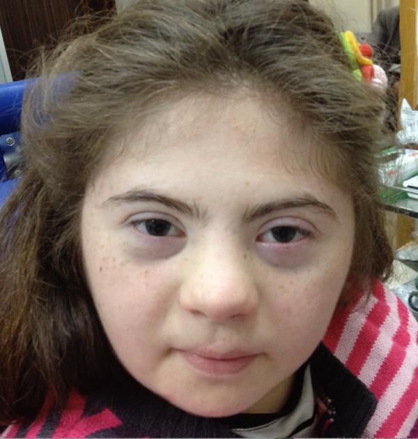
Clinical photograph of a 15-year-old female patient exhibiting phenotypic facial features characteristic of Down syndrome (Trisomy 21). The image demonstrates a flattened facial profile with a notably low-set and depressed nasal bridge. Ocular findings include upslanting palpebral fissures and prominent bilateral epicanthic folds (medial epicanthal skin folds) covering the inner canthi. Additional craniofacial features include a relatively small oral cavity and mild brachycephaly. The patient has light skin with fine freckling across the midface. This image serves as a teaching tool for medical genetics and clinical pediatrics, illustrating the classic dysmorphic facial features associated with Trisomy 21 in an adolescent. The clinical context suggests an association with Morning Glory Syndrome (MGS), though the internal ocular pathologies are not visible in this external facial view.
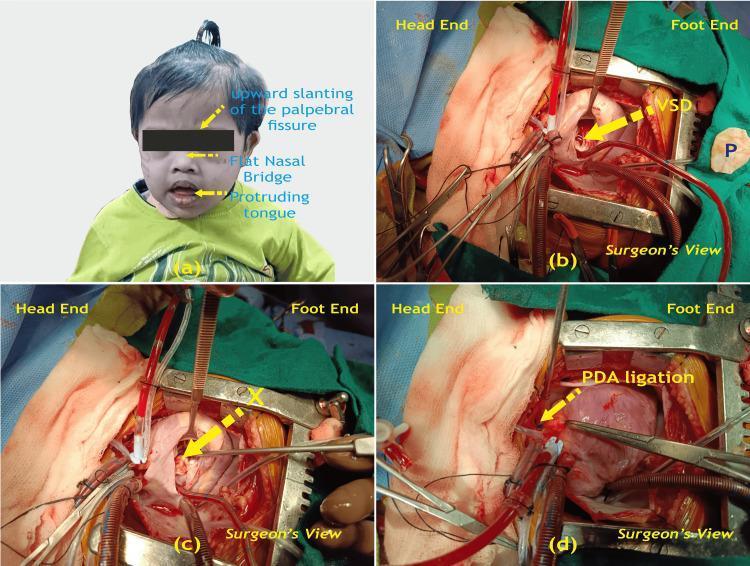
A multi-panel medical image illustrating the clinical presentation and surgical management of congenital heart disease in a pediatric patient with Trisomy 21 (Down Syndrome). Panel (a) is a clinical photograph of a child displaying characteristic dysmorphic facial features, including upward slanting palpebral fissures, a flat nasal bridge, and a protruding tongue. Panels (b), (c), and (d) provide intraoperative views during a median sternotomy. Panel (b) shows the exposed heart before surgical repair, identifying a ventricular septal defect (VSD) and a glutaraldehyde-treated pericardial patch (P) ready for use. Panel (c) demonstrates the intraoperative site after the VSD has been successfully closed with the patch (marked X). Panel (d) displays the ligation of a patent ductus arteriosus (PDA). The surgical images are labeled from the 'Surgeon's View' with 'Head End' and 'Foot End' orientations provided. This composite image serves to correlate the phenotypic features of Trisomy 21 with common associated cardiac anomalies (VSD and PDA) and their definitive surgical correction.
Turner syndrome facial features webbed neck
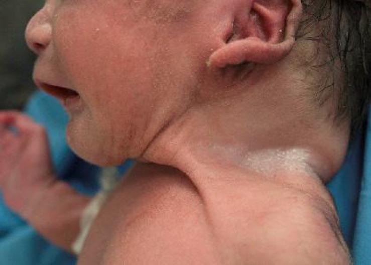
This clinical photograph shows a side profile of a neonate, highlighting characteristic dysmorphic features of the head and neck. The most prominent finding is a short, webbed neck (pterygium colli), with a redundant fold of skin extending from the mastoid area down to the acromion of the shoulder. The infant's ear is posteriorly rotated and positioned low relative to the facial features, with a small preauricular skin tag or appendage visible near the superior helix. The skin on the face and neck appears erythematous and exhibits fine desquamation, typical of a newborn or preterm infant. Additionally, the posterior skull appears slightly flattened, and the neck region shows increased subcutaneous tissue or skin laxity. These visual signs are clinically significant in the evaluation of genetic syndromes, most notably Turner syndrome or Noonan syndrome, where webbed neck and ear malformations are hallmark physical findings.
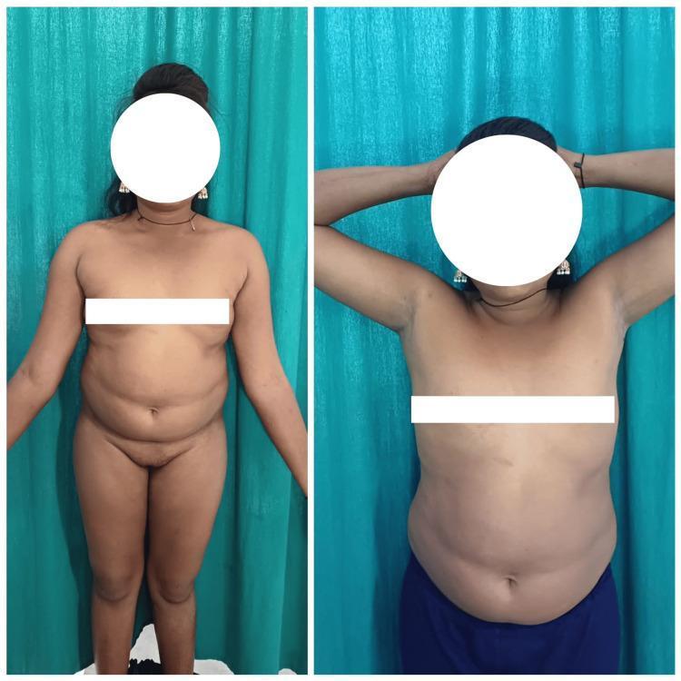
Two clinical photographs show the anterior trunk of a female patient, illustrating characteristic physical features of Turner syndrome (45,XO). The images demonstrate a broad, shield-shaped chest (pectus excavatum/carinatum tendency) with widely spaced (hyperteloric) nipples. The patient exhibits a webbed neck (pterygium colli), where folds of skin extend from the mastoid process to the acromion. The photographs also depict short stature and a lack of secondary sexual characteristic development consistent with primary amenorrhea and ovarian dysgenesis. These visual findings serve as clinical markers for genetic screening in reproductive medicine and endocrinology. Patient identity is protected by facial masking.
Marfan syndrome tall stature arachnodactyly features

A clinical photograph displaying the posterior view of a pediatric patient, illustrating a marfanoid habitus characterized by a tall stature, slender build, and arachnodactyly with disproportionately long limbs relative to the torso. The primary musculoskeletal finding is a visible scoliosis, presenting as a lateral curvature of the spine. Key clinical features include asymmetrical waist triangles, trunk shift, and slight pelvic obliquity. This visual is highly representative of connective tissue disorders such as Marfan syndrome, where skeletal abnormalities including progressive spinal curvature and elongated extremities are common. The image serves as an educational resource for identifying marfanoid body habitus and secondary orthopedic manifestations like scoliosis during a clinical physical examination.
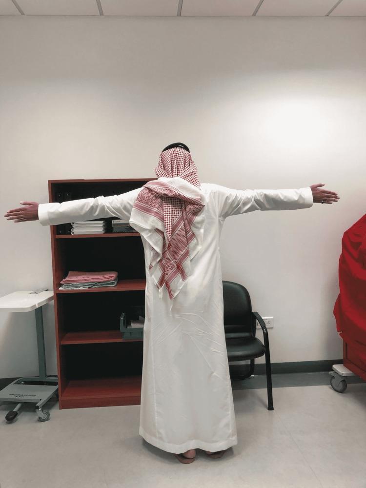
Clinical photograph of a patient viewed from behind, demonstrating Marfanoid habitus. The subject is standing with arms abducted to 90 degrees, highlighting a disproportionately long arm span in relation to total height. Key anatomical features include tall stature (dolichostenomelia) and long upper extremities, which are hallmarks of connective tissue disorders such as Marfan syndrome. The patient is wearing traditional Middle Eastern attire, including a white thobe and a red-and-white checkered ghutrah. This visual serves to illustrate the clinical assessment of body proportions and skeletal manifestations in medical genetics and internal medicine. The image emphasizes the increased arm-span-to-height ratio, an important clinical diagnostic criterion for identifying tall stature syndromes.
Noonan syndrome facial features hypertelorism ptosis child
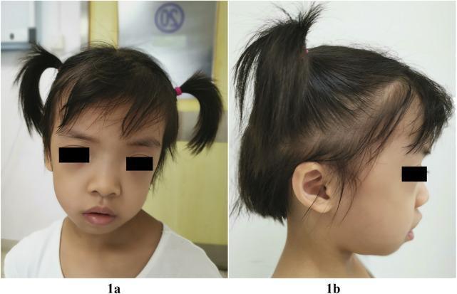
Clinical photograph of a 5-year-old child presenting with dysmorphic facial features characteristic of Noonan syndrome-like disorder with loose anagen hair (NSLAH) associated with a SHOC2 variant. Panel 1a (frontal view) demonstrates macrocephaly, a broad square forehead with frontal bossing, hypertelorism, and downslanting palpebral fissures. A unilateral left-sided ptosis and long eyelashes are also visible. Panel 1b (lateral view) illustrates low-set, posteriorly angulated ears with overfolded pinnae. Both views highlight hair anomalies, specifically sparse, thin, and slow-growing scalp hair with a high frontal hairline. The educational focus is on the recognition of the phenotypic manifestations of RASopathies, aiding in the clinical differentiation of syndromic macrocephaly and ectodermal dysplasia-like features.
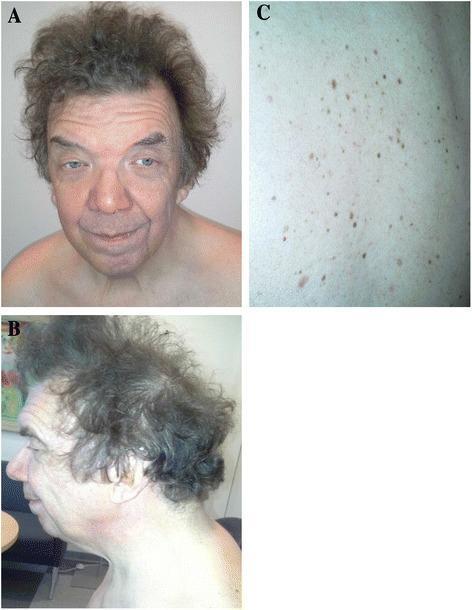
This composite of three clinical photographs displays the phenotypic manifestations of Noonan Syndrome in an adult male. Panel A (frontal view) and Panel B (lateral profile) demonstrate characteristic dysmorphic facial features, including hypertelorism, down-slanting palpebral fissures, and bilateral ptosis (more pronounced on the left). The individual exhibits a broad forehead, a short, widened neck with a low posterior hairline, and distinctively curly, coarse hair. Panel C provides a close-up view of the skin on the back, showing a high density of multiple lentigines. These are small, discrete, pigmented macules ranging in color from light to dark brown, appearing as a scattered distribution of 'freckle-like' lesions. The images collectively illustrate the clinical presentation of a RASopathy, specifically associated with an NRAS mutation, highlighting the multi-systemic involvement of craniofacial morphology and cutaneous pigmentation.
Williams syndrome elfin facies periorbital fullness child
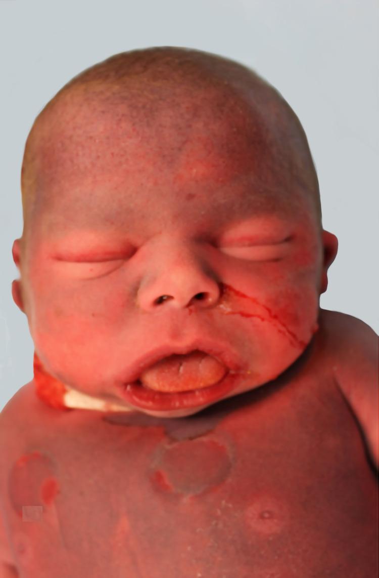
This clinical photograph displays the facial and upper torso characteristics of a neonate with Williams Syndrome (7q11.23 deletion). The image illustrates classic 'elfin' facies, including periorbital puffiness (soft tissue fullness around the eyes), a short nose with a depressed nasal bridge and anteverted nares, and midface fullness. The mouth is wide with thick, prominent lips and a slightly protruding, hypertrophied tongue. Additional findings include macrocephaly and a shortened neck. The skin exhibits a generalized reddish-purple hue, likely post-mortem or related to systemic congestion, with visible circular lesions on the upper chest. Clinical context suggests these features are associated with multisystemic manifestations such as supravalvular aortic stenosis. The photograph serves as an educational resource for identifying dysmorphic features in genetic syndromes and neonatal pathology.
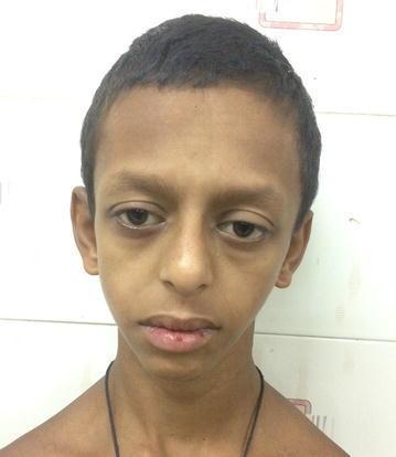
Clinical photograph showing the facial features of a pediatric patient exhibiting 'elfin facies,' a dysmorphic appearance characteristic of Williams syndrome. Key visible features include a broad forehead and prominent, low-set ears. The midface is characterized by a flat nasal bridge with a bulbous nasal tip and periorbital puffiness (soft tissue fullness around the eyes). The mouth shows a long philtrum and a wide, prominent mouth with a relatively thin upper lip. These phenotypic characteristics are associated with a 7q11.23 microdeletion. The image serves as an educational reference for medical genetics and pediatrics to identify the clinical manifestations of this chromosomal disorder, which often correlates with systemic findings such as supravalvular aortic stenosis and hypercalcemia.
Treacher Collins syndrome malar hypoplasia coloboma micrognathia
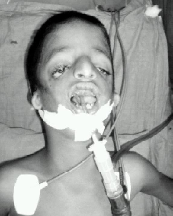
This clinical photograph shows a pediatric patient with craniofacial features characteristic of Treacher Collins syndrome (mandibulofacial dysostosis), specifically demonstrating downsloping palpebral fissures, coloboma of the lower eyelids, and hypoplasia of the malar and mandibular regions. The patient is undergoing airway management, with a size 4 uncuffed RAE (Ring-Adair-Elwyn) endotracheal tube in situ. The tube is properly secured using adhesive white tape across the mandible and maxilla. Connected to the endotracheal tube is a standard ventilation circuit adapter. Additional clinical monitoring is visible, including an ECG electrode placed on the upper left chest wall. This image serves as a clinical example of a 'difficult airway' scenario due to micrognathia and retrognathia in a patient scheduled for cleft palate repair. The educational focus is on the anatomical challenges of intubation in syndromic patients and the application of specialized endotracheal tubes to maintain a clear surgical field.
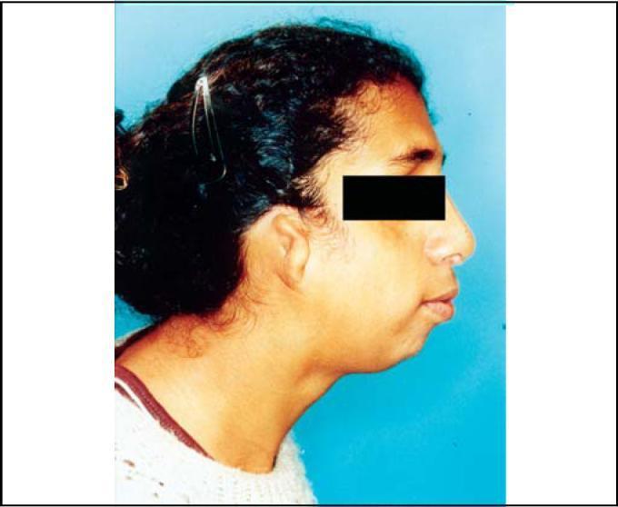
This clinical photograph shows a right-side profile view of an individual demonstrating the craniofacial manifestations of Treacher Collins Syndrome (mandibulofacial dysostosis). Key visible features include significant malar hypoplasia (underdevelopment of the zygomatic arch), which contributes to a flattened midface appearance. Marked mandibular hypoplasia (micrognathia) is evident, resulting in a receding chin and an altered facial profile. The external ear exhibits malformation of the auricular pinna (microtia), and while the eyes are partially obscured for privacy, the visible periocular region suggests an antimongoloid (downward) slanting of the palpebral fissures. These combined features are classic phenotypic markers of TCOF1, POLR1C, or POLR1D gene mutations. The photograph serves as an educational resource for identifying autosomal dominant disorders affecting the first and second branchial arch development, highlighting typical facial dysmorphism and associated skeletal abnormalities.
Pierre Robin sequence micrognathia cleft palate glossoptosis infant
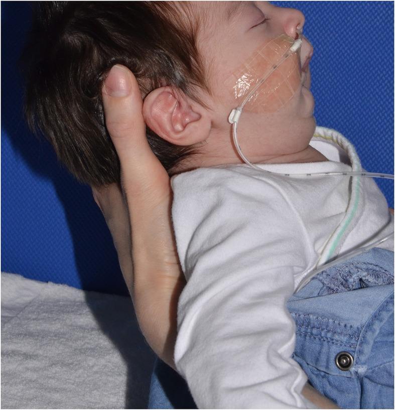
This clinical photograph shows a lateral (profile) view of an infant held in a half-sitting position, illustrating characteristic craniofacial features of Pierre Robin sequence (PRS). The primary finding is significant micrognathia and retrognathia, evidenced by a posteriorly displaced mandible. This results in the lower lip appearing recessed relative to the upper lip, a key clinical marker for assessing the severity of mandibular hypoplasia. A clear nasogastric or feeding tube is visible, entering through the nostril and secured to the right cheek with a transparent adhesive patch; numerical markings on the tube are visible, likely indicating insertion depth. The image demonstrates the neonatal presentation of PRS, which is typically characterized by the triad of micrognathia, glossoptosis, and often a cleft palate, potentially leading to airway obstruction and feeding difficulties. This visual is used in pediatrics and neonatology to teach physical examination techniques and the grading of mandibular recession in craniofacial anomalies.
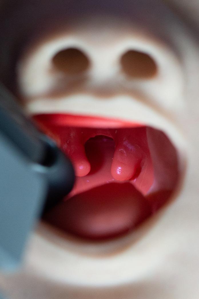
A clinical photograph of a specialized airway manikin representing the oropharyngeal anatomy of an infant with Pierre Robin Sequence (PRS). The image provides a direct view into the oral cavity, highlighting several key congenital anomalies. Centrally, a significant midline cleft palate is visible, characterized by the failure of the palatal shelves to fuse. Suspended from the margins of the soft palate is a prominent bifid uvula, which appears as two distinct, fleshy lobes. The external facial features of the manikin also demonstrate micrognathia (a small, recessed mandible), which is a hallmark of PRS and contributes to glossoptosis (posterior displacement of the tongue), potentially complicating airway management. This educational tool is used to simulate difficult pediatric intubation scenarios, allowing clinicians to practice identifying and navigating anatomical obstructions related to craniofacial microsomia and palatal defects. The image is highly relevant for training in neonatology, anesthesiology, and pediatric otolaryngology.
Angelman syndrome happy puppet syndrome child features
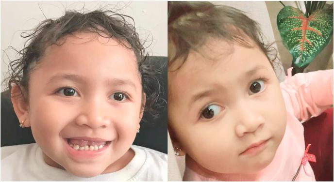
Two clinical photographs show the facial features of a 3-year-old child with Angelman Syndrome. Key diagnostic features include a characteristically wide mouth and an exuberant, happy facial expression. Dental examination reveals widely spaced teeth (diastema). Ocular findings demonstrate divergent strabismus, visible as a misalignment of the visual axes. The child exhibits generalized hypopigmentation of the skin and curly hair, which appears lighter than expected for her genetic background. Additionally, the head shape is suggestive of microcephaly. These visual findings represent the typical craniofacial phenotype of Angelman Syndrome, often associated with intellectual disability and ataxia. The images serve as an educational example of the neurodevelopmental and dermatological manifestations of this genetic condition, highlighting the importance of recognizing the 'happy puppet' disposition and associated dysmorphic features in pediatric clinical diagnosis.
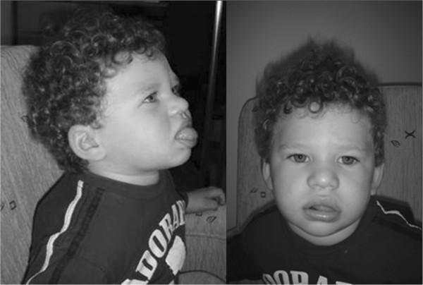
This clinical photograph consists of a lateral profile (left) and a frontal view (right) of a 16-month-old child exhibiting dysmorphic features characteristic of Angelman syndrome. The patient displays a wide nasal bridge, low-set ears, and a wide mouth with notably thick lips. A key clinical finding illustrated is macroglossia with a protruding tongue (tongue thrust), visible in both views but most prominent in the profile. The facial structure is rounded, and the patient has distinctive curly hair. This image serves as a clinical reference for the phenotypic manifestations of Angelman syndrome (AS) resulting from paternal uniparental disomy (UPD) of chromosome 15. The photograph is an educational resource for medical students and geneticists to identify craniofacial markers of genomic imprinting disorders, emphasizing the characteristic 'happy puppet' facies often associated with the condition.
Prader-Willi syndrome almond shaped eyes obesity child
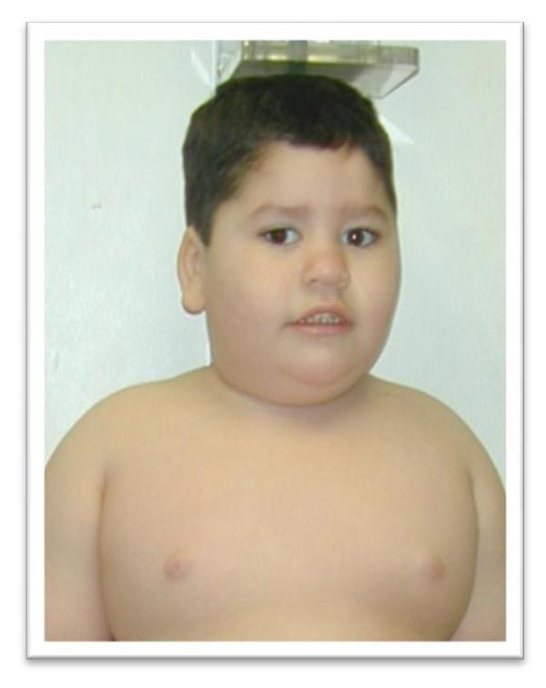
A clinical photograph of a young child illustrating the characteristic physical phenotype of Prader-Willi Syndrome (PWS). The image displays typical facial dysmorphism, including almond-shaped eyes, a narrow bifrontal diameter, and a downturned angle of the mouth with a thin upper lip. The subject exhibits a round facial contour and a notably short neck. Below the head, the photograph demonstrates a morbidly obese body habitus, characterized by excess adipose tissue distribution in the trunk and chest areas. This visual serves as an educational reference for the diagnostic clinical features of PWS in the pediatric population, emphasizing the transition from neonatal hypotonia to the childhood phase of hyperphagia and subsequent obesity. Key concepts shown include genetic syndrome recognition and characteristic craniofacial morphology.
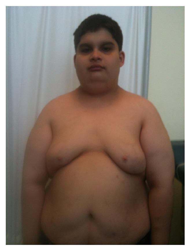
This clinical photograph shows the torso and head of a male teenager demonstrating the characteristic physical phenotype of Prader-Willi syndrome (PWS). Facial features include a narrow bifrontal diameter (narrow forehead), almond-shaped eyes with slightly upslanted palpebral fissures, and a thin upper lip. The body composition is characterized by generalized obesity with significant central (truncal) adiposity, notably involving the abdominal area and chest (gynecomastia or pseudogynecomastia). The skin appears fair. This visual presentation is educationally significant for medical students and clinicians as a textbook illustration of the dysmorphic features and metabolic manifestations (morbid obesity) associated with chromosomal deletions or uniparental disomy at the 15q11-q13 locus. Key educational concepts illustrated include phenotypic recognition of genetic obesity syndromes and dysmorphology assessment.
Cri du chat syndrome cat cry microcephaly hypertelorism infant
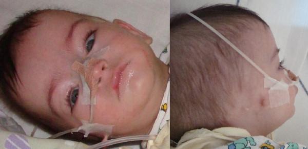
This clinical photograph displays a 5-month-old infant in two views (frontal and profile) illustrating dysmorphic facial features associated with a complex chromosome 5 rearrangement (Cri-du-Chat syndrome variant). In the frontal view, there is evidence of hypertelorism (increased interpupillary distance), convergent strabismus, and notably long eyelashes. The skull shape exhibits dolichocephaly (elongation) and relative macrocephaly. The lower face shows microretrognathia, characterized by a small, receding mandible. The lateral profile photograph highlights significant auricular abnormalities on the right side, including ear agenesis (anotia) with two visible preauricular skin tags. Medical equipment, including a nasogastric tube and adhesive dressings on the nose and cheek, is present, indicating ongoing clinical care for feeding difficulties or airway management. These visible phenotypic markers are critical for the clinical diagnosis of genetic syndromes involving the 5p chromosomal region.
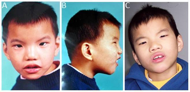
Clinical photographs of a male pediatric patient with 5p deletion syndrome (Cri-du-chat syndrome) at age 4.5 years (panels A and B) and 11.75 years (panel C). The frontal views (A, C) demonstrate characteristic dysmorphic features including a round facial profile, microcephaly, and pronounced hypertelorism (widely spaced eyes). Notable ocular findings include an upslanting palpebral fissure (external canthus upslope) and esotropia. Midface features include a broad, large nasal bridge and a short philtrum. The profile view (B) highlights micrognathia (a small, receding jaw), midfacial hypoplasia, and low-set ears. Additionally, the images show evidence of plagiocephaly (asymmetric skull distortion). These visual findings are consistent with the phenotypic manifestations of interstitial deletion of chromosome 5p13.3p15.3, used here for medical education on genetic dysmorphology and developmental delay syndromes.
👶 Paediatric Facies & Associated Syndromes
Visual Learning Guide — "See It, Know It"
How to use this guide: Each syndrome has a "FACE CODE" — a short visual memory hook linking the face features to the diagnosis. Learn the image → learn the code → know the syndrome.
1. 🧬 DOWN SYNDROME (Trisomy 21)
🔑 FACE CODE: "FLAT FACE, HAPPY EYES, TONGUE OUT"
| Feature | Detail |
|---|---|
| Genetics | Trisomy 21 (95% non-disjunction, 4% Robertsonian translocation, 1% mosaic) |
| Upslanting palpebral fissures | Eyes slant upward (opposite of Noonan/Treacher Collins) |
| Epicanthic folds | Skin fold at inner corner of eyes |
| Brushfield spots | White/grey spots on iris periphery |
| Flat nasal bridge | Depressed, broad nasal root |
| Protruding tongue | Macroglossia + small mouth (relative) |
| Single palmar crease | "Simian crease" — single transverse palmar crease |
| Sandal gap | Wide gap between 1st and 2nd toes |
| Low-set ears | Small, dysplastic |
| Brachycephaly | Short, broad skull |
| Hypotonia | "Floppy baby" at birth |
- Heart: AVSD / VSD / PDA (40-50%)
- GI: Duodenal atresia ("double bubble"), Hirschsprung's
- Eyes: Brushfield spots, cataracts, nystagmus
- Hypothyroidism
- Atlanto-axial instability
- Intellectual disability (mild-moderate)
- Leukaemia risk (20x ALL, 50x AML M7)
🧠 Memory trick: "Down = Depressed bridge, Downward fold inner eye, Double bubble duodenum, Duodenal atresia"
2. 🎀 TURNER SYNDROME (45,X)
🔑 FACE CODE: "SHORT GIRL, WEB NECK, WIDE CHEST"
| Feature | Detail |
|---|---|
| Genetics | 45,X (or mosaics 45X/46XX) |
| Short stature | Most consistent feature |
| Webbed neck (pterygium colli) | Excess skin folds on neck |
| Low posterior hairline | Hair extends down the back of neck |
| Shield chest | Broad chest, widely spaced nipples |
| Cubitus valgus | Increased carrying angle at elbows |
| Lymphoedema | Hands and feet at birth |
| Low-set ears | Slightly low and posteriorly rotated |
- Heart: Bicuspid aortic valve, coarctation of aorta (most common cardiac defect)
- Kidneys: Horseshoe kidney (most common renal anomaly)
- Primary amenorrhoea, streak gonads, infertility
- Normal intelligence (except visuospatial tasks)
- No virilisation
🧠 Memory trick: "Turner = Tiny height, Thick neck webbing, Two-horseshoe kidney, Testosterone = none"
3. ♥️ NOONAN SYNDROME
🔑 FACE CODE: "MALE TURNER — WEBBED NECK + DOWN-SLANTING EYES"
| Feature | Detail |
|---|---|
| Genetics | PTPN11 mutation (50%), also SOS1, RAF1, KRAS — RASopathy |
| Hypertelorism | Wide-set eyes |
| Down-slanting palpebral fissures | Eyes slant DOWNWARD (opposite of Down syndrome) |
| Ptosis | Drooping eyelids |
| Webbed neck | Like Turner, but occurs in BOTH sexes |
| Low posterior hairline | Like Turner |
| Low-set ears | Posteriorly rotated, thick helices |
| Short stature | Common |
- Heart: Pulmonary stenosis (most common, 50-80%) — key differentiator from Turner (coarctation)
- HCM (hypertrophic cardiomyopathy)
- Cryptorchidism in males (fertility affected)
- Bleeding tendency (factor XI deficiency, platelet abnormalities)
- Normal or mild ID
🧠 Memory trick: "Noonan = Normal chromosomes (46XX or 46XY), Pulmonary stenosis (vs Coarctation in Turner), PTPNN11 gene"Turner vs Noonan:
Turner Noonan Sex Female only Both sexes Karyotype 45,X Normal (46XX/46XY) Heart Coarctation Pulmonary stenosis Gene Chromosomal PTPN11
4. 🧝 WILLIAMS SYNDROME (Elfin Facies)
🔑 FACE CODE: "ELF FACE — WIDE MOUTH, PUFFY EYES, BUTTON NOSE"
| Feature | Detail |
|---|---|
| Genetics | 7q11.23 deletion (includes elastin gene ELN) |
| "Elfin" facies | Classic description |
| Periorbital fullness | Puffy skin around eyes |
| Flat nasal bridge + anteverted nares | Upturned nose ("button nose") |
| Wide mouth with thick lips | "Cupid's bow" appearance |
| Long philtrum | Lengthened groove between nose and lip |
| Dental anomalies | Widely spaced, small teeth |
| Stellate iris pattern | Star-like appearance of iris |
- Heart: Supravalvular aortic stenosis (SVAS) — most characteristic
- Hypercalcaemia in infancy
- Intellectual disability (mild-moderate)
- "Cocktail party personality" — very sociable, friendly, overly talkative
- Hypersensitivity to sound (hyperacusis)
🧠 Memory trick: "Williams = Wide mouth elf, William Tell's aorta (SVAS), Warm personality"
5. 🦋 MARFAN SYNDROME
🔑 FACE CODE: "TALL THIN SPIDER FINGERS, LONG FACE"
| Feature | Detail |
|---|---|
| Genetics | FBN1 gene (fibrillin-1), chromosome 15; autosomal dominant |
| Dolichocephaly | Long, narrow skull and face |
| High arched palate | "Gothic" arch palate |
| Deep-set eyes | Enophthalmos |
| Arachnodactyly | Long spider fingers |
| Tall stature | Disproportionately long limbs (dolichostenomelia) |
| Arm span > height | Key clinical sign |
| Pectus excavatum/carinatum | Chest wall deformity |
| Scoliosis / kyphosis | Spinal curvature |
| Thumb sign (Steinberg) | Thumb protrudes beyond ulnar border of closed fist |
| Wrist sign (Walker-Murdoch) | 1st and 5th finger overlap around opposite wrist |
- Eyes: Ectopia lentis (upward lens dislocation — vs. down in homocystinuria)
- Aorta: Aortic root dilatation → aortic regurgitation, aortic dissection
- Mitral valve prolapse
- Pneumothorax (tall thin build)
🧠 Memory trick: "Marfan = Microfibrils broken, Mitral prolapse, Myopia, aortic dilatation, Measure arm span"Marfan vs Homocystinuria lens dislocation:
- Marfan → lens dislocates UP
- Homocystinuria → lens dislocates DOWN
6. 😢 TREACHER COLLINS SYNDROME (Mandibulofacial Dysostosis)
🔑 FACE CODE: "DIAGONAL FACE — EVERYTHING GOES DOWN AND BACK"
| Feature | Detail |
|---|---|
| Genetics | TCOF1 gene; autosomal dominant |
| Downslanting palpebral fissures | Eyes slope downward laterally |
| Coloboma of lower eyelids | Notch or absence of lower lid lashes |
| Malar hypoplasia | Underdeveloped cheekbones (zygomatic arch) |
| Micrognathia | Very small chin/jaw |
| Microtia | Small, malformed ears |
| Conductive hearing loss | External/middle ear anomalies |
| Absent/hypoplastic zygomatic arch | "Sunken" cheeks |
| Cleft palate | May be present |
- Intelligence is normal
- Major concern: airway management (difficult intubation)
- Bilateral and symmetric (unlike hemifacial microsomia)
🧠 Memory trick: "Treacher Collins = Two Cs — Cheekbones absent, Coloboma lower lid; diagonal face = 'downhill' features"
7. 👶 PIERRE ROBIN SEQUENCE
🔑 FACE CODE: "TINY JAW → TONGUE FALLS BACK → CLEFT PALATE"
| Feature | Detail |
|---|---|
| Sequence (not syndrome) | One anomaly causes a cascade |
| Micrognathia | Primary defect — very small mandible |
| Glossoptosis | Tongue displaced backward, causing airway obstruction |
| Cleft palate | U-shaped cleft (tongue prevents palatal shelf fusion) |
Micrognathia → Glossoptosis → U-shaped cleft palate
     ↑
PRIMARY DEFECT
- Prone positioning (tongue falls forward)
- Nasopharyngeal airway
- Surgery (jaw distraction osteogenesis, tongue-lip adhesion)
🧠 Memory trick: "Pierre Robin = Poor Retracted jaw → tongue Rolls back → Roof of mouth clefts (U-shaped)"Pierre Robin vs Treacher Collins:
Pierre Robin Treacher Collins Type Sequence Syndrome Ear Normal Microtia Cheekbones Normal Hypoplastic Cleft U-shaped May be present
8. 😊 ANGELMAN SYNDROME ("Happy Puppet")
🔑 FACE CODE: "HAPPY, WIDE MOUTH, BLONDE, JERKY MOVEMENTS"

| Feature | Detail |
|---|---|
| Genetics | Chromosome 15q11-13 deletion — MATERNAL copy (UBE3A gene) |
| Imprinting | Only maternal allele expressed; paternal = silent |
| Happy, frequent laughter | "Happy puppet" — inappropriate, frequent laughter |
| Wide mouth, widely spaced teeth | Characteristic grin |
| Microcephaly | Small head circumference |
| Hypopigmentation | Fair skin and hair (melanin-related gene also deleted) |
| Strabismus | Common ocular finding |
| Ataxic gait | Jerky, "puppet-like" walking movements |
- Severe intellectual disability
- No or minimal speech (receptive > expressive)
- Seizures (EEG: large amplitude slow spike-and-wave)
- EEG is highly abnormal even before clinical seizures
🧠 Memory trick: "Angelman = Absence of speech, Abnormal EEG, Ataxia, always Amused"Angelman vs Prader-Willi — SAME REGION, DIFFERENT PARENT:
Angelman Prader-Willi Chromosome 15q11-13 15q11-13 Deleted parent MATERNAL PATERNAL Body Thin, ataxic Obese, hypotonic Behaviour Happy, laughter Obsessive, food-seeking Speech Absent Present but impaired Seizures Common Rare
9. 🍔 PRADER-WILLI SYNDROME
🔑 FACE CODE: "ALMOND EYES, FAT BODY, OBSESSED WITH FOOD"
| Feature | Detail |
|---|---|
| Genetics | Chromosome 15q11-13 deletion — PATERNAL copy |
| Almond-shaped eyes | Upslanting, narrow |
| Narrow bifrontal diameter | Narrow forehead |
| Downturned mouth, thin upper lip | Characteristic expression |
| Short stature | Growth hormone deficiency |
| Hypotonia at birth | "Floppy" infant; poor suck |
| Hyperphagia | Insatiable appetite → morbid obesity in childhood |
| Hypogonadism | Undescended testes, small genitalia |
| Fair skin | Relative hypopigmentation |
Phase 1 (Infancy): Severe hypotonia + poor feeding + failure to thrive
             ↓
Phase 2 (2-6 years): Hyperphagia begins → rapid weight gain → obesity
- Type 2 diabetes (obesity-related)
- Sleep apnoea
- Scoliosis
- Obsessive-compulsive behaviours
- Mild intellectual disability
- Growth hormone deficiency → GH therapy is first-line treatment
🧠 Memory trick: "Prader-Willi = Paternal deletion, Poor suck then Pig-like eating, Penis small"
10. 😿 CRI-DU-CHAT SYNDROME (5p- Syndrome)
🔑 FACE CODE: "CAT CRY BABY — WIDE EYES, SMALL HEAD, HIGH-PITCHED CRY"
| Feature | Detail |
|---|---|
| Genetics | Deletion of chromosome 5p (short arm) |
| High-pitched cat-like cry | Due to laryngeal/neurological abnormality — pathognomonic in infancy |
| Microcephaly | Small head circumference |
| Hypertelorism | Widely spaced eyes |
| Downward slanting palpebral fissures | Eyes slope down (like Treacher Collins) |
| Low birth weight | Intrauterine growth restriction |
| Low-set ears | Posteriorly rotated |
| Micrognathia | Small chin |
| Broad, flat nasal bridge |
- Severe intellectual disability
- Hypotonia
- Feeding difficulties
- Heart defects (VSD, ASD in some)
- Short stature
🧠 Memory trick: "Cri-du-Chat = Chromosome 5 deletion, Cat-like Cry, Compact small head"
📊 Master Comparison Table — All Syndromes at a Glance
| Syndrome | Key Facial Feature | Genetics | Pathognomonic Fact | Heart |
|---|---|---|---|---|
| Down | Upslanting eyes, flat bridge, protruding tongue | Trisomy 21 | Simian crease, Brushfield spots | AVSD |
| Turner | Webbed neck, low hairline | 45,X | Horseshoe kidney, coarctation | Coarctation of aorta |
| Noonan | Downslanting eyes, ptosis, webbed neck | PTPN11 mutation | Normal karyotype in phenotypic Turner | Pulmonary stenosis |
| Williams | Elfin face, periorbital puffiness | 7q11.23 deletion | "Cocktail party" personality | SVAS |
| Marfan | Long face, high arch palate | FBN1 (chr 15) | Lens up, aorta dilatation | Aortic root dilatation |
| Treacher Collins | Downslanting, malar hypoplasia, coloboma | TCOF1 | Normal intelligence | None specific |
| Pierre Robin | Micrognathia | Sequence | U-shaped cleft palate | None specific |
| Angelman | Wide grin, happy expression | Chr 15 maternal | No speech, seizures, EEG abnormal | None |
| Prader-Willi | Almond eyes, obesity | Chr 15 paternal | Hyperphagia phase 2 | None specific |
| Cri-du-Chat | Hypertelorism, microcephaly | 5p deletion | Cat-like cry | VSD/ASD |
🔁 Eyes Direction Quick Memory
Eyes Slant UPWARD (↗):
  ✅ Down syndrome
  ✅ Prader-Willi

Eyes Slant DOWNWARD (↘):
  ✅ Noonan syndrome
  ✅ Treacher Collins
  ✅ Cri-du-Chat
🧠 Quick Mnemonics Summary
| Syndrome | Mnemonic |
|---|---|
| Down | FLAT — Flat face, Low-set ears, Almond fold (epicanthic), Tongue out |
| Turner | COWL — Coarctation, Ovarian failure, Webbed neck, Low hairline |
| Noonan | HELP — Hypertelorism, Ears low-set, Low hairline, Pulmonary stenosis |
| Williams | SELVES — Stellate iris, Elfin face, Lively personality, SVAS, Elastin gene, hypercalcaemia |
| Marfan | MALES — Mitral prolapse, Aorta dilated, Lens up, Ectopia lentis, Scoliosis |
| Angelman | ASHES — Ataxia, Speech absent, Happy always, EEG abnormal, Seizures |
| Prader-Willi | PHAT — Paternal deletion, Hyperphagia, Almond eyes, Tight (small) genitalia |
| Cri-du-Chat | CHIME — Cry cat-like, Hypertelorism, Intellectual disability, Microcephaly, 5p dEleted |
⚡ Top Quiz Facts
- Upslanting palpebral fissures = Down syndrome; Downslanting = Noonan/Treacher Collins
- Webbed neck occurs in both Turner AND Noonan — differentiate by karyotype and heart defect
- Coarctation = Turner; Pulmonary stenosis = Noonan; SVAS = Williams; AVSD = Down
- Chromosome 15q11 deletion: Maternal = Angelman; Paternal = Prader-Willi
- Pierre Robin is a SEQUENCE (cascade), not a syndrome
- FBN1 = Marfan; lens dislocates up (homocystinuria = down)
- Cat-like cry at birth = Cri-du-Chat (5p deletion)
- Williams syndrome "elfin facies" + very friendly personality + supravalvular aortic stenosis
- Treacher Collins = normal intelligence despite severe craniofacial abnormalities
- EEG is grossly abnormal in Angelman syndrome even before clinical seizures
Generate x ray pediatrics quiz with options
pediatric chest X-ray hyaline membrane disease respiratory distress syndrome newborn ground glass
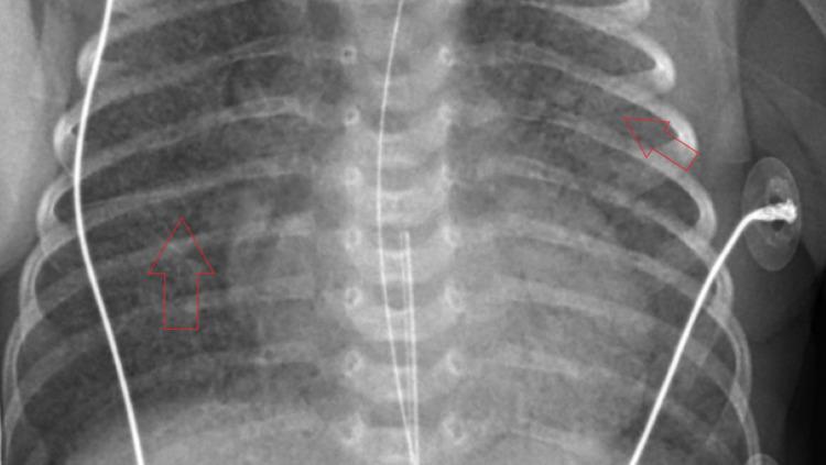
Anteroposterior (AP) chest X-ray of a neonate demonstrating classic radiographic features of neonatal respiratory distress syndrome, also known as hyaline membrane disease. The image shows diffuse, bilateral, and symmetric reticulogranular (ground-glass) opacities throughout both lung fields. These granular infiltrates, highlighted by red arrows, represent widespread alveolar atelectasis and result in reduced lung volumes and obscured vascular markings. The cardiomediastinal silhouette is visible but slightly blurred by the surrounding parenchymal opacification. Several medical devices are present, including an endotracheal tube, an umbilical catheter, and external leads, consistent with a patient receiving intensive care in a neonatal NICU setting. This imaging is characteristic of surfactant deficiency in premature infants, where collapsed alveoli create a fine, stippled appearance across the thorax.
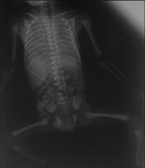
**Imaging Modality:** Anteroposterior (AP) conventional radiograph (X-ray). **Anatomical Region:** Neonatal chest and abdomen. **Observed Pathology:** The image demonstrates classic findings of Respiratory Distress Syndrome (RDS), also known as Hyaline Membrane Disease. The lung fields exhibit diffuse, bilateral, and symmetric "ground-glass" opacification with a fine reticulogranular pattern. There is a notable loss of lung volume (low lung volumes). **Characteristic Visual Features:** * **Parenchymal Pattern:** Diffuse reticulogranular densities (micranodular mottling) throughout both lungs. * **Air Bronchograms:** Prominent lucent tubular structures representing air-filled bronchi outlined by non-aerated alveoli, extending peripherally. * **Cardiac Silhouette:** The borders of the heart and diaphragm are obscured (silhouette sign) due to the extensive alveolar collapse and opacification. * **Support Devices:** An endotracheal tube and an umbilical venous catheter are visible, consistent with neonatal intensive care management. **Differentiating Features:** The combination of low lung volumes, bell-shaped thorax, and diffuse ground-glass appearance distinguishes this from neonatal pneumonia or transient tachypnea of the newborn.
pediatric X-ray intussusception abdominal bowel obstruction child
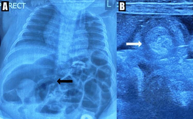
This composite diagnostic image demonstrates a case of pediatric small bowel obstruction caused by intussusception. Panel A shows an anteroposterior (AP) erect abdominal X-ray of a child. It reveals multiple dilated, air-filled loops of small bowel throughout the abdomen, indicative of a distal obstruction. A black arrow points to a particularly distended segment in the right upper quadrant. Panel B shows a corresponding abdominal ultrasound in transverse section. A white arrow highlights the 'target' or 'donut' sign, which is a pathognomonic finding for intussusception. This sign appears as a concentric, multilayered mass representing the intussusceptum within the intussuscipiens. These imaging modalities together illustrate the classic radiological workup for pediatric abdominal emergencies, showcasing the bowel gas patterns seen on radiography and the specific sonographic features used to confirm the diagnosis of intussusception.
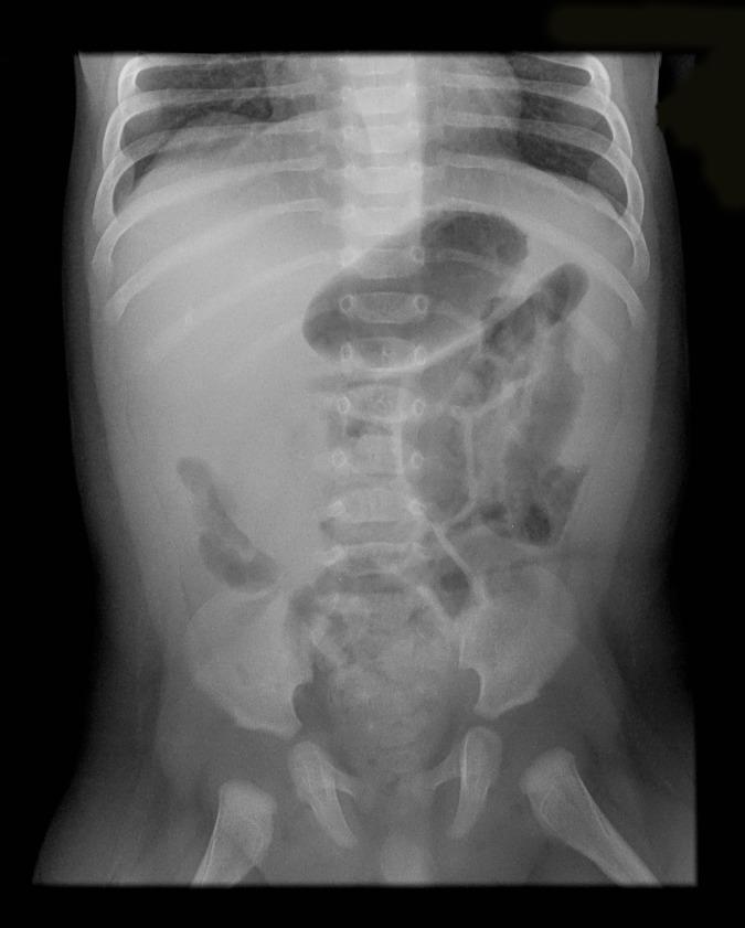
This diagnostic image is an anteroposterior (AP) supine abdominal radiograph of a pediatric patient. The X-ray demonstrates classic signs of a bowel obstruction, likely secondary to intussusception. Visible features include several dilated, gas-filled proximal small bowel loops concentrated in the upper and central abdomen. There is a notable paucity of distal bowel gas in the lower abdominal quadrants and pelvis. In the right hypochondrium, a subtle soft tissue mass effect is present, which can represent the intussusceptum. Bony structures including the lower ribcage, spine, iliac wings, and proximal femurs appear normal for the patient's developmental age. This clinical photograph serves as a primary educational example for identifying small bowel obstruction patterns and localizing pathology in pediatric emergency radiology.
pediatric epiglottitis thumbprint sign X-ray lateral neck child
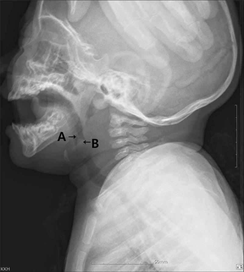
This lateral soft tissue X-ray of a pediatric neck demonstrates a significant upper airway obstruction caused by a mass at the level of the oropharynx. Label 'A' points to an elongated uvula, while label 'B' indicates a well-defined, radiopaque mass identified as a uvular epidermoid cyst. The mass occupies a substantial portion of the airway lumen, mimicking the 'thumbprint sign' typically associated with epiglottitis, although the epiglottis itself remains non-swollen. Secondary findings include diffuse thickening of the prevertebral soft tissues and anterior displacement of the airway column. This diagnostic image illustrates a rare cause of neonatal/infant respiratory distress, highlighting the importance of radiographic evaluation in pediatric airway management to differentiate between infectious etiologies like epiglottitis and mechanical obstructions such as benign keratinous cysts.
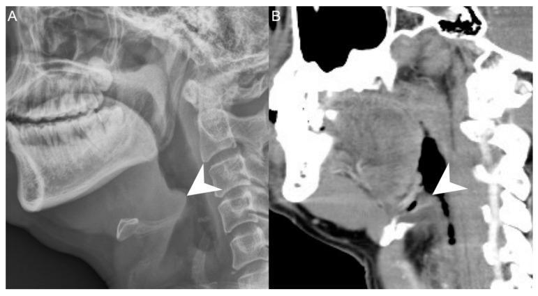
This dual-panel diagnostic image compares a lateral neck radiograph (A) with a sagittal computed tomography (CT) scan (B) of a patient with acute epiglottitis. In panel A, the lateral X-ray demonstrates a classic 'thumbprint sign,' where the arrowhead indicates a severely swollen, radiopaque epiglottis protruding into the airway. The surrounding soft tissues show decreased clarity compared to the dense cervical vertebrae. Panel B provides a sagittal CT reconstruction of the same anatomical region, offering superior soft tissue contrast and spatial resolution. The arrowhead in the CT scan highlights the thickened, edematous epiglottis and aryepiglottic folds, which significantly narrow the laryngeal inlet. The CT modality also better delineates the surrounding deep neck spaces, aiding in the assessment of concurrent deep neck infections. These images serve as a clinical comparison of imaging modalities used in diagnosing upper airway emergencies and illustrating the pathological morphology of epiglottic inflammation.
croup steeple sign subglottic narrowing X-ray child airway
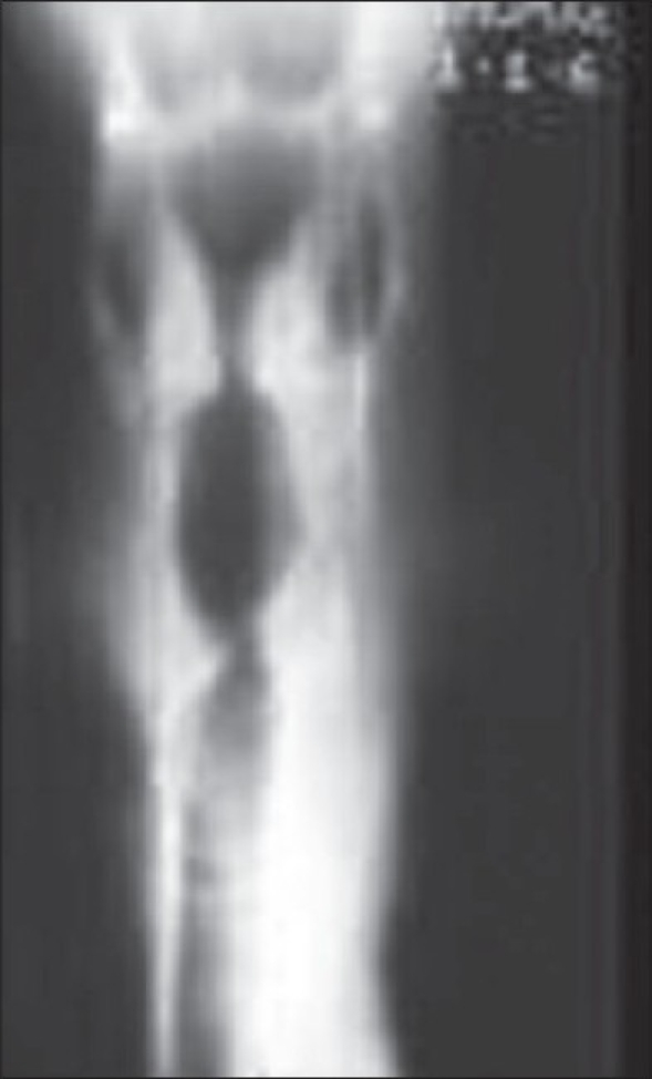
**Imaging Modality:** Plain radiograph (X-ray) **Anatomical Region:** Neck and upper airway (Anteroposterior view) **Observed Pathology:** The image demonstrates a characteristic narrowing of the subglottic airway, appearing as a tapered, inverted "V" or "church steeple" shape. **Characteristic Visual Features:** * **Steeple Sign:** Symmetric subglottic narrowing due to inflammatory edema in the subglottic region. * **Airway Patency:** Contrast between the radiolucent (dark) air-filled column and the surrounding radiopaque (light) soft tissue highlights the funnel-shaped tapering. * **Landmarks:** The narrowing begins below the level of the vocal cords and extends into the upper trachea. **Relevant Clinical Context:** This radiologic finding is classically associated with Croup (laryngotracheobronchitis), a common cause of upper airway obstruction in pediatric patients. **Key Diagnostic Features:** The symmetric, progressive tapering of the subglottic air column (Steeple Sign) distinguishes this condition from epiglottitis, which typically shows a "thumb sign" on lateral neck radiographs rather than subglottic narrowing on the anteroposterior view.
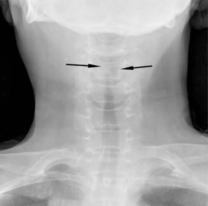
**Imaging Modality:** Anteroposterior (AP) plain film radiograph (X-ray). **Anatomical Region:** Soft tissue of the neck and upper respiratory tract. **Observed Pathology:** Subglottic narrowing of the airway column. **Characteristic Visual Features:** The image demonstrates a characteristic "steeple sign" (or "pencil point sign"), characterized by symmetrical, subglottic narrowing of the tracheal air column. This narrowing is caused by inflammatory soft tissue edema in the subglottic region. The proximal tracheal lumen is significantly constricted compared to the distal segment. **Key Diagnostic Features:** The tapering of the upper airway on an AP view is a classic radiologic hallmark of Croup (laryngotracheobronchitis). This feature helps differentiate the condition from epiglottitis, which typically presents with a "thumbprint sign" on a lateral neck radiograph. **Clinical Context:** The findings are highly suggestive of acute inflammatory airway obstruction in the subglottic space, typically seen in pediatric populations presenting with respiratory distress or stridor.
pediatric chest X-ray pneumonia lobar consolidation child
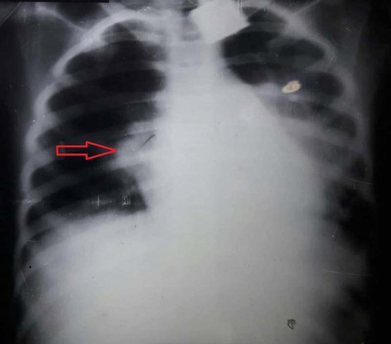
This diagnostic image is an anteroposterior (AP) chest X-ray of a pediatric patient demonstrating classic findings of lobar pneumonia. A prominent, dense, and homogenous opacification is localized to the right upper lobe, consistent with pulmonary consolidation. The opacification has a well-defined inferior border along the horizontal fissure, indicated by a red arrow, which is a hallmark of lobar involvement. Normal lung markings in this region are obscured, while the left lung field appears relatively clear and well-aerated. The cardiac silhouette and mediastinal structures appear within normal limits for this projection, with no significant shift or widening. The diaphragm and costophrenic angles are visible, though the right side is partially obscured by the adjacent consolidation. This image serves as an educational example of bacterial pneumonia presentation in a clinical setting, highlighting the radiographic appearance of alveolar space filling.
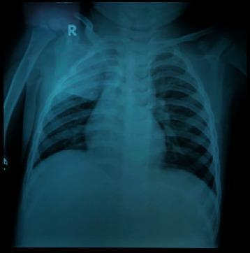
This diagnostic image is a posterior-anterior (PA) pediatric chest X-ray. The primary finding is a dense, homogeneous opacity located in the right upper lobe, characterized by a well-defined inferior border along the minor fissure, which is highly consistent with lobar consolidation. Bilateral perihilar and parenchymal spotting (reticulonodular opacities) are also visible, suggesting an associated bronchopneumonia component. The cardiac silhouette and mediastinal structures appear within normal limits for a pediatric patient, though the bony structures of the rib cage and vertebrae are visible with moderate clarity. This imaging demonstrates typical radiological signs of community-acquired or atypical pneumonia, such as right superior lobar pneumonia. Clinically, this serves as an educational example of differentiating focal lobar consolidation from generalized patchy bronchopneumonia patterns in a pediatric context.
rickets X-ray cupping fraying metaphysis child long bones
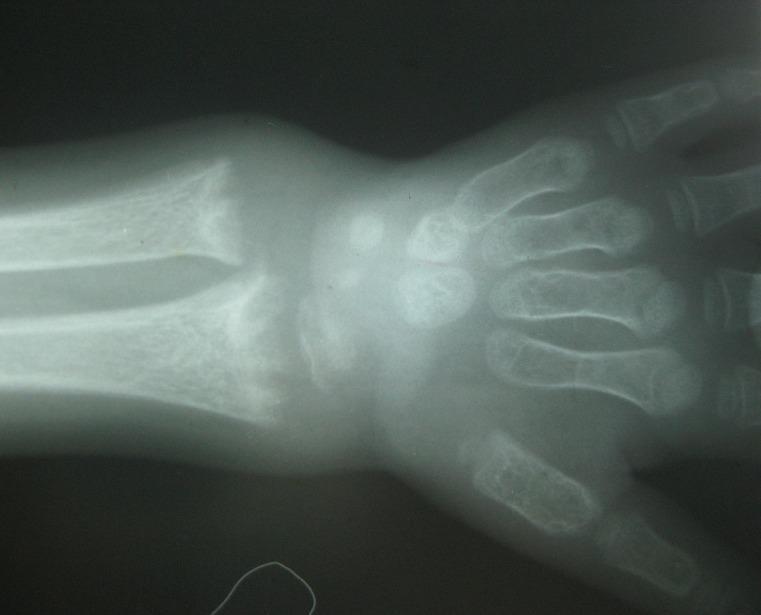
This diagnostic radiograph displays the wrist and hand of a pediatric patient, highlighting characteristic skeletal features of rickets. The imaging modality is conventional X-ray. The distal metaphyses of the radius and ulna exhibit significant pathological changes, including transverse widening (flaring), distinctive concave incurvation known as 'cupping,' and irregular, frayed margins referred to as 'fraying.' There is a visible increase in the radiolucent space between the metaphysis and the epiphysis, indicating growth plate widening. Additionally, the radiograph demonstrates generalized osteopenia or diffuse demineralization, characterized by thinned cortical bone and reduced trabecular density throughout the visualized long bones and metacarpals. These findings are clinically significant for vitamin D deficiency or other metabolic bone diseases affecting mineralized cartilage. This material is suitable for intermediate medical education focusing on pediatric orthopedics or endocrinology.
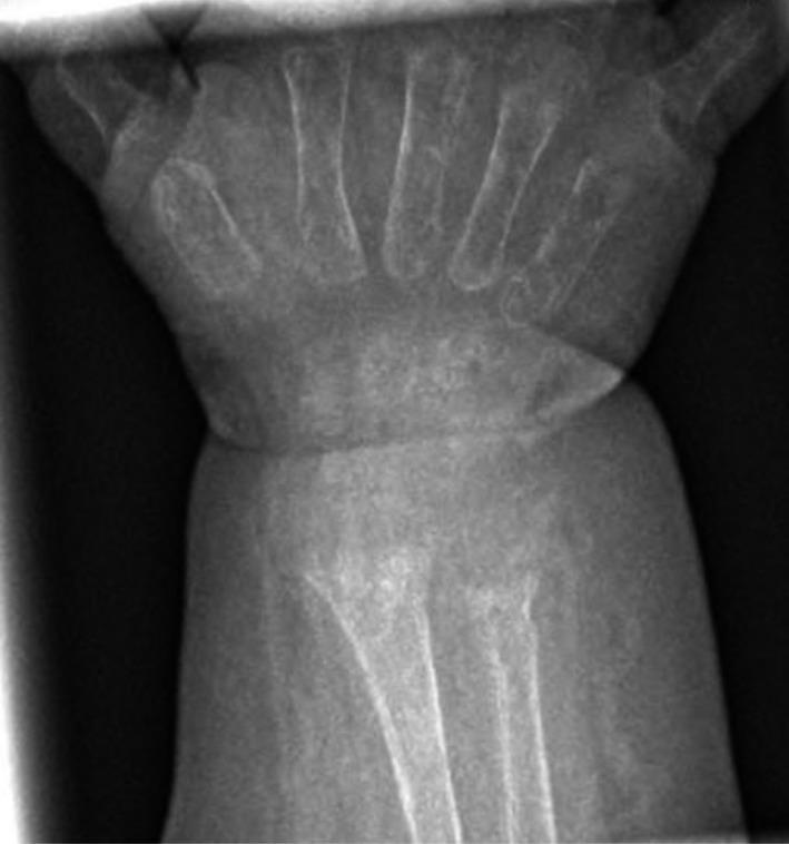
This diagnostic image is an anteroposterior (AP) x-ray radiograph of a pediatric wrist and hand. The imaging demonstrates hallmark signs of rickets, most notably at the distal radius and ulna. Key radiographic findings include generalized osteopenia, characterized by reduced bone mineral density, and significant metaphyseal irregularities. Specifically, the metaphyses of the radius and ulna show prominent 'cupping' (a concave deformity of the distal ends) and 'fraying' (a ragged, brush-like appearance of the mineralizing margin). The physis (growth plate) appears widened, and the demarcation between the metaphysis and the carpal region is ill-defined. These features are classic manifestations of impaired bone mineralization typical of nutritional or hypophosphatemic rickets. The image serves as an educational example of metabolic bone disease in pediatric patients, illustrating the structural alterations that occur in rapidly growing long bones during states of vitamin D deficiency or mineral imbalance.
congenital heart disease VSD ASD pediatric chest X-ray cardiomegaly
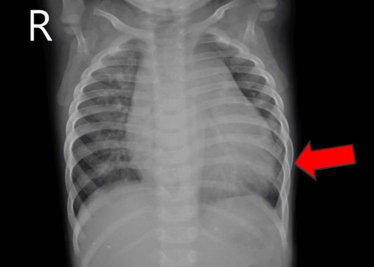
A frontal (anteroposterior) pediatric chest X-ray demonstrating significant cardiomegaly. The image shows an enlarged cardiac silhouette that occupies more than 50% of the thoracic diameter, with a prominent left heart border indicated by a red arrow. The skeletal structures, including the ribs and clavicles, are clearly visible, consistent with a pediatric patient's anatomy. A lateral 'R' marker is present in the upper right field (patient's right). This diagnostic image illustrates the radiological manifestation of heart enlargement, which in this clinical context is associated with congenital heart disease, such as a ventricular septal defect (VSD) and patent ductus arteriosus (PDA). The lung fields appear relatively clear, though there is potential for increased pulmonary vascular markings. This image serves as a teaching tool for identifying cardiomegaly and assessing cardiac size in a pediatric population.
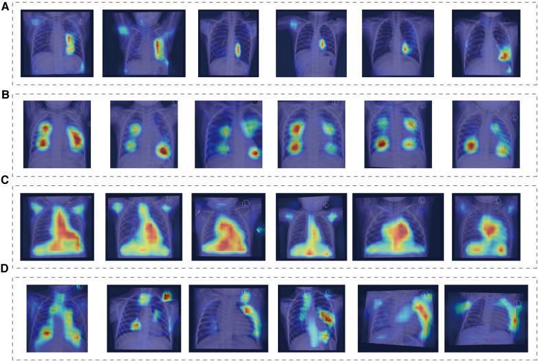
This diagnostic image displays a series of pediatric chest X-rays overlaid with Class Activation Mapping (CAM) heatmaps, used to visualize deep learning model attention in classifying congenital heart diseases. The figure is organized into four rows corresponding to specific diagnostic groups: (A) Normal, (B) Atrial Septal Defect (ASD), (C) Ventricular Septal Defect (VSD), and (D) Patent Ductus Arteriosus (PDA). In the Normal group, heatmap intensity is low and localized primarily near the cardiac silhouette or upper mediastinum. The ASD group shows moderate-intensity foci distributed across the bilateral lung fields, particularly in the hilar and upper lung regions. The VSD group exhibits the most intense and confluent heatmaps, heavily concentrated over the enlarged heart and central thoracic structures, reflecting cardiomegaly and increased pulmonary blood flow features. The PDA group displays a more scattered, diffuse pattern of moderate intensity along the pulmonary vasculature and cardiac borders. This visualization demonstrates how AI models prioritize different anatomical and pathological features, such as cardiac size and pulmonary vascular markings, to differentiate between shunting lesions in congenital heart disease.
neonatal necrotizing enterocolitis NEC X-ray pneumatosis intestinalis
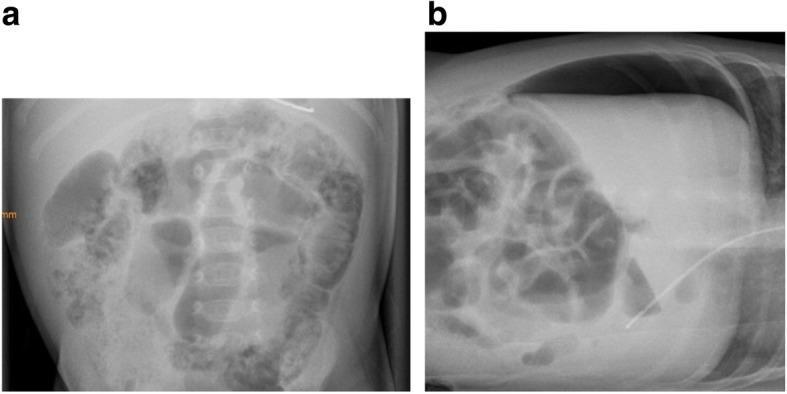
Two radiographic panels illustrating the progression of necrotizing enterocolitis (NEC) in a pediatric patient. Panel A is a supine abdominal X-ray demonstrating pneumatosis intestinalis. This is characterized by the presence of intramural gas, visualized as linear and curvilinear radiolucencies (bubbles) outlining the walls of the large bowel loops. Panel B is a left lateral decubitus X-ray from the same patient eight days later, showing progression to intestinal perforation. The image clearly depicts a large volume of pneumoperitoneum (free intraperitoneal air), most evident as a prominent crescentic radiolucency in the suprahepatic space, situated between the liver surface, the anterior abdominal wall, and the right hemidiaphragm. These findings represent classic radiographic stages of NEC (Bell Stages IIa and IIIb), critical for diagnosing bowel ischemia and subsequent surgical emergency in neonatal and pediatric populations.
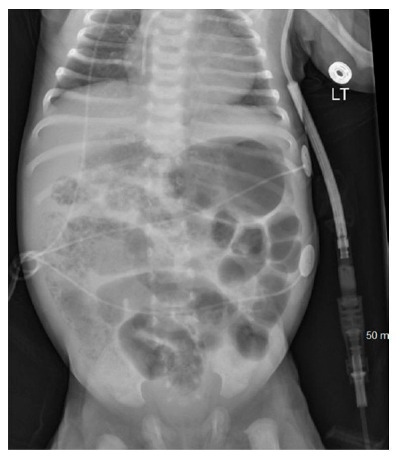
This diagnostic image is an anteroposterior (AP) abdominal X-ray of a neonate. The primary finding is diffuse dilation of multiple bowel loops throughout the abdominal cavity. Notably, the radiograph demonstrates classic signs of pneumatosis intestinalis, characterized by linear and curvilinear radiolucent areas (intramural air) within the walls of the dilated intestinal loops. This radiographic finding is highly suggestive of Necrotizing Enterocolitis (NEC) in this clinical context. There is no clear evidence of pneumoperitoneum visible in this view. Externally, several medical devices are present, including an umbilical catheter extending superiorly, ECG leads on the lateral abdominal walls, and a large-bore drainage or monitoring tube on the right side marked with '50 m' and secured with an adhesive disc labeled 'LT'. The image serves as a critical educational example of pediatric gastrointestinal pathology and neonatal intensive care monitoring.
Tetralogy of Fallot boot shaped heart X-ray pediatric
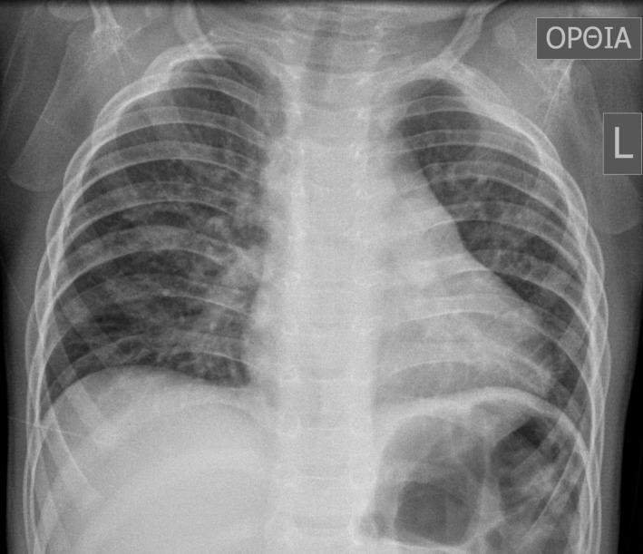
This diagnostic image is an anteroposterior (AP) chest X-ray of a pediatric patient, specifically a 16-month-old male. The image demonstrates a classic 'boot-shaped' heart (coeur en sabot), a hallmark radiographic sign of Tetralogy of Fallot. The cardiac silhouette shows an upturned apex due to right ventricular hypertrophy and a concave main pulmonary artery segment. The lung fields exhibit increased, patchy opacities and markings, particularly prominent in the right lung, which may suggest compensatory pulmonary blood flow or associated bronchial anomalies. The mediastinum appears slightly shifted, and the overall lung volume is clear enough to evaluate the bronchovascular markings. This radiograph serves as a primary educational example of congenital heart disease (CHD) manifestations in pediatric radiology, illustrating the morphological changes to the heart and the secondary effects on pulmonary vascular distribution.
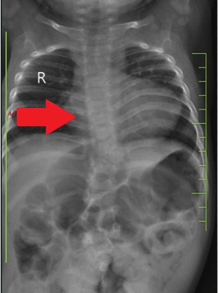
This diagnostic image is a posteroanterior (PA) chest X-ray of a pediatric patient demonstrating classic radiographic signs of Tetralogy of Fallot (TOF). The most prominent finding is a 'boot-shaped' heart (coeur en sabot), characterized by an upturned cardiac apex due to right ventricular hypertrophy and a concave pulmonary artery segment (pulmonary conus). The lung fields appear relatively oligemic, consistent with decreased pulmonary blood flow. The mediastinum is centrally located, and the diaphragm is well-defined, with the right hemidiaphragm slightly superior to the left. The skeletal structures show visible ribs and a vertebral column that appears to have mild alignment irregularities, potentially correlating with lumbar kyphoscoliosis mentioned in clinical context. The lower portion of the radiograph captures the upper abdomen, showing normal bowel gas patterns. This imaging is a classic educational example for pediatric cardiology and radiology, illustrating the morphological changes in the heart and great vessels associated with cyanotic congenital heart disease.
foreign body aspiration right lower lobe hyperinflation pediatric chest X-ray
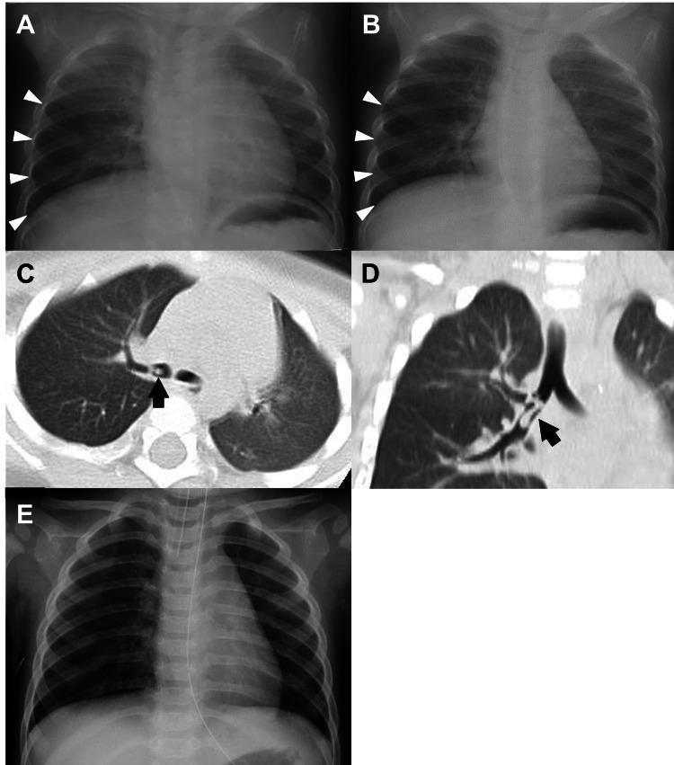
This composite figure illustrates a pediatric case of airway foreign body aspiration through chest X-rays (CXR) and computed tomography (CT) scans. (A, B) Serial frontal chest radiographs compare expiratory and inspiratory phases; white arrowheads indicate localized air trapping and hyperinflation in the right lower lung field, characteristic of a check-valve obstruction. (C) Axial chest CT image demonstrates a small, low-density, cylindrical foreign body (black arrow) lodged within the right inferior lobar bronchus, partially obstructing the lumen. (D) Coronal CT reconstruction provides a longitudinal view of the 'seven-shaped' plastic foreign body (black arrow) within the bronchial tree, highlighting its anatomical relationship to the branching airways. (E) Post-procedural frontal CXR shows resolution of the right-sided emphysema and normal lung expansion following the successful removal of the object via bronchoscopy. This series demonstrates the diagnostic utility of dynamic expiratory radiography and CT in identifying radiolucent foreign bodies in pediatric patients.
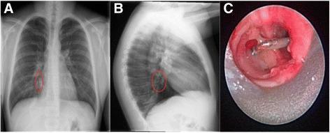
This composite figure illustrates a case of tracheobronchial foreign body aspiration and its secondary pulmonary complications. Panel A presents a posteroanterior (PA) chest x-ray showing a radio-opaque, vertically oriented metallic foreign body located in the right lower lobe bronchus, demarcated by a red circle. Panel B, a lateral chest x-ray, confirms the location of the metallic object and reveals focal airspace consolidation in the posterior basal segment, indicating secondary post-obstructive pneumonia. Panel C provides a bronchoscopic endoscopic view, demonstrating the impacted metallic foreign body within the bronchial lumen. The airway is significantly narrowed by surrounding erythematous, irregular granulation tissue and diffuse mucosal inflammation, representing a chronic inflammatory response to the foreign object. This educational material highlights the correlation between radiological findings (radio-opaque foreign body and consolidation) and direct endoscopic visualization of airway pathology including granulation and edema, relevant for pediatric pulmonology and otolaryngology.
🩻 Paediatric X-Ray Quiz
10 Image-Based Questions with Options & Explanations
Instructions: Look at each X-ray image carefully, read the clinical scenario, then attempt the question before revealing the answer below.
❓ Question 1
- A. Transient Tachypnoea of the Newborn (TTN)
- B. Neonatal pneumonia
- C. Hyaline Membrane Disease (RDS)
- D. Meconium aspiration syndrome
- E. Congenital diaphragmatic hernia
🟢 Reveal Answer & Explanation
✅ Answer: C — Hyaline Membrane Disease (Respiratory Distress Syndrome)
| Finding | Significance |
|---|---|
| Bilateral, symmetric ground-glass opacification | Widespread alveolar collapse (atelectasis) |
| Reticulogranular (granular) pattern | Air bronchograms against collapsed alveoli |
| Prominent air bronchograms | Air in bronchi against non-aerated alveoli |
| Low lung volumes | Surfactant deficiency → alveoli collapse |
| Bell-shaped thorax | Hallmark of RDS |
- TTN: Also premature, but clears within 24-48h; streaky perihilar pattern, NOT granular
- Neonatal pneumonia: Patchy asymmetric consolidation; history of chorioamnionitis
- Meconium aspiration: Post-term infant; patchy, coarse, asymmetric opacities; hyperinflation
- CDH: Bowel loops in chest, mediastinal shift, absent bowel gas in abdomen
🧠 "GRANULAR + LOW VOLUME + PREMATURE = RDS/HMD"
❓ Question 2
- A. Thumbprint sign — Epiglottitis
- B. Steeple sign — Croup (Laryngotracheobronchitis)
- C. Sail sign — Thymic shadow
- D. Double bubble sign — Duodenal atresia
- E. String sign — Pyloric stenosis
🟢 Reveal Answer & Explanation
✅ Answer: B — Steeple Sign — Croup (Laryngotracheobronchitis)
| Finding | Significance |
|---|---|
| Symmetric subglottic narrowing | Inflammatory oedema narrows subglottic airway |
| "Steeple" or "church steeple" or "pencil point" shape | Inverted V narrowing of tracheal air column |
| AP view | Best view to see steeple sign |
| Feature | Croup | Epiglottitis |
|---|---|---|
| X-ray view | AP neck | Lateral neck |
| Sign | Steeple sign | Thumbprint sign |
| Age | 6 months – 3 years | 2-7 years (older) |
| Onset | Gradual, nights | Rapid, toxic-looking |
| Cause | Parainfluenza virus | H. influenzae type b |
| Position | Prefers lying down | Tripod position, drooling |
| Stridor | Inspiratory | Both inspiratory + expiratory |
- Mild: Humidified air, single dose oral dexamethasone
- Moderate/Severe: Nebulised adrenaline (epinephrine) + dexamethasone
- DO NOT examine throat in epiglottitis (may cause laryngospasm)
🧠 "Steeple = Subglottic = Sealing bark cough"
❓ Question 3
- A. Prescribe oral antibiotics and send home
- B. Take throat swab culture immediately
- C. Secure airway in operating theatre with ENT/anaesthetics present
- D. Start nebulised adrenaline and dexamethasone
- E. Perform lateral neck X-ray urgently
🟢 Reveal Answer & Explanation
✅ Answer: C — Secure airway in operating theatre with ENT/anaesthetics present
| Finding | Significance |
|---|---|
| Thumbprint sign | Swollen epiglottis looks like a thumb pressing on the airway |
| Widened epiglottis | Normal epiglottis is thin and pencil-like |
| Aryepiglottic fold thickening | Further airway narrowing |
- Cause: Haemophilus influenzae type b (HiB) — now rare due to vaccination
- DO NOT: examine throat, lie the child down, take throat swabs, or perform investigations that distress the child
- PRIORITY: Airway, Airway, Airway
- Keep child calm, in position of comfort (tripod), with parent
- Senior anaesthetist + ENT surgeon at bedside
- Transfer to OT → intubation under anaesthesia
- IV antibiotics: Ceftriaxone (3rd-generation cephalosporin)
- Extubate after 24-48h when oedema resolves
🧠 "Epiglottitis = Emergency airway, Epipen not needed — Endotracheal tube needed"
❓ Question 4
- A. Double bubble sign
- B. Ground-glass abdomen
- C. Gasless abdomen distally + single large gastric bubble
- D. Pneumatosis intestinalis
- E. Free air under diaphragm
🟢 Reveal Answer & Explanation
✅ Answer: C — Gasless abdomen distally + single large gastric bubble
| Finding | Significance |
|---|---|
| Large, distended stomach bubble | Stomach filled with air/milk, unable to empty |
| Paucity of distal bowel gas | No gas passing pylorus |
| "Caterpillar sign" | Visible gastric peristaltic waves on abdomen |
- Pyloric muscle thickness > 4 mm
- Pyloric channel length > 17 mm
- Hypochloraemic, hypokalaemic metabolic ALKALOSIS
- Mechanism: Loss of HCl in vomit → metabolic alkalosis → kidneys compensate by excreting K+ to retain H+
- Rehydrate and correct electrolytes FIRST (never operate on an alkalotic baby)
- Then: Ramstedt's pyloromyotomy (surgical splitting of pyloric muscle)
| Feature | HPS | Duodenal Atresia |
|---|---|---|
| Age | 3-6 weeks | Day 1-2 of life |
| Vomiting | Non-bilious (projectile) | Bilious |
| X-ray | Single bubble | Double bubble |
| Electrolytes | Alkalosis | Normal |
| Sex | Males >> females (4:1) | Equal |
🧠 "Pyloric stenosis = Projectile vomiting, Palpable olive, Potassium low"
❓ Question 5
- A. Emergency laparotomy
- B. Oral rehydration and watchful waiting
- C. IV antibiotics and NGT decompression
- D. Air (pneumatic) or hydrostatic (barium/saline) enema reduction
- E. Ramstedt's pyloromyotomy
🟢 Reveal Answer & Explanation
✅ Answer: D — Air (pneumatic) or hydrostatic enema reduction
| Finding | Significance |
|---|---|
| Dilated proximal small bowel loops | Obstruction distal to lead point |
| Paucity of gas in right iliac fossa | Bowel has telescoped away |
| Soft tissue mass | The intussusceptum visible as soft tissue density |
- "Target sign" / "Donut sign" (transverse view) — concentric rings of bowel
- "Pseudokidney sign" (longitudinal view) — telescoped bowel resembles kidney
Colicky abdominal pain + Redcurrant jelly stool + Palpable sausage-shaped mass (RUQ)
- Resuscitation (IV fluids)
- Air enema (first-line, 80-90% success rate, less radiation)
- Surgery if enema fails, peritonitis, or perforation
🧠 "Intussusception = Infant 6-12mo, Into itself (telescoping), Ileo-colic, Inflate with air to fix"
❓ Question 6
- A. Left lower lobe collapse — Mycobacterium tuberculosis
- B. Right upper lobe consolidation — Streptococcus pneumoniae
- C. Bilateral perihilar infiltrates — Mycoplasma pneumoniae
- D. Right middle lobe syndrome — Staphylococcus aureus
- E. Pleural effusion — Group B Streptococcus
🟢 Reveal Answer & Explanation
✅ Answer: B — Right upper lobe consolidation — Streptococcus pneumoniae
| Finding | Significance |
|---|---|
| Dense homogeneous opacity | Alveolar space filling (consolidation) |
| Confined to right upper lobe | Well-defined border at horizontal fissure |
| Air bronchograms | Air-filled bronchi within consolidated lung |
| No mediastinal shift | Consolidation, not collapse |
| Consolidation | Collapse (Atelectasis) | |
|---|---|---|
| Volume | Normal/increased | Decreased |
| Mediastinal shift | Away from lesion | Toward lesion |
| Air bronchograms | Present | Absent (usually) |
| Fissure | Normal position | Displaced toward lesion |
| Age | Common organism |
|---|---|
| Neonate | Group B Strep, E. coli |
| 1 month – 5 years | Streptococcus pneumoniae (most common), Haemophilus influenzae |
| 5-15 years | Mycoplasma pneumoniae ("walking pneumonia"), S. pneumoniae |
🧠 "Consolidation = Cloudy white, Confined to lobe, Cause = S. pneumo in children"
❓ Question 7
- A. ↑ Ca, ↑ PO₄, ↑ PTH, ↑ ALP
- B. ↓ Ca, ↓ PO₄, ↑ PTH, ↑ ALP
- C. Normal Ca, ↓ PO₄, Normal PTH, Normal ALP
- D. ↑ Ca, ↓ PO₄, ↓ PTH, Normal ALP
- E. ↓ Ca, ↑ PO₄, ↓ PTH, ↓ ALP
🟢 Reveal Answer & Explanation
✅ Answer: B — ↓ Ca, ↓ PO₄, ↑ PTH, ↑ ALP
| Finding | Description |
|---|---|
| Cupping | Concave deformity of metaphysis |
| Fraying | Ragged, frayed appearance of metaphyseal margin |
| Widened growth plate | Unmineralised osteoid at physis |
| Osteopenia | Generalised reduced bone density |
| Splaying/flaring of metaphysis | Widened end of long bone |
↓ Vitamin D
    ↓
↓ Intestinal Ca/PO₄ absorption
    ↓
↓ Serum Ca → ↑ PTH (secondary hyperparathyroidism)
    ↓
PTH → ↑ Ca reabsorption (kidneys), ↑ PO₄ excretion (kidneys)
    ↓
Still ↓ PO₄ in serum → poor bone mineralisation → ↑ ALP (osteoblast activity)
- Skeletal: Bow legs (genu varum), knock knees, Rachitic rosary (costochondral bead-like swellings), Harrison sulcus (groove on rib cage), frontal bossing, Craniotabes (soft skull)
- Dental: Delayed dentition, enamel defects
🧠 "RICKETS BIOCHEMISTRY = LOW Ca, LOW PO4, HIGH PTH, HIGH ALP"
❓ Question 8
- A. Pneumoperitoneum — gastric perforation
- B. Pneumatosis intestinalis — gas within bowel wall (necrotising enterocolitis)
- C. Dilated bowel loops only — small bowel obstruction
- D. Portal venous gas — hepatic vein thrombosis
- E. Calcification — meconium peritonitis
🟢 Reveal Answer & Explanation
✅ Answer: B — Pneumatosis intestinalis — Necrotising Enterocolitis (NEC)
| Finding | Bell Stage | Significance |
|---|---|---|
| Dilated bowel loops | I | Ileus |
| Pneumatosis intestinalis | IIa | Gas in bowel wall — pathognomonic |
| Portal venous gas | IIb | Severe, gas tracking to liver |
| Pneumoperitoneum | IIIb | Perforation — surgical emergency |
- Linear (subserosal) gas — along the bowel wall
- Curvilinear/bubbly (submucosal) gas — within mucosa
Stage I: Suspected NEC → Dilated loops, feeding intolerance
Stage II: Definite NEC → PNEUMATOSIS intestinalis
Stage III: Advanced NEC → Perforation, pneumoperitoneum
- Stage I-II: NBM (nil by mouth), NG decompression, IV antibiotics (ampicillin + gentamicin + metronidazole), TPN
- Stage III: Urgent surgery — laparotomy, bowel resection, stoma
🧠 "NEC = Bubbles in the wall (pneumatosis) = Dangerous = NBM + Antibiotics"
❓ Question 9
- A. ASD, PDA, Tricuspid atresia, Pulmonary hypertension
- B. VSD, Pulmonary stenosis, Overriding aorta, Right ventricular hypertrophy
- C. VSD, ASD, Aortic stenosis, Left ventricular hypertrophy
- D. Pulmonary atresia, VSD, Right aortic arch, PDA
- E. Transposition of great arteries, VSD, ASD, PDA
🟢 Reveal Answer & Explanation
✅ Answer: B — VSD, Pulmonary stenosis, Overriding aorta, Right ventricular hypertrophy
| Finding | Significance |
|---|---|
| "Boot-shaped" heart (coeur en sabot) | Upturned apex due to RV hypertrophy + concave pulmonary bay |
| Oligaemic lung fields | Reduced pulmonary blood flow (pulmonary stenosis) |
| Normal/small heart size | Not enlarged (unlike ASD/VSD with L→R shunt) |
| Concave pulmonary artery segment | Pulmonary trunk hypoplasia |
P — Pulmonary stenosis (outflow obstruction)
R — Right ventricular hypertrophy
O — Overriding aorta (straddles VSD)
V — VSD (large)
E — (Everything causes cyanosis — R→L shunt)
- Triggered by crying, feeding, defecation
- ↑ pulmonary resistance → ↑ R→L shunt → sudden cyanosis
- Squatting relieves it: ↑ systemic vascular resistance → ↓ R→L shunt
- Knee-chest position (squatting equivalent)
- Oxygen
- Morphine (↓ sympathetic tone)
- IV propranolol or phenylephrine
- IV fluids
🧠 "TOF = PROVE (Pulmonary stenosis, RVH, Overriding aorta, VSD, Equals cyanosis)" "BOOT = Boot kicks Right (RVH makes upturned apex)"
❓ Question 10
- A. Bilateral consolidation
- B. Mediastinal shift TOWARD the affected side on inspiration
- C. Unilateral hyperinflation / air trapping on the affected side on EXPIRATION
- D. Ground-glass opacity with air bronchograms
- E. Pneumothorax with absent lung markings
🟢 Reveal Answer & Explanation
✅ Answer: C — Unilateral hyperinflation on EXPIRATORY film
| Finding | Significance |
|---|---|
| Unilateral hyperinflation (expiratory film) | Ball-valve (check-valve) mechanism — air enters on inspiration but cannot exit |
| Mediastinal shift AWAY from affected side on expiration | Trapped air pushes mediastinum |
| Radiolucent (dark) affected side | Hyperinflated lung |
| Radiopaque foreign body | Only if radio-opaque (bones, coins, metal) |
Inspiration → airway opens slightly → air passes PAST object ✓
Expiration → airway narrows → object blocks → air TRAPPED ✗
→ Progressive hyperinflation on affected side
- Most FBs are radiolucent (organic material) → not seen on plain X-ray
- Expiratory film or fluoroscopy reveals the air trapping
- Diagnosis confirmed by rigid bronchoscopy (also therapeutic)
- Stable → Rigid bronchoscopy (OT setting)
- Acute asphyxiation → Back blows and abdominal thrusts (Heimlich in >1 year)
🧠 "FB in Right bronchus = Right side brighter (hyperinflated) on EXPIRAtory film" "The air CANNOT ESCAPE — like blowing into a one-way valve"
📊 Quiz Summary Table
| Q | Topic | Diagnosis | Key Sign |
|---|---|---|---|
| 1 | Neonatal respiratory distress | Hyaline Membrane Disease / RDS | Ground-glass + reticulogranular pattern |
| 2 | Stridor + barking cough | Croup (Laryngotracheobronchitis) | Steeple sign (AP neck) |
| 3 | Drooling + tripod position | Epiglottitis | Thumbprint sign (lateral neck) |
| 4 | Projectile non-bilious vomiting | Hypertrophic Pyloric Stenosis | Single gastric bubble, gasless distally |
| 5 | Colicky pain + redcurrant jelly stool | Intussusception | Target/donut sign (USS); dilated loops (X-ray) |
| 6 | Fever + cough + consolidation | Lobar Pneumonia (S. pneumoniae) | Right upper lobe opacification |
| 7 | Bow legs + wrist swelling | Nutritional Rickets | Cupping + Fraying of metaphysis |
| 8 | Premature + bloody stool + distension | Necrotising Enterocolitis (NEC) | Pneumatosis intestinalis |
| 9 | Cyanosis + squatting + Tet spells | Tetralogy of Fallot | Boot-shaped heart + oligaemic fields |
| 10 | Sudden choke + unilateral wheeze | Foreign Body Aspiration | Unilateral hyperinflation on expiratory film |
⚡ Key Radiological Signs — Rapid Recall
| Sign | X-ray | Diagnosis |
|---|---|---|
| Steeple sign | AP neck | Croup |
| Thumbprint sign | Lateral neck | Epiglottitis |
| Boot-shaped heart | CXR PA | Tetralogy of Fallot |
| Ground-glass + low volume | CXR AP neonatal | RDS / HMD |
| Cupping + fraying | Wrist X-ray | Rickets |
| Pneumatosis intestinalis | Abdominal X-ray | NEC |
| Double bubble | Abdominal X-ray | Duodenal atresia |
| Gasless abdomen + large stomach | Abdominal X-ray | Pyloric stenosis |
| Target/donut sign | Ultrasound | Intussusception |
| Unilateral hyperinflation | CXR expiratory | Foreign body aspiration |