
Cellulitis
cellulitis skin infection clinical photo
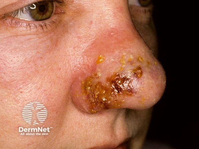
Clinical photography of the external nose shows unilateral cellulitis with thick, adherent crusts and yellow-brown exudate on the nasal dorsum and ala. The image captures erythematous, swollen skin with crusted lesions and surrounding desquamation. Modality is close-up color photography without contrast, suitable for documenting cutaneous infection and supporting differential diagnoses such as impetigo, erysipelas, and nasal cellulitis. The clinical impression references PVL-producing Staphylococcus aureus with potential necrotizing features; PVL infections are notable for rapid progression and tissue destruction, underscoring the need for culture-directed therapy and infection-control measures. This image is relevant for medical education, dermatology and infectious disease research on PVL staph infections, and case-based learning in clinical rounds. Relevance extends to wound care planning, antibiotic stewardship, and monitoring for progression to abscess or deeper facial space involvement. Overall, the photo emphasizes external nasal involvement, crusting, edema, and the importance of prompt clinical assessment and targeted management in suspected PVL-associated skin infections.
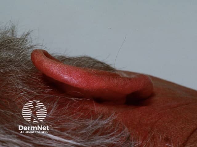
Imaging modality: Clinical photography of the left external ear (pinna) using high-resolution digital capture. Anatomical site: auricle, helix, antihelix, and surrounding periauricular skin; cutaneous tissue overlying auricular cartilage. Visual features: a broad-based, well-demarcated erythematous patch on the superior and lateral aspects of the pinna with uniform red coloration, no focal vesiculation or crusting, mild edema of the surrounding skin, and intact skin surface. Pinna appears inflamed but without purulent drainage or deep abscess signs. The background skin shows no concurrent rash elsewhere. There is no visible piercing or trauma in image. The observation suggests an inflammatory dermatosis of the ear, such as contact dermatitis or photodermatitis, rather than infection, given absence of warmth, warmth indistinct. Clinical significance: skin inflammation limited to the auricle may reflect sun exposure, allergen exposure, or irritant contact dermatitis; differential includes eczema, seborrheic dermatitis, cellulitis with fever, or less likely vasculitis. This image provides educational value for dermatology teaching, primary care evaluation of auricular dermatitis, and photo-documentation. Potential clinical use cases: triage for dermatitis vs infection, monitoring response to topical corticosteroids, sun protection counseling, documentation for derm exam. Researchers may study lesion distribution on ear skin and sun exposure effects.
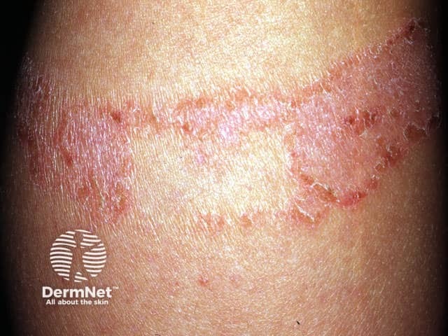
Clinical photograph depicting an adhesive plaster reaction (adhesive tape dermatitis) on exposed forearm skin. The image shows a linear, well-demarcated erythematous plaque with scaling and mild edema along the bandage outline. Peripheral papules and crusting are present, reflecting an inflammatory skin response to adhesive components. The pattern is predominantly contact-mediated and follows the applicator margin, consistent with irritant contact dermatitis or allergic contact dermatitis to plaster ingredients such as acrylates, rubber additives, or latex. There is no purulence or overt infection; warmth and systemic signs are absent. The lesion is superficial, mainly involving the epidermis with possible shallow dermal involvement, and may improve with removal of the offending adhesive. This image is diagnostically helpful for recognizing contact dermatitis from dressings, differentiating from infection (cellulitis), chronic eczema, or psoriasiform eruptions, and guiding patch testing decisions. Clinically relevant terms include contact dermatitis, irritant dermatitis, allergic dermatitis, adhesive allergy, bandage reaction, and dermatitis limited to a band. Educationally, the photo illustrates lesion morphology, distribution pattern, and resolution trajectory after allergen avoidance. In practice, this aids patient counseling, selection of hypoallergenic tapes, topical corticosteroids, and barrier protection to prevent recurrence in future dressings. Careful adhesive avoidance reduces recurrence. Follow-up with moisturizers and barrier films may promote healing.
cellulitis skin AND soft tissue infection management
erysipelas cellulitis lower leg erythema
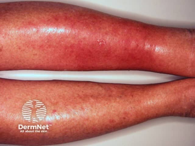
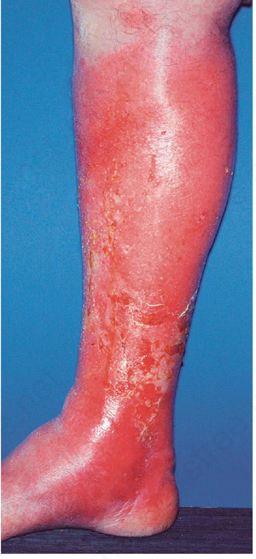
This is a clinical photograph of the left lower leg demonstrating features compatible with acute bacterial cellulitis, most likely streptococcal in origin. Imaging modality: clinical photography of the skin and soft tissues; technique: standard dermatology photography of the extremity with even, diffuse illumination to assess color, warmth, and edema. Anatomical location: left leg, lower leg region involving the skin and subcutaneous tissue; body region: integumentary system of the left limb. Visual features: diffuse, confluent erythema with pronounced warmth and edema; tender, swollen, and hot to palpation as described; margins are ill-defined; skin surface appears taut and glossy, with possible mild vesiculation or surface desquamation not clearly seen; no obvious abscess or necrosis identified; no obvious lymphangitic streaking in the visible field. Pathology/etiology: non-purulent, spreading infection of cutaneous and subcutaneous tissues due to Streptococcus species; neutrophilic inflammation with edema and vascular congestion may be inferred; potential risk of cellulitis-related complications if untreated (lymphangitis, bacteremia). Diagnostic significance and clinical correlation: supports clinical diagnosis of acute cellulitis in the appropriate context (fever, leukocytosis, localized warmth, swelling); useful for documentation, education, and telemedicine triage. Differential considerations: stasis dermatitis, dermatitis, deep vein thrombosis, erysipelas, necrotizing fasciitis (less likely). Clinical use cases: dermatology education, infectious disease review, antibiotic selection guidance, follow-up assessment, patient counseling.
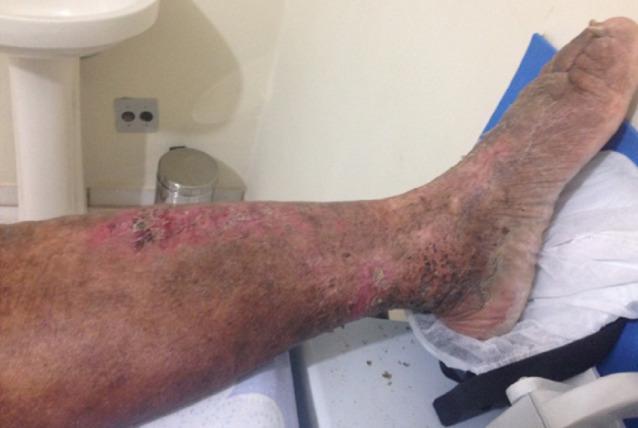
Content Type: Clinical Photograph showing skin manifestations in a patient with chronic lymphedema and recurrent erysipelas. Anatomical Region: Anterior lower leg, ankle, and foot. Findings: The image reveals widespread integumentary changes including diffuse hyperpigmentation (stasis-related browning), patchy erythema (redness), and significant texture irregularities. The skin surface exhibits extensive scaling, dryness, and areas of lichenification or thickening. Specific lesions include a cluster of eroded, raw areas on the mid-anterior leg with associated reddish inflammation and focal dark crusting, suggesting secondary infection or chronic irritation. The distribution of these findings is circumferential and continuous from the mid-calf down to the dorsum of the foot. Clinical Significance: These findings illustrate common dermatological complications of chronic lymphedema, such as stasis dermatitis, fibrosclerosis, and impaired barrier integrity leading to increased risk of recurrent cellulitis. Target Audience: Medical students and dermatology residents focusing on chronic limb edema complications.
Cellulitis
Definition and Pathology
- Andrews' Diseases of the Skin, p. 14
- Tietz Textbook of Laboratory Medicine, 7th Edition, p. 2405
Clinical Photograph
Etiology and Microbiology
| Setting | Common Organism(s) |
|---|---|
| Non-purulent cellulitis (most common) | Streptococcus spp. (~75%), especially Group A |
| Purulent cellulitis / abscess | Staphylococcus aureus (including CA-MRSA) |
| Erysipelas | S. pyogenes (Groups B, C, G also) |
| Animal bite (cat/dog) | Pasteurella multocida, Capnocytophaga canimorsus |
| Human bite | Eikenella corrodens, oral anaerobes |
| Freshwater exposure | Aeromonas hydrophila |
| Saltwater exposure | Vibrio vulnificus |
| Animal exposure (swine/fish handlers) | Erysipelothrix rhusiopathiae |
| Periorbital/orbital | S. pneumoniae, H. influenzae |
| Buccal cellulitis (children) | H. influenzae type b |
| Puncture through shoe sole | Pseudomonas aeruginosa |
| Black necrotic eschar | Bacillus anthracis |
- Rosen's Emergency Medicine, Table 126.1
- Tietz Textbook of Laboratory Medicine, p. 2405
Risk Factors
- Tinea pedis (common portal of entry on legs)
- Impaired lymphatic drainage / lymphedema
- Venous insufficiency and edema
- Obesity (2024 systematic review [PMID: 39163102] confirms significant association)
- Diabetes mellitus
- Disruption of the skin barrier (wounds, ulcers, eczema)
- Immunodeficiency / immunosuppression
- Prior saphenous phlebectomy, lymphadenectomy, or irradiation
- Alcoholism
Clinical Features
- Erythema - confluent, spreading; borders are typically poorly defined and irregular
- Warmth and tenderness (may be variable, especially in neuropathy)
- Edema - may pit on pressure; taut, shiny skin
- Vesicles or bullae - form when severe localized edema separates epidermal layers
- Lymphangitis - streaks extending proximally from the main area along vascular tracts, commonly seen with streptococcal infections and Pasteurella multocida
- Fever, chills, malaise - systemic features may or may not be present
- In severe cases: central nodularity, vesicle formation, discharge of pus and necrotic material
- Rosen's Emergency Medicine, p. 594-598
- Andrews' Diseases of the Skin, p. 47
Differential Diagnosis ("Pseudocellulitis")
| Mimic | Distinguishing Features |
|---|---|
| Venous stasis dermatitis | Afebrile, circumferential or centered over medial malleoli, usually bilateral, no pain |
| Allergic contact dermatitis | Itchy, not painful; well-defined pattern matching allergen exposure |
| Deep vein thrombosis | Asymmetric swelling, no erythema spreading, confirmed by Doppler |
| Erythema migrans (Lyme disease) | Bright red, round >5 cm, target/bull's-eye appearance, central clearing, painless; tick exposure |
| Eosinophilic cellulitis | Insect bite history; less pain; eosinophilia (not neutrophilia) |
| Gout / crystal arthropathy | Joint-centered, podagra pattern |
| Necrotizing fasciitis | Pain out of proportion, rapid progression, crepitus, skin color changes (red → purple → blue), systemic shock |
- Rosen's Emergency Medicine, p. 635-637
Diagnosis
-
Wound cultures: Not recommended for routine non-purulent cellulitis (needle aspiration, swabs, and biopsies have low yield). Indicated for purulent cellulitis, immunocompromised patients, animal bites, and immersion injuries.
-
Blood cultures: Not indicated in uncomplicated cellulitis. Recommended if sepsis or immunocompromise is present.
-
CBC: May show leukocytosis (used in ALT-70 scoring).
-
Ultrasound: Useful to differentiate abscess (requiring I&D) from cellulitis. Point-of-care ultrasound (POCUS) has strong utility per a 2025 systematic review ([PMID: 40274497]).
-
Plain radiograph: To detect foreign body or soft tissue gas (necrotizing fasciitis).
-
CT scan: When deeper extension suspected (post-surgical, orbital cellulitis, necrotizing fasciitis).
-
MRI: Most sensitive for osteomyelitis when bony involvement is suspected.
-
Rosen's Emergency Medicine, p. 645-664
Management
Outpatient (Non-Purulent, No Systemic Toxicity)
- First-line: Dicloxacillin or cephalexin (covers streptococci and MSSA), 5 days
- Alternative: Clindamycin
- Elevation of the affected limb reduces edema and speeds recovery
Purulent Cellulitis (MRSA suspected)
- TMP-SMX (trimethoprim-sulfamethoxazole) or doxycycline monotherapy
- Incision and drainage for frank abscesses; consider antibiotics adjunctively
Inpatient / Systemic Toxicity
- IV antibiotics - vancomycin for MRSA coverage
- Hospitalize: fever >38.5°C, rapid spread, systemic sepsis, failure of outpatient therapy, immunocompromise
Special Situations (per Rosen's Table 126.1)
| Situation | Antibiotic |
|---|---|
| Diabetic foot (mild-moderate) | Amoxicillin-clavulanic acid + TMP-SMX |
| Animal bite | Amoxicillin-clavulanic acid |
| Human bite | Amoxicillin-clavulanic acid |
| Saltwater exposure (Vibrio) | Doxycycline |
| Freshwater exposure (Aeromonas) | Ciprofloxacin |
| Animal handler (Erysipelothrix) | Amoxicillin |
| Shoe puncture (Pseudomonas) | Levofloxacin |
| Buccal cellulitis (H. influenzae) | Ceftriaxone |
| Anthrax eschar | Ciprofloxacin |
- Andrews' Diseases of the Skin, p. 58
- Rosen's Emergency Medicine, Table 126.1
Recurrent Cellulitis
-
Long-term prophylactic antibiotics (low-dose) reduce recurrence in high-risk patients.
-
Compression therapy to address underlying lymphedema helps prevent recurrence.
-
A 2025 systematic review ([PMID: 40530782]) highlights the importance of non-pharmacological management (compression, skin hygiene, weight management) in cellulitis recurrence prevention.
-
Untreated or repeatedly infected lymphatics can lead to permanent fibrotic lymphedema - elephantiasis nostras.
-
Andrews' Diseases of the Skin, p. 63-65
Complications
- Lymphangitis and regional lymphadenitis
- Bacteremia and sepsis
- Metastatic abscesses
- Gangrene
- Progression to necrotizing fasciitis - a surgical emergency (extensive debridement + IV antibiotics; see below)
- Chronic lymphedema / elephantiasis nostras (from recurrent episodes)
Necrotizing Fasciitis - The Key "Do Not Miss" Complication
-
Rapidly spreading necrosis along fascial planes; can appear deceptively normal on the overlying skin early on
-
Red flags: pain out of proportion to exam, fever, crepitus, rapid progression, skin color change (red → purple → blue → black)
-
Two types:
- Type I (polymicrobial): anaerobes + gram-negatives; older patients with diabetes/PVD
- Type II (monomicrobial): usually S. pyogenes; any age
-
Treatment: emergency surgical debridement + IV antibiotics (including vancomycin)
-
Goldman-Cecil Medicine, p. 249-255
-
Tietz Textbook of Laboratory Medicine, p. 2412
Recent Evidence Update
- POCUS for abscess vs. cellulitis ([PMID: 40274497], 2025 Systematic Review): Point-of-care ultrasound is highly useful in the ED for distinguishing cellulitis from abscess, guiding I&D decisions.
- Non-pharmacological prevention of recurrence ([PMID: 40530782], 2025 Systematic Review): Guidelines consistently recommend compression and skin care alongside antibiotics for recurrence prevention.
- Obesity as a risk factor ([PMID: 39163102], 2024 Meta-Analysis): Obesity is significantly associated with cellulitis risk.