
Treatment of lumpy jaw
- Penicillin is first-line: high-dose IV penicillin G (18-24 million units/day) for 2-6 weeks, followed by oral penicillin or amoxicillin for 6-12 months
- Surgical drainage of abscesses and debridement of necrotic bone (sequestrectomy)
- Alternatives: doxycycline, erythromycin, clindamycin, or tetracycline for penicillin-allergic patients
- Long treatment duration is key - relapse is common with short courses
lumpy jaw actinomycosis treatment guidelines 2024
cervicofacial actinomycosis lumpy jaw draining sinus sulfur granules
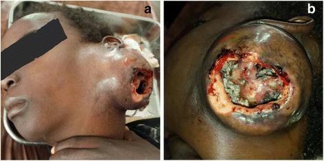
Clinical photograph of a patient with a giant, disfiguring cervicofacial mass (Panel A) and a detailed view of its ulcerated surface (Panel B). The lesion is located on the left cervical region, extending from the infra-auricular area down the neck. Panel A illustrates the mass's significant size and expansive growth, which distorts the normal facial contours. The skin surrounding the lesion appears indurated and irregular. Panel B shows a large, central ulceration with irregular, erythematous, and inflamed borders. The base of the ulcer contains yellowish purulent exudate and granular, necrotic-appearing tissue. This presentation is characteristic of advanced cervicofacial actinomycosis, an indolent bacterial infection that typically presents with 'lumpy jaw' features, abscess formation, and draining sinus tracts that may contain characteristic sulfur granules. The image demonstrates the severe tissue destruction and chronic inflammatory response associated with untreated or late-stage anaerobic infections of the head and neck.
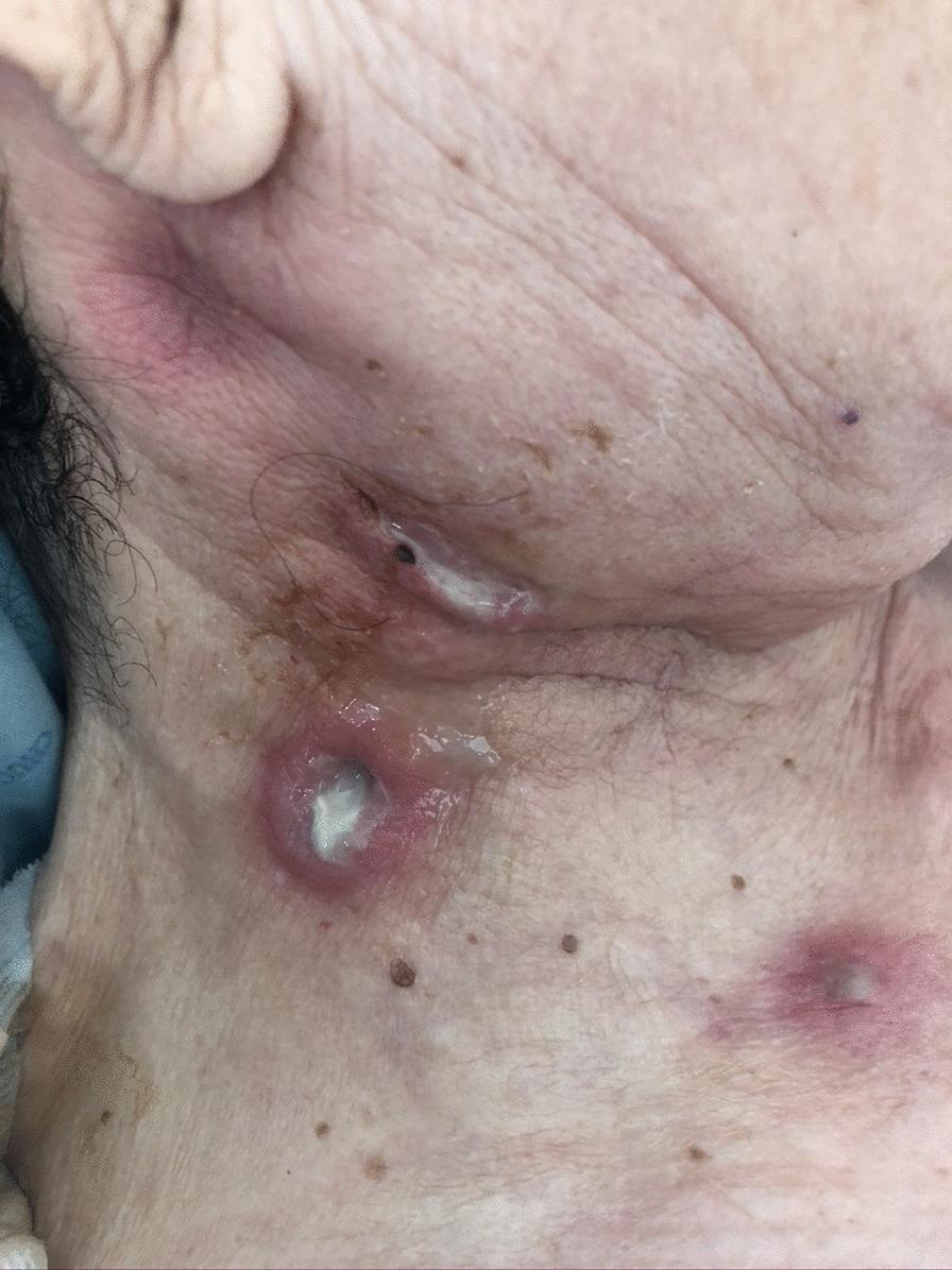
Clinical photograph of the right cervical and submandibular region of an elderly patient, showing multiple suppurative cutaneous lesions. The primary pathology consists of several tender, erythematous nodules and abscesses with central ulceration and spontaneous drainage. A prominent lesion in the lower cervical area exhibits a well-defined, raised inflammatory border surrounding a crateriform defect filled with thick, whitish-yellow purulent material. A second elongated lesion near the jawline shows similar purulent discharge and moisture, suggesting active sinus tract formation. The surrounding skin displays significant erythema and edema, consistent with localized infection. Benign seborrheic keratoses and skin tags are also visible in the periphery. Clinically, these findings represent a case of cervicofacial actinomycosis, characterized by 'lumpy jaw' syndrome with chronic abscesses and draining sinuses that may contain characteristic sulfur granules.
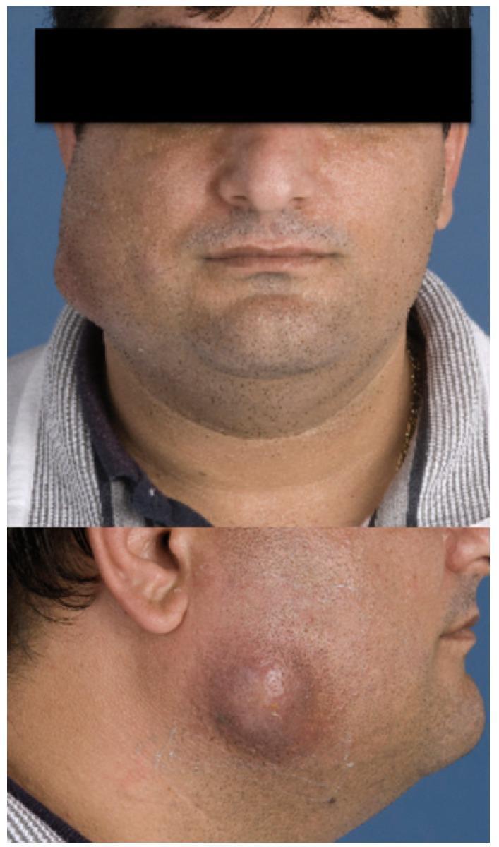
This clinical photograph consists of two views (frontal and profile) demonstrating a significant soft tissue swelling on the left cheek of an adult patient. The swelling is localized to the buccal and parotid-masseteric regions, appearing as a rounded, protruding mass. In the profile view, the lesion exhibits characteristic signs of cervicofacial actinomycosis, including erythematous to violaceous skin discoloration and a central indurated area with an incipient sinus tract formation. This pinpoint area of skin breakdown suggests the classic 'lumpy jaw' presentation where chronic granulomatous inflammation lead to the formation of abscesses and draining fistulas. The educational focus is on identifying the clinical morphology of cervicofacial actinomycosis, specifically the presence of a chronic, painless or slightly tender swelling that may eventually develop external drainage pathways. This visual is typical for dental or oral surgery curricula regarding deep space neck infections and chronic bacterial infections.
Lumpy Jaw (Cervicofacial Actinomycosis) - Treatment
Clinical Picture Before Treating
| Feature | Detail |
|---|---|
| Causative organism | A. israelii, A. gerencseriae, and others |
| Risk factors | Poor dental hygiene, tooth extraction, alcoholism, diabetes, immunosuppression |
| Classic sign | Indurated, non-tender jaw mass with draining sinuses + sulfur granules |
| Bone involvement | Mandible 4x more often than maxilla; periostitis or osteomyelitis may occur |
| Note | Acid-fast stain is negative (distinguishes from Nocardia, which is variably acid-fast positive) |
Treatment
1. First-Line: Penicillin
| Phase | Drug | Dose/Duration |
|---|---|---|
| Initial (IV) - severe/deep disease | Penicillin G IV | 10-24 million units/day for 4-6 weeks |
| Consolidation (oral) | Oral penicillin VK or amoxicillin | Up to 2 g/day, continued for 6-12 months total |
| Mild disease | Oral penicillin from the start | 2-3 weeks minimum; extend beyond symptom resolution |
- For mild/early disease: oral therapy from the start is acceptable.
- Duration depends on extent of disease and whether surgical drainage was performed.
- Therapy must continue beyond resolution of symptoms to prevent relapse.
2. Surgical Management
3. Alternative Agents (Penicillin-Allergic Patients)
| Drug | Notes |
|---|---|
| Doxycycline / minocycline | Acceptable alternatives; avoid doxycycline in children <8 years if course >21 days |
| Clindamycin | Effective |
| Erythromycin | Effective (older studies) |
| Tetracyclines | Effective alternative |
| Ceftriaxone | High in vitro activity |
| Amoxicillin/clavulanate, piperacillin/tazobactam | Broad spectrum; reserve for complicated cases |
| Imipenem/meropenem | Effective for treatment-resistant cases |
4. Special Considerations
- Polymicrobial infection is common - co-pathogens (e.g. Aggregatibacter actinomycetemcomitans, other anaerobes) may need to be covered.
- No person-to-person spread - standard precautions only.
- Prevention: good oral hygiene, regular dental care, and proper wound care after dental procedures.
Clinical Images
Summary
- Drug of choice: Penicillin G IV x 4-6 weeks → oral penicillin/amoxicillin for 6-12 months total
- Surgery: Drainage and debridement of sinuses/necrotic bone is mandatory for most cases
- Penicillin allergy: Use doxycycline, clindamycin, or minocycline
- Resistant cases: Imipenem
- Avoid: Ciprofloxacin, metronidazole (intrinsically resistant)
- Key principle: Long treatment duration is essential - relapse is common if therapy is stopped too early