
tekl me the procedure of wet thick thin film preparation
- Thin films (nanometers to a few microns): spin coating, dip coating, spray coating
- Thick films (microns to hundreds of microns): doctor blade/tape casting, screen printing, slot-die coating
- Both involve depositing a liquid precursor/suspension onto a substrate, then drying/sintering
wet process thin film thick film preparation procedure techniques
wet chemical thin film deposition methods spin coating dip coating sol-gel procedure
in case if filariasis
microfilariae blood film filariasis Wuchereria bancrofti thick thin smear
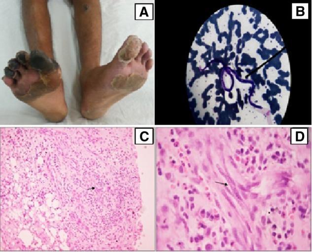
Educational multi-panel figure illustrating the clinical and pathological findings of lymphatic filariasis (Wuchereria bancrofti) masquerading as vasculitis. Panel A (Clinical Photograph): Displays the lower extremities of a patient with dry gangrene of the right hallux and lesser digits, alongside an ulcerated nodule on the left shin and diffuse skin excoriation, indicating severe peripheral vascular compromise. Panel B (Peripheral Blood Film): Giemsa-stained blood smear showing a characteristic microfilaria (approximately 240-250 um long) with identifiable micromorphological features. Panels C and D (Histopathology): Haematoxylin and Eosin (H&E) stained skin biopsy sections from the shin nodule. Low-power magnification (C, 100x) reveals dense inflammatory infiltrates spanning the dermis and subcutaneous fat with capillary proliferation. High-power magnification (D, 1000x) demonstrates a microfilaria (arrow) localized within a capillary lumen, surrounded by a mixed inflammatory cell population consisting of plasma cells, lymphocytes, and neutrophils. This visual set demonstrates the importance of parasitic evaluation in cases of atypical limb ischemia and nodular skin lesions.
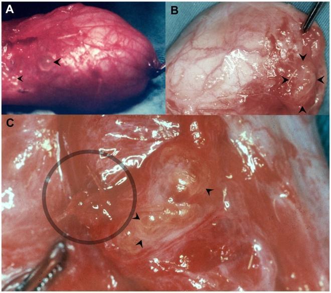
This composite of three clinical photographs (A, B, and C) illustrates surgical findings of scrotal filariasis caused by Wuchereria bancrofti. Image A shows an emptied hydrocele sac with multiple small, translucent, round dilations of the lymphatic vessels (lymphangiectasias) visible on the outer serosal surface. Image B demonstrates similar lymphangiectasias in the retrotesticular area near the excision margin following a hydrocelectomy. Image C provides a high-magnification view of a cluster of these dilated lymphatics. Through the semi-transparent walls of the largest lymphangiectasia, elongated, filiform (thread-like) white structures are visible, representing adult W. bancrofti worms. These findings highlight the characteristic lymphatic pathology of bancroftian filariasis, where adult parasites reside within and cause focal dilation of the scrotal lymphatic system. The images serve as an educational resource for identifying gross pathological features of parasitic lymphatic disease during surgical procedures for hydrocele.
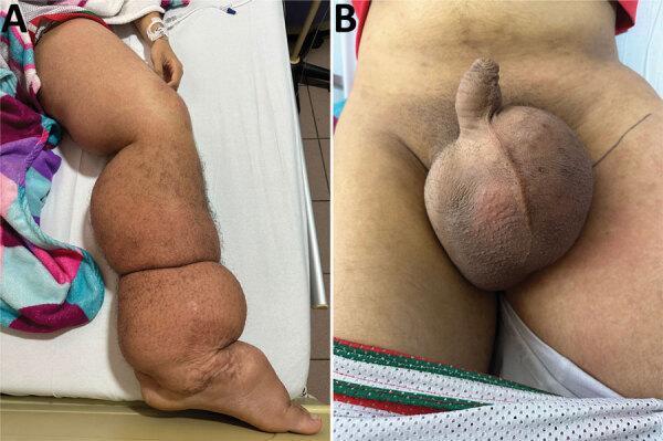
Two-panel clinical photograph illustrating the manifestations of lymphatic filariasis (elephantiasis) caused by Wuchereria bancrofti. Image A displays a side profile of the left lower limb exhibiting severe, generalized lymphedema from the foot to the groin. There is significant swelling and skin folds, erythematous patches on the thigh, and trophic skin changes including perimaleolar hypocromia and hypercromia. Image B provides a frontal view of the pelvic region, showing massive, bulbous scrotal edema (hydrocele) and induration. The skin of the scrotum appears thickened and tense. These findings are characteristic of chronic lymphatic obstruction leading to massive tissue enlargement and secondary dermatological changes. The images are highly relevant for tropical medicine and infectious disease specialties, demonstrating the late-stage physical sequelae of nematode-induced lymphatic damage.
Wet Thick & Thin Blood Film Preparation for Filariasis
Step 0 - Timing of Blood Collection (Critical!)
| Species | Periodicity | Blood Collection Time |
|---|---|---|
| W. bancrofti | Nocturnal periodic | 10 PM - 4 AM |
| Brugia malayi | Nocturnal periodic | 10 PM - 4 AM |
| Brugia timori | Nocturnal periodic | 10 PM - 4 AM |
| Loa loa | Diurnal periodic | Around noon |
Wet Mount (Quick Screening)
- Place 1-2 drops of fresh blood on a clean glass slide
- Apply a coverslip immediately
- Examine under low power (10x) microscopy within minutes
- Look for actively motile microfilariae wriggling among RBCs
- This detects live microfilariae of W. bancrofti and trypanosomes
- Limitation: motility ceases quickly as blood cools; not species-specific
Thin Blood Film Preparation
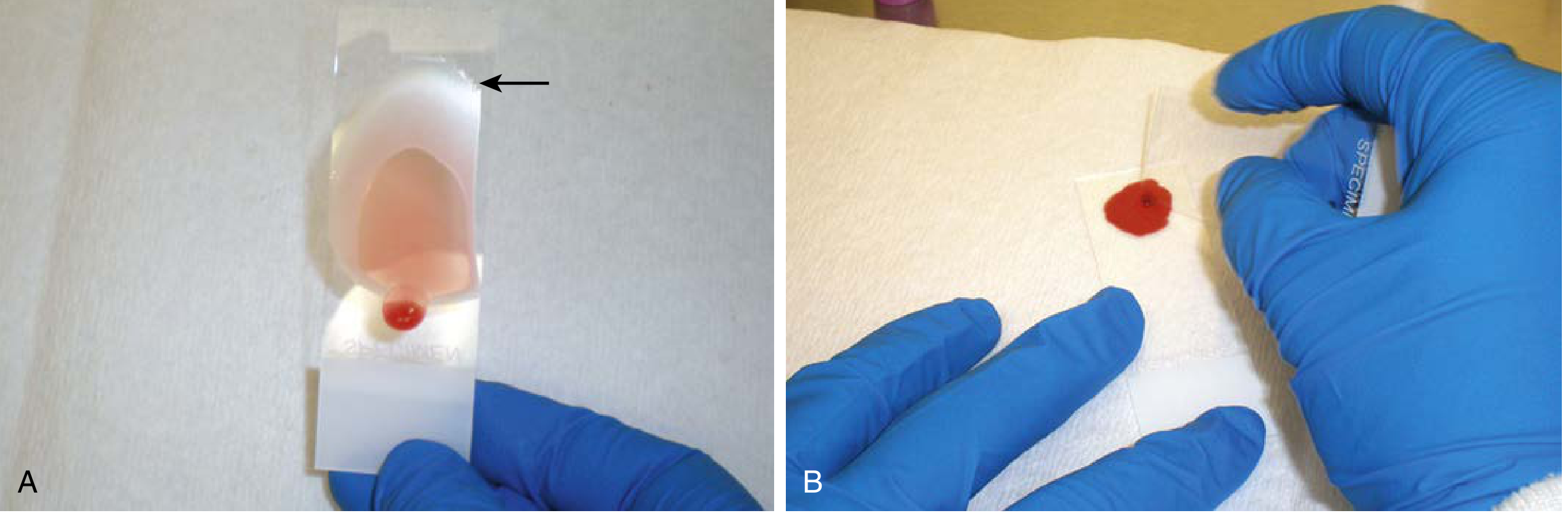
Procedure:
- Place a small drop of blood (about 2-3 µL) near one end of a clean, grease-free glass slide
- Place a spreader slide (at a 30-45° angle) and draw it back to touch the blood drop
- Push the spreader slide forward in a smooth, steady motion across the length of the carrier slide
- The resulting film should end in a feathered edge - thin, well-separated cells toward the tail end
- Allow to air dry completely (do NOT blow on it)
- Fix with methanol for 1-2 minutes (fixes RBCs intact)
- Stain with Giemsa (buffered to pH 7.0-7.2), typically 10% Giemsa for 10-15 minutes, or 3% Giemsa for 30-45 minutes
- Rinse gently with buffered water, air dry, and examine
Thick Blood Film Preparation
Procedure:
- Place a larger drop of blood (~5-10 µL, or 3 drops) in the center of the slide
- Using the corner of a spreader slide, spread the blood in circular/scratch motions into a film about 1.5-2.0 cm in diameter
- Apply gentle pressure to create minute scratches in the carrier slide - this improves adherence and prevents the thick film from detaching during staining
- Allow to dry completely for at least 30-60 minutes (ideally overnight or 1-2 hours minimum)
- Do NOT fix with methanol - the thick film must remain unfixed so RBCs lyse during staining
- Stain with Giemsa (buffered pH 7.0-7.2): 3% Giemsa for 30-45 min or 10% for 10-15 min
- The water-based stain lyses the unfixed RBCs, leaving only WBCs, platelets, and any parasites
- Rinse gently with buffered water, allow to air dry, then examine
Staining Details for Filariasis Specifically
- Giemsa stain (pH 7.0-7.2): Standard, good for general morphology
- Hematoxylin-based stains (e.g., Mayer's or iron hematoxylin): Preferred when the sheath of microfilariae needs to be visualized - the sheath may not stain well with Giemsa in some species (this is key for differentiating W. bancrofti from Brugia spp.)
- Sheaths stain differently: B. malayi sheath stains pink with Giemsa; W. bancrofti sheath does not stain
Concentration Methods for Light Infections
1. Knott Concentration Technique
- Mix 1 mL blood with 9 mL of 2% formalin (1:9 ratio)
- Centrifuge at 500g for 5 minutes
- Discard supernatant; take the sediment
- Prepare a wet mount or make a smear of the sediment
- Stain with Giemsa or hematoxylin
- The formalin lyses RBCs and kills/fixes microfilariae (they lose motility but remain identifiable)
2. Membrane Filtration Technique
- Lyse blood with a lytic agent or distilled water
- Pass through a 5-µm polycarbonate membrane filter
- Remove the filter, place on a slide
- Stain with hematoxylin to reveal microfilariae trapped on the filter
- More sensitive than Knott for very low parasitemias
3. Buffy Coat Preparation
- Centrifuge anticoagulated blood
- Draw off the buffy coat layer (between plasma and packed RBCs)
- Prepare a smear or wet mount
- Examine for motile microfilariae or stain with Giemsa
- Microfilariae concentrate in the buffy coat due to their size
Microscopy Protocol
- Scan entire thin film and thick film under 10x first (microfilariae are large - ~200-300 µm)
- Pay special attention to the feathered edge of the thin film
- Examine at 50x or 100x oil immersion for species identification
- An experienced microscopist should examine at least 100 oil immersion fields on both films; up to 300 fields for immunologically naïve patients
Peripheral Blood Film - Microfilariae (Giemsa stain):
- Medical Microbiology 9e (Sherris & Ryan) - Blood Films section
- Henry's Clinical Diagnosis and Management by Laboratory Methods - Blood Concentration Techniques, p. 1496