
Management of TB spine
"spinal tuberculosis" AND management
tuberculosis spine Pott disease MRI X-ray
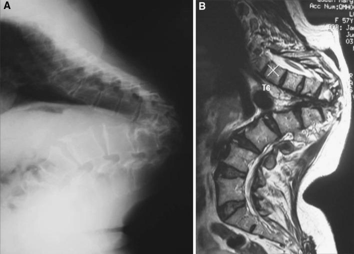
This composite educational graphic features two lateral diagnostic images of the thoracic spine illustrating late-onset Pott's paraplegia in healed disease. Image A is a lateral plain film X-ray showing severe, sharp-angled thoracic kyphosis (gibbus deformity). There is evidence of significant vertebral body destruction and anterior wedging at the lower thoracic level, leading to collapse and a prominent sagittal deformity. Image B is a sagittal T2-weighted MRI scan of the same patient. The MRI demonstrates the acute angulation of the spinal column and the proximity of the spinal cord to the apex of the internal kyphus. Notably, the vertebral bodies show uniform marrow signal intensity with no evidence of marrow edema, soft tissue abscesses, or gadolinium enhancement, which indicates that the underlying tuberculous infection is healed and inactive. The educational focus is on distinguishing structural mechanical complications (kyphosis causing cord kinking) from active spinal tuberculosis (Pott's disease).
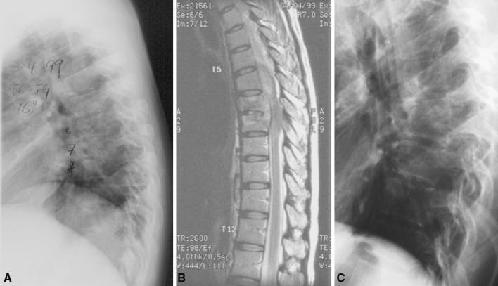
This composite diagnostic image series displays the clinical progression of thoracic spinal tuberculosis (Pott's disease) in a pregnant patient. Panel A is a baseline lateral X-ray of the thoracic spine showing vertebral body endplate irregularity and mild narrowing of intervertebral disc spaces, particularly around levels T6-T9. Panel B is a sagittal T2-weighted MRI at presentation, illustrating marrow edema, disc space involvement, and early inflammatory changes. Panel C is a follow-up lateral X-ray taken one year after a successful course of anti-tuberculous chemotherapy. It demonstrates spontaneous bony fusion of the affected vertebrae, characterized by the complete loss of intervertebral disc spaces and the development of a localized angular kyphosis. The series highlights the natural history of spinal TB where medical management leads to stabilization via auto-fusion, while also documenting the resultant structural deformity common in healed tuberculous spondylitis.
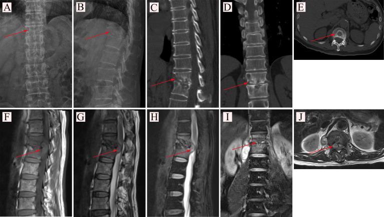
This composite figure presents a multi-modal imaging evaluation of spinal tuberculosis (Pott's disease) in the lower thoracic and upper lumbar spine. (A-B) X-ray radiography in PA and lateral views showing disc space narrowing and endplate irregularities. (C-E) CT images in sagittal, coronal, and axial planes provide high-resolution visualization of bony destruction, vertebral body erosion, and sequestration. Red arrows highlight the focal bone loss. (F-J) MRI sequences offer superior soft-tissue characterization. (F) T1-weighted sagittal view shows low signal intensity in affected vertebrae. (G) T2-weighted sagittal and (H) T2-weighted fat-suppressed (lipid compression) images demonstrate hyperintense signals consistent with inflammatory edema and a prevertebral/paravertebral abscess (red arrows). (I-J) Coronal and axial MRI slices further delineate the extent of the abscess and its relationship to the spinal canal and paraspinal muscles. The collection serves as an educational comparison of imaging modalities (X-ray, CT, MRI) in detecting infectious spondylodiscitis and associated complications like abscess formation.
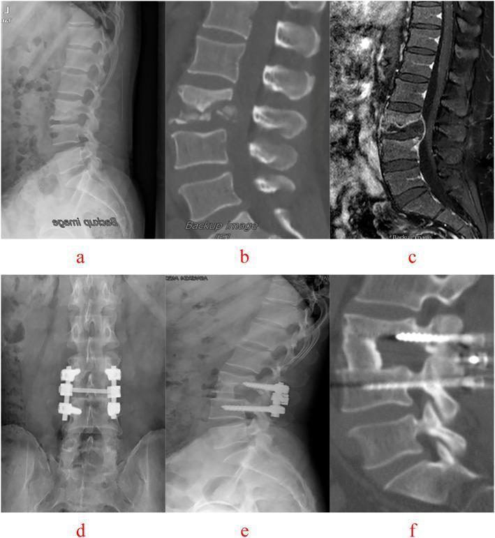
This composite of diagnostic imaging illustrates the preoperative state and postoperative outcome of lumbar spine tuberculosis (Pott's disease) at the L3-L4 level in a 47-year-old male. The top row (a-c) displays preoperative findings: (a) lateral X-ray showing collapsed intervertebral space; (b) sagittal CT reconstruction revealing significant osseous destruction of the L3-L4 vertebral bodies; and (c) T2-weighted sagittal MRI demonstrating an epidural abscess and disc space destruction causing spinal cord/nerve root compression. The bottom row (d-f) shows the 64-month postoperative status: (d) anteroposterior and (e) lateral X-rays visualize a diseased intervertebral (single-segment) fixation involving short pedicle screws and connecting rods at the L3-L4 level. (f) Sagittal CT reconstruction confirms successful interbody bone graft fusion, resolution of the tuberculous lesion, and maintenance of spinal alignment without significant loss of the correction angle. The case highlights the surgical management of spinal tuberculosis through lesion debridement, internal fixation, and fusion.
Management of Tuberculosis of the Spine (Pott Disease)
Pathology
- Infection begins as hematogenous spread from a primary (usually pulmonary) source
- Characterized by acid-fast positive, caseating granulomas with or without purulence
- In the spine, infection spares the intervertebral discs in adults (unlike pyogenic infection), spreading under the anterior and posterior longitudinal ligaments
- Anterior vertebral body is involved first; progressive destruction leads to the characteristic gibbus deformity
- Epidural infection carries a higher risk of permanent neurologic damage
Clinical Features
| Feature | Detail |
|---|---|
| Constitutional symptoms | Weakness, malaise, night sweats, fever, weight loss (early) |
| Back pain | Present in ~70% at presentation |
| Onset | Can be months to years after initial infection |
| Neurologic deficit | ~30% in developing countries; less common in developed nations |
| Cervical disease | Hoarseness (recurrent laryngeal nerve), dysphagia, stridor, rarely sudden death from great vessel erosion |
| Cold abscess / sinus | Present in many cases; can be the presenting sign |
Diagnosis
Laboratory
- Anemia, hypoproteinemia, mildly elevated ESR and CRP
- ESR is normal in >50% of patients - do not rely on it alone
- Tuberculin skin test is contraindicated in prior TB (risk of skin slough from intense reaction)
- Definitive diagnosis: culture of organism via biopsy, or rapid molecular testing (e.g., GeneXpert MTB/RIF) which also detects drug resistance
Imaging
- Plain X-ray: subtle decrease in disc space, localized osteopenia; gibbus deformity in late disease
- CT: bone involvement and paraspinal abscess detail
- MRI with and without contrast (preferred): best for epidural abscess, cord compression, early marrow changes, and soft tissue extent
Biopsy
- Percutaneous needle biopsy under CT/fluoroscopic guidance is usually adequate
- Alternatives: percutaneous thoracoscopic, laparoscopic, or endoscopic biopsy
- Open biopsy if needle biopsy is nonproductive or dangerous
Medical Management
Standard ATT Regimen (Drug-Sensitive TB)
- Rifampicin (R)
- Isoniazid (H)
- Pyrazinamide (Z)
- Ethambutol (E)
Total duration for spinal TB is typically 12 months (longer than pulmonary TB), though some guidelines extend to 18 months for extensive disease or drug resistance.
Drug-Resistant TB (MDR/XDR)
- MDR-TB: resistant to rifampicin + isoniazid; requires injectable agents (amikacin, kanamycin, or capreomycin) plus oral agents chosen by susceptibility (cycloserine, ethionamide, PAS, fluoroquinolones, linezolid)
- Therapy is prolonged; drug-resistant cases benefit more from surgical intervention
Baseline Monitoring (Multidisciplinary)
- HIV screening and other blood-borne viruses
- Baseline + interval renal and liver function tests (drug-induced hepatitis is the most serious side effect)
- Hearing tests if injectable agents used; color vision if ethambutol used
- Drug interaction review (rifampicin is a potent CYP450 inducer - interacts with anticonvulsants, antiretrovirals, anticoagulants)
- Infection control and contact tracing
- DOT (Directly Observed Therapy) to optimize adherence
Bracing / Immobilization
- Bed rest and external orthoses (TLSO or cervical collar) can help control pain and prevent progressive deformity
- Particularly important in children who are at higher risk of deformity because of physis destruction
- Deformity is more significant in children
Surgical Management
Indications for Surgery
- Neurologic deficit - cord/root compression with deficit or progressive deficit despite medical treatment
- Diagnostic uncertainty - biopsy needed when percutaneous techniques are nonproductive
- Spinal instability - significant bone destruction with risk of collapse
- Failure of medical management - persistent or worsening disease
- Significant kyphotic deformity (especially >40°) - correction and stabilization
- Large abscess - particularly cervical or compressive abscesses
- Drug-resistant TB - surgery reduces bacterial burden and improves antibiotic efficacy
Principles of Surgical Treatment
- Debridement of necrotic tissue, caseous material, and bone
- Decompression of neural elements
- Reconstruction of the anterior column (bone grafting)
- Stabilization (instrumentation and fusion) to restore alignment
Surgical Approaches by Region
Cervical Spine
- Anterior approach via transverse incision (2-3 vertebrae) or longitudinal incision along medial border of sternocleidomastoid (longer exposure)
- Allows abscess drainage with bony reconstruction at the same sitting
- Posterior triangle approach for retropharyngeal abscess
Thoracic (Dorsal) Spine
- Costotransversectomy (Ménard, 1894) - most abscesses of the dorsal spine can be drained via this approach
- Resect transverse process, disarticulate rib head, drain abscess via paravertebral approach
- Hemilaminectomy/laminectomy added if epidural component present
- Thoracotomy/anterior approach for major debridement and reconstruction
Lumbar Spine
- Retroperitoneal approach for paravertebral abscess drainage and vertebral debridement
- Posterior pedicle screw fixation (posterior approach) offers one-stage debridement, stabilization, and deformity correction
Bone Grafting Options
- Autograft (structural or non-structural - iliac crest, rib) remains the gold standard
- Allograft
- Titanium mesh cages filled with bone graft
- Recent meta-analysis (PMID 37202780) comparing bone graft types shows comparable outcomes between autograft and titanium mesh cage
Instrumentation
- Anterior instrumentation - direct
- Posterior pedicle screw-rod fixation - preferred by many because it avoids the infected field; provides stronger fixation
- Combined anterior-posterior - used for severe kyphosis or multi-level disease
Complications
| Complication | Notes |
|---|---|
| Pott paraplegia | Cord compression from abscess, granulation tissue, or bony deformity |
| Kyphosis (gibbus deformity) | Can progress even after infection is controlled; more severe in children |
| Late-onset paraplegia | Can occur in healed disease from cord kinking at the kyphotic apex - no active infection |
| Sinus tract formation | Cold abscess tracking to skin |
| Drug-induced hepatitis | Most serious medical complication; requires monitoring |
| Deformity progression | Especially in children with physis involvement |
Outcomes and Prognosis
- With early diagnosis and drug-sensitive disease, prognosis is generally good
- Spontaneous bony fusion occurs in ~80% with medical management
- Neurologic deficits often improve with both medical and surgical treatment
- Drug-resistant TB and late presentations carry worse outcomes
- Motor function and rectal tone are the best prognostic predictors for neurologic recovery
Key Recent Evidence (2023-2026)
- A 2024 systematic review and meta-analysis (PMID 37975989) on surgical strategy for spinal TB confirmed good clinical outcomes with surgery, supporting operative intervention for appropriate indications
- A 2024 systematic review on healing assessment (PMID 38367856) highlighted MRI as the best modality for monitoring response to ATT
- A 2024 bibliometric systematic review (PMID 39925700) on surgical management confirms growing consensus around posterior-only and combined approaches for thoracolumbar disease
TB HIP LEFT SACROILITIS
tuberculosis hip joint X-ray MRI sacroiliitis
"tuberculosis hip" AND management