
Factors that maintain liver in its anatomical position
liver ligaments falciform coronary triangular anatomy diagram
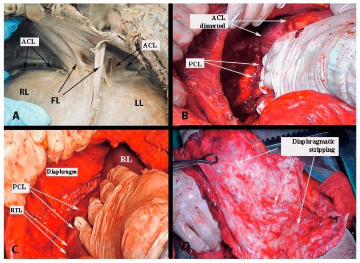
This composite of clinical and cadaveric photographs illustrates the surgical anatomy and mobilization of the liver. Panel A is a cadaveric dissection showing the right lobe (RL) and left lobe (LL) of the liver, with the falciform ligament (FL) and the anterior layer of the coronary ligament (ACL) highlighted. Panels B, C, and D demonstrate steps of liver mobilization and diaphragmatic stripping during open surgery, likely for oncological cytoreduction. Panel B shows the dissection of the ACL and the posterior layer of the coronary ligament (PCL). Panel C visualizes the relationship between the diaphragm, the right lobe (RL), PCL, and the right triangular ligament (RTL) during mobilization. Panel D displays the final appearance of diaphragmatic stripping, characterized by a raw, roughened, and hyperemic surface following the removal of the diaphragmatic peritoneum, often performed in cases of massive metastatic disease. The collection serves as an educational guide for surgeons to identify hepatic ligaments and safely mobilize the liver to access the retroperitoneal space and diaphragm.
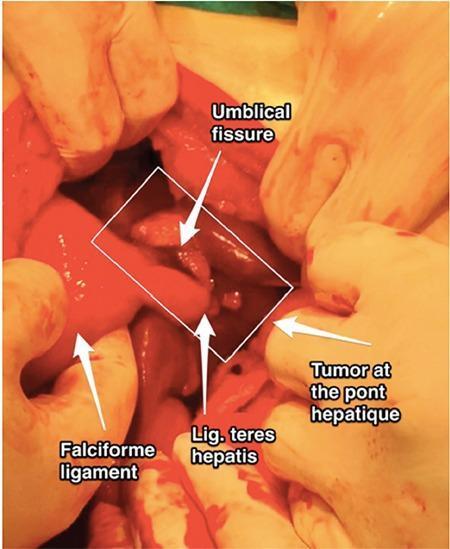
An intraoperative clinical photograph displaying an open abdominal surgical field focused on the liver and its supporting ligaments. The image features several anatomical landmarks and pathological findings labeled with arrows and a white bounding box. On the left, the falciform ligament is identified as it attaches to the liver surface. Centrally, within the white box, the umbilical fissure and the ligamentum teres hepatis (round ligament of the liver) are visible. To the right of these structures, a tumor implant is identified at the pont hépatique (hepatic bridge), appearing as an irregular, nodular tissue mass distinct from the surrounding smooth hepatic parenchyma. The tissues exhibit a glossy, reddish-pink appearance typical of live visceral organs during laparotomy. This image serves as a clinical example of hepatobiliary oncology and surgical anatomy, illustrating metastatic or primary tumor distribution within the hepatic fissures and along ligamentous attachments.
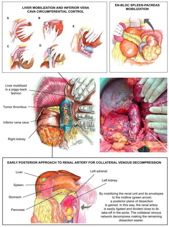
A multi-panel medical illustration and clinical photograph detailing the transplant-based surgical approach for radical nephroureterectomy with tumor thrombectomy. The upper panels (A–E) demonstrate liver mobilization steps: division of left and right triangular ligaments, division of the coronary ligament, rolling the right hepatic lobe to the midline, 'piggy-back' dissection of short hepatic veins, and circumferential control of the infrahepatic/retrohepatic inferior vena cava (IVC). A central anatomical diagram and corresponding intraoperative clinical photograph show a right kidney with a level IIIa tumor thrombus extending into the IVC. The lower section illustrates 'En-bloc spleen-pancreas mobilization' and the 'early posterior approach to the renal artery,' showing the medial displacement of the renal unit (green arrow) to facilitate ligation of the renal artery near the aorta. This technique aims to decompress collateral venous networks and provide exposure of the retroperitoneal space for oncological resection.
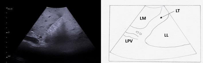
This composite image consists of an intraoperative ultrasound (IOUS) of the liver (left) and a corresponding anatomical line diagram (right) illustrating segmental liver anatomy and vascular landmarks. The ultrasound displays the ligamentum teres (LT) as a distinct, hyperechoic (bright) linear zone located at the free edge of the falciform ligament. This ligament serves as a key surgical landmark, separating the left medial (LM) segment (Segment 4b) from the left lateral (LL) segment (Segment 3). The umbilical portion of the left portal vein (LPV) is visualized as an anechoic (black) tubular structure terminating at the level of the ligamentum teres. The LPV is shown giving off its terminal segmental branches to supply segments 3 and 4b. The educational focus is on the use of IOUS for precise intrahepatic anatomical localization, identification of the Couinaud segments, and tracking the portal venous pedicles during hepatobiliary surgery.
Factors That Maintain the Liver in Its Anatomical Position
1. Peritoneal Ligaments (Peritoneal Reflections)
a. Falciform Ligament
- A sickle-shaped double fold of peritoneum that runs from the umbilicus and anterior abdominal wall to the superior surface of the liver
- It attaches the liver to the anterior abdominal wall and the diaphragm
- Its free lower edge contains the ligamentum teres hepatis (round ligament), the fibrous remnant of the left umbilical vein
- Dividing it during surgery exposes the suprahepatic IVC
b. Coronary Ligament
- Formed by the anterior and posterior reflections of peritoneum from the diaphragm onto the liver
- Consists of an anterior (superior) layer and a posterior (inferior) layer
- The two layers enclose the bare area of the liver - the region where the liver directly contacts the diaphragm without intervening peritoneum
- This bare area itself creates a direct adhesion between liver and diaphragm
c. Right and Left Triangular Ligaments
- Formed where the anterior and posterior leaves of the coronary ligament meet laterally on each side
- Right triangular ligament: fixes the right lobe to the undersurface of the right hemidiaphragm; division mobilises the right lobe to allow rotation to the left
- Left triangular ligament: attaches the left lobe to the diaphragm; division exposes the left lateral wall of the IVC
d. Lesser Omentum (Hepatogastric and Hepatoduodenal Ligaments)
- The hepatogastric ligament connects the liver to the lesser curvature of the stomach
- The hepatoduodenal ligament connects the liver to the duodenum; its right free edge (the free edge of the lesser omentum) contains the portal triad - portal vein, hepatic artery, and common bile duct
- Together they form the lesser omentum, anchoring the inferior aspect of the liver
2. Hepatic Veins and the Inferior Vena Cava (IVC)
- The three hepatic veins (right, middle, and left) drain directly into the IVC at the level of the diaphragm
- This creates a strong vascular tether between the liver and the IVC/diaphragm
- The bare area, where the liver lies directly against the diaphragm adjacent to the IVC, provides additional fixation
- The groove for the IVC on the posterior surface of the liver is a particularly firm attachment point
3. Intra-abdominal Pressure and Diaphragmatic Support
- The diaphragm directly supports the liver from above, especially through the bare area where there is direct contact between the two organs
- Positive intra-abdominal pressure from surrounding viscera and the abdominal wall musculature helps cradle the liver in its position
- The liver is suspended partly by the negative intrathoracic pressure transmitted through the diaphragm
4. Glisson's Capsule and Supporting Connective Tissue
- The liver is enclosed in a fibrous capsule - Glisson's capsule (tunica fibrosa) - covered by visceral peritoneum (serosa)
- At the porta hepatis, the capsule extends inward along the portal tracts, contributing to structural integrity
- The connective tissue framework within and around the liver contributes to its overall cohesion
Summary Table
| Factor | Structure | Attachment |
|---|---|---|
| Peritoneal ligament | Falciform ligament | Anterior abdominal wall + diaphragm |
| Peritoneal ligament | Coronary ligament | Diaphragm (encloses bare area) |
| Peritoneal ligament | Right triangular ligament | Right hemidiaphragm |
| Peritoneal ligament | Left triangular ligament | Left hemidiaphragm / IVC |
| Peritoneal ligament | Lesser omentum | Stomach + duodenum |
| Vascular | Hepatic veins draining into IVC | Diaphragm via IVC |
| Direct contact | Bare area | Diaphragm (no peritoneum) |
| Pressure | Intra-abdominal pressure | Surrounding viscera |