
Eczema for 25 marks
atopic dermatitis AND treatment
atopic eczema NICE guideline diagnosis management topical corticosteroids emollients
atopic dermatitis eczema flexural distribution clinical photograph lichenification
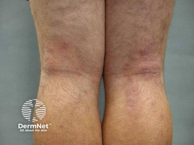
This clinical photograph depicts chronic eczematous changes consistent with atopic dermatitis affecting the bilateral lower legs. Acquired with standard color-calibrated dermatology photography (clinical photography), the image shows low-magnification frontal views of the flexural surfaces around the knees and ankles. The skin is dry (xerosis) with diffuse erythema and pronounced lichenification characterized by thickened plaques with accentuated vertical and horizontal wrinkling. Plaques display scaling and subtle fissuring within creases, and there is episodic excoriation from pruritus. The distribution is bilateral and symmetric, with accentuation in flexural regions and relative sparing of extensor surfaces. The surrounding skin shows mottled hyperpigmentation and mild edema. These features reflect a chronic inflammatory dermatosis with pruritic dominance, often linked to personal or family atopy. Clinically, this pattern supports the diagnosis of atopic dermatitis, versus differential diagnoses such as contact dermatitis, asteatotic eczema, nummular eczema, or psoriasis in atypical locations. Recognizing lichenification, xerosis, and cycled flares is essential for guiding treatment, including regular emollients, barrier repair, topical corticosteroids, calcineurin inhibitors, and anti-pruritic strategies. The image serves educational and diagnostic use for dermatology trainees and researchers studying chronic eczema phenotype, disease severity, and response to therapy. This image enables comparative analysis across eczema severities and informs treatment planning.
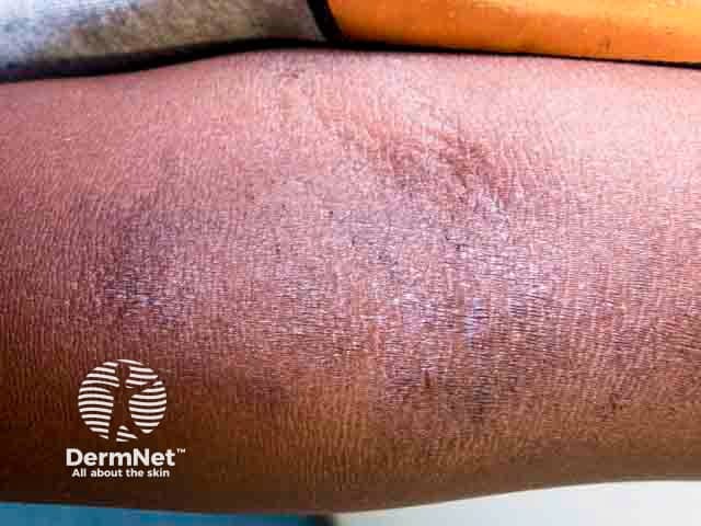
Imaging modality: Clinical photography of in vivo skin. Technique: macro, close-up photograph of the antecubital fossa taken in standard lighting to document chronic eczema. Location: flexural surface of the forearm, antecubital fossa, anterior aspect; laterality not specified. Visual features: accentuation of skin creases (lichenification) with thickened, leathery plaques, diffuse mottling, hyperpigmentation, and subtle surface scale or excoriation from pruritus. Distribution: flexural predominance in an atopic dermatitis context. Pathological correlation: features consistent with chronic inflammatory dermatosis; underlying histology would show epidermal hyperplasia (acanthosis), hyperkeratosis, and dermal chronic inflammatory infiltrate if biopsy were performed. Diagnostic significance: photodocumentation of lichenified flexural dermatitis supports a history of chronic atopic dermatitis and helps monitor treatment response and disease severity. Differential considerations: lichen simplex chronicus, chronic contact dermatitis, nummular eczema, psoriasis with flexural involvement. Clinical correlation: commonly linked with atopy, itch-scratch cycle, and environmental triggers; management implications include intensified emollients, topical corticosteroids or calcineurin inhibitors, and barrier care. Clinical use cases: baseline severity assessment, therapy response monitoring, patient education, teledermatology image reference, and research on pruritus-driven skin changes. This image serves as a reference for longitudinal studies of disease progression, aids in patient counseling about prognosis, and supports educational demonstrations of flexural dermatitis morphology in dermatology training.
Eczema (Atopic Dermatitis) - 25 Marks
Definition
Etiopathogenesis
1. Genetic predisposition
- Family history of atopy is common.
- Mutations affecting filaggrin and other epidermal-barrier proteins cause increased transepidermal water loss, dry skin, and entry of irritants/allergens.
2. Skin-barrier defect
- Reduced lipids and defective stratum corneum lead to xerosis.
- Damaged skin permits penetration of allergens, microbes, and irritants.
3. Immune dysregulation
- Predominantly type 2 immune response, especially in acute disease.
- Cytokines such as IL-4 and IL-13 promote inflammation, IgE production, and impaired barrier function.
- Chronic lesions show additional Th1-mediated inflammatory activity.
4. Environmental and aggravating factors
- Soaps, detergents, fragrances, wool, rough clothing.
- Heat, sweating, low humidity, stress.
- House-dust mite and other aeroallergens may aggravate selected patients.
- Secondary bacterial infection, especially Staphylococcus aureus.
- Food allergy should be considered only when there is a convincing temporal relationship, particularly in young children with severe or refractory eczema. Routine food-allergy testing is not indicated.
Itch-scratch cycle
Clinical Features
Symptoms
- Intense pruritus is the cardinal symptom.
- Dryness, burning, soreness, sleep disturbance, irritability in children.
- Relapsing course with flares and remissions.
Morphology according to stage
| Stage | Clinical lesions |
|---|---|
| Acute | Erythema, edema, papules, vesicles, oozing, crusting |
| Subacute | Erythematous scaly plaques with excoriations |
| Chronic | Dryness, scaling, fissures, hyperpigmentation or hypopigmentation, and lichenification |
Age-related distribution
- Usually begins at 2-6 months.
- Cheeks, forehead, scalp, trunk, and extensor surfaces.
- Napkin area is relatively spared.
- Flexural eczema is typical.
- Antecubital fossae, popliteal fossae, wrists, ankles, neck, and eyelids.
- Flexures, hands, feet, face, neck, eyelids, and nipple areolae.
- Chronic hand eczema and lichenified plaques are common.
Associated features
- Personal or family history of asthma, allergic rhinitis, or food allergy.
- Xerosis, ichthyosis vulgaris, keratosis pilaris.
- Palmar hyperlinearity.
- Dennie-Morgan infraorbital folds.
- Pityriasis alba.
- Elevated serum IgE or eosinophilia may occur, but neither is diagnostic.
Diagnosis
Essential diagnostic points
- Persistent or recurrent itchy dermatitis.
- Typical morphology and age-related distribution.
- Chronic-relapsing course.
- Personal or family history of atopy supports the diagnosis.
Investigations
- Skin swab/culture: if infection is suspected.
- Patch test: if allergic contact dermatitis is suspected, especially in persistent hand, facial, or localized eczema.
- KOH examination: to exclude dermatophytosis.
- Skin biopsy: only in atypical, severe, or treatment-resistant lesions.
- Serum total IgE and allergy tests are not routinely diagnostic.
Histopathology
- Acute eczema: spongiosis, intraepidermal vesicles, superficial perivascular inflammatory infiltrate.
- Chronic eczema: acanthosis, hyperkeratosis, parakeratosis, and dermal fibrosis.
Differential Diagnosis
- Seborrheic dermatitis
- Irritant or allergic contact dermatitis
- Scabies
- Psoriasis
- Tinea corporis
- Nummular eczema
- Lichen simplex chronicus
- Ichthyosis vulgaris
- Cutaneous T-cell lymphoma in persistent atypical adult disease
Complications
- Secondary bacterial infection: impetiginization, oozing, crusting, pustules, pain, fever.
- Eczema herpeticum: sudden painful monomorphic vesicles, punched-out erosions, fever, malaise. This is an urgent condition requiring systemic antiviral treatment.
- Sleep deprivation and impaired school or work performance.
- Anxiety, depression, social stigma, and poor quality of life.
- Adverse effects from inappropriate steroid use, including skin atrophy.
Management
A. General measures and patient education
- Explain the chronic relapsing nature of disease and the need for regular maintenance therapy.
- Avoid known irritants: harsh soaps, detergents, perfumes, wool, overheating, and scratching.
- Keep nails short.
- Wear soft cotton clothing.
- Use lukewarm, brief baths or showers and apply moisturizer immediately afterward.
- Do not recommend broad dietary restriction without evidence of food allergy.
- Identify exacerbating contact allergens where appropriate.
B. Emollients
- Foundation of treatment in all patients, including during remission.
- Use liberal quantities of fragrance-free creams or ointments several times daily.
- Apply immediately after bathing to reduce transepidermal water loss.
- Ointments are useful for very dry skin; lotions may be less effective because of high water content.
- Emollients should continue even when topical anti-inflammatory therapy is stopped.
C. Topical corticosteroids
- Apply to areas of active eczema once or twice daily.
- Select potency according to site, age, and severity:
- Mild potency for face, eyelids, flexures, and infants.
- Moderate potency for trunk and limbs in moderate disease.
- Potent preparations for short periods in severe thickened lesions, preferably under medical supervision.
- Use the lowest effective potency for the shortest duration required.
- Teach fingertip-unit application to improve adherence and prevent undertreatment.
D. Topical calcineurin inhibitors
- Tacrolimus and pimecrolimus are useful steroid-sparing agents.
- Particularly useful for face, eyelids, neck, and flexural areas where prolonged corticosteroid use is undesirable.
- Transient burning or stinging may occur initially.
- Can be used intermittently as proactive maintenance in recurrent localized disease.
E. Wet-wrap therapy and dressings
- Useful for severe acute flares, especially in children.
- Wet dressing over emollient, with or without an appropriately prescribed topical corticosteroid, improves hydration and reduces scratching.
- Should be supervised in extensive disease because occlusion may increase corticosteroid absorption.
F. Antihistamines
- Non-sedating antihistamines do not routinely control eczema itch.
- A short course of sedating antihistamine may occasionally help disturbed sleep, under clinical supervision.
G. Treatment of infection
- Treat clinical bacterial infection with appropriate topical or systemic antibiotics based on extent and local guidance.
- Do not prescribe antibiotics merely for colonization or uncomplicated eczema.
- Suspected eczema herpeticum requires urgent assessment and systemic antiviral treatment.
H. Phototherapy
- Narrowband UVB may be used for chronic moderate-to-severe disease not controlled with topical treatment.
I. Systemic therapy
- Biologic agents, for example dupilumab and selected IL-13-targeted drugs.
- Oral JAK inhibitors in appropriately selected patients.
- Conventional immunosuppressants such as ciclosporin, methotrexate, azathioprine, or mycophenolate mofetil.
Prevention of Relapse
- Continue daily emollient therapy even after lesions settle.
- Avoid individual triggers.
- Treat early signs of flare promptly.
- In frequent relapses, intermittent proactive topical corticosteroid or calcineurin-inhibitor therapy to previously affected areas may reduce recurrence.
- Regular review of technique, adherence, and steroid concerns is important.
Conclusion
Eczema 25 marks please
atopic dermatitis AND treatment
NICE atopic eczema diagnosis management emollients topical corticosteroid guideline
Eczema (Atopic Dermatitis) - 25 Marks
Definition
Etiology and Pathogenesis
1. Genetic factors
- Family history of eczema, asthma, allergic rhinitis, or food allergy is common.
- Defects in filaggrin and other skin-barrier proteins cause dry skin and increased penetration of irritants and allergens.
2. Epidermal barrier dysfunction
- Increased transepidermal water loss causes xerosis.
- Entry of irritants, microbes, and allergens produces inflammation.
3. Immunological factors
- Acute eczema is predominantly associated with a type 2 immune response.
- Cytokines such as IL-4 and IL-13 promote inflammation, pruritus, and IgE production.
- Chronic lesions develop additional cell-mediated inflammatory changes.
4. Triggering and aggravating factors
- Soaps, detergents, perfumes, cosmetics, wool, and synthetic clothing.
- Heat, sweating, low humidity, stress.
- Dust, smoke, and environmental allergens in susceptible persons.
- Bacterial colonization or infection, especially Staphylococcus aureus.
- Food allergy in a small, selected group of children.
5. Itch-scratch cycle
Clinical Features
Symptoms
- Pruritus is the most important symptom.
- Dry skin, soreness, burning, and disturbed sleep.
- Disease follows a chronic relapsing course with remissions and exacerbations.
Lesions
| Stage | Lesions |
|---|---|
| Acute eczema | Erythema, edema, papules, vesicles, oozing, crusting |
| Subacute eczema | Erythematous scaly papules and plaques, excoriations |
| Chronic eczema | Dryness, thickening, fissures, pigmentation, lichenification |
Distribution according to age
- Begins commonly at 2-6 months.
- Face, cheeks, scalp, trunk, and extensor aspects of limbs.
- Napkin area is usually spared.
- Flexural distribution is typical.
- Antecubital fossae, popliteal fossae, wrists, ankles, neck, and eyelids.
- Flexures, hands, face, neck, eyelids, nipples, and feet.
- Chronic hand eczema and lichenified plaques may occur.
Associated signs
- Xerosis.
- Keratosis pilaris.
- Palmar hyperlinearity.
- Dennie-Morgan infraorbital folds.
- Pityriasis alba.
- Personal or family history of asthma, allergic rhinitis, or food allergy.
Diagnosis
Diagnostic criteria
- Pruritus.
- Typical morphology and distribution for age.
- Chronic or chronically relapsing course.
- Personal or family history of atopy supports the diagnosis.
Investigations
- Skin swab/culture if secondary bacterial infection is suspected.
- KOH mount to exclude dermatophytosis.
- Patch testing when allergic contact dermatitis is suspected.
- Skin biopsy in atypical or treatment-resistant cases.
- Serum IgE and eosinophil counts may be raised but are not diagnostic.
Histopathology
- Acute eczema: spongiosis, intraepidermal vesicles, superficial perivascular infiltrate.
- Chronic eczema: acanthosis, hyperkeratosis, parakeratosis, and dermal fibrosis.
Differential Diagnosis
- Seborrheic dermatitis
- Irritant contact dermatitis
- Allergic contact dermatitis
- Scabies
- Psoriasis
- Tinea corporis
- Nummular eczema
- Lichen simplex chronicus
- Ichthyosis vulgaris
Complications
- Secondary bacterial infection: impetigo, oozing, yellow crusting, pustules.
- Eczema herpeticum: painful monomorphic vesicles, punched-out erosions, fever, and malaise. This requires urgent treatment.
- Sleep disturbance and poor school or work performance.
- Emotional distress, anxiety, depression, and reduced quality of life.
- Steroid-related adverse effects from incorrect prolonged use.
Management
A. General measures
- Explain chronic relapsing nature and need for regular skin care.
- Avoid irritants such as harsh soaps, detergents, perfumes, wool, and overheating.
- Use mild soap substitutes or non-soap cleansers.
- Take short lukewarm baths and gently pat skin dry.
- Keep nails short and discourage scratching.
- Wear loose, soft cotton garments.
- Avoid unnecessary food restriction.
B. Emollients
- Emollients are the foundation of treatment and should be used even when the skin is clear.
- Apply liberally and frequently, especially immediately after bathing.
- Use fragrance-free creams or ointments.
- Ointments are preferable in markedly dry or chronic eczema.
C. Topical corticosteroids
- Apply once or twice daily to active eczema.
- Use potency according to severity and site:
- Mild potency: face, eyelids, flexures, and infants.
- Moderate potency: trunk and limbs.
- Potent preparations: severe, thickened lesions for a short course under supervision.
- Avoid unnecessarily potent or prolonged treatment, especially on thin skin.
D. Topical calcineurin inhibitors
- Tacrolimus and pimecrolimus are steroid-sparing agents.
- Useful for face, eyelids, neck, and flexures.
- May cause transient burning or stinging.
- Useful for recurrent disease and maintenance therapy.
E. Wet-wrap therapy and dressings
- Helpful in severe acute eczema, especially in children.
- A wet layer over emollient or prescribed topical corticosteroid reduces itching, improves hydration, and limits scratching.
- Use for a limited period and preferably with specialist advice in extensive disease.
F. Antihistamines
- Do not routinely treat eczema itself.
- Sedating antihistamines may be considered briefly when severe itch disrupts sleep.
G. Treatment of infection
- Treat clinically evident bacterial infection with appropriate topical or oral antibiotics.
- Do not use antibiotics routinely in uncomplicated eczema.
- Suspected eczema herpeticum requires urgent systemic antiviral treatment.
H. Phototherapy
- Narrow-band UVB may be used in chronic moderate-to-severe refractory eczema.
I. Systemic treatment
- Dupilumab and other biologics.
- Oral JAK inhibitors in selected patients.
- Ciclosporin, methotrexate, azathioprine, or mycophenolate mofetil.
- Long-term systemic corticosteroids should generally be avoided due to adverse effects and rebound flares.
Stepwise Treatment Plan
| Severity | Treatment |
|---|---|
| Mild | Emollients + mild topical corticosteroid during flare |
| Moderate | Emollients + moderate topical corticosteroid; consider calcineurin inhibitor and bandages |
| Severe | Potent topical corticosteroid for limited periods, calcineurin inhibitor, wet wraps, phototherapy, systemic therapy, and specialist referral |
Prevention of Relapse
- Regular emollient use, including during remission.
- Avoid known triggers.
- Early treatment of flares.
- Intermittent proactive topical corticosteroid or calcineurin inhibitor on frequently recurring sites.
- Review adherence and correct use of topical treatment.
Conclusion
Make a comparison table of eczema types
dermatitis classification
DermNet eczema types atopic contact discoid dyshidrotic seborrhoeic stasis asteatotic
Comparison of Common Types of Eczema
| Type | Main cause / association | Typical age | Common sites | Characteristic features | Key management |
|---|---|---|---|---|---|
| Atopic eczema | Genetic barrier defect and atopy. Associated with asthma, allergic rhinitis, and sometimes food allergy | Usually begins in infancy or childhood, but may persist into adulthood | Infants: cheeks and extensor surfaces. Children/adults: flexures, neck, eyelids, hands | Intense itch, xerosis, recurrent flares, excoriations and lichenification in chronic disease | Regular emollients, topical corticosteroids for flares, calcineurin inhibitors for sensitive sites, avoid irritants |
| Irritant contact dermatitis | Direct skin injury from repeated exposure to soaps, detergents, water, chemicals, friction, or saliva | Any age, especially occupational exposure | Hands, finger webs, forearms; perioral area in children | Burning or stinging may exceed itch; dry, fissured, erythematous skin confined to exposure area | Avoid or reduce irritant exposure, gloves/barrier protection, emollients, topical corticosteroids for inflammation |
| Allergic contact dermatitis | Delayed type IV hypersensitivity to allergen, for example nickel, fragrance, rubber, hair dye, cosmetics | Any age after sensitization | Area of contact, often hands, earlobes, eyelids, face, or feet | Marked itch; acute vesicles and oozing; may spread beyond contact area | Identify and strictly avoid allergen, patch testing, emollient and topical corticosteroid |
| Seborrhoeic dermatitis | Inflammatory reaction in sebaceous areas, associated with Malassezia yeast | Infants and adults | Scalp, eyebrows, nasolabial folds, ears, presternal area, skin folds | Greasy yellow scales with erythema; itch is mild to moderate | Antifungal shampoo or cream, short course mild topical corticosteroid, calcineurin inhibitor on face |
| Nummular eczema or discoid eczema | Often associated with dry skin; may be triggered by irritants or contact allergy | More common in middle-aged or older adults | Limbs, especially legs; may affect trunk and hands | Multiple well-defined round or coin-shaped itchy, scaly or oozing plaques | Emollients, potent topical corticosteroid for active plaques, treat infection if present, consider patch testing if persistent |
| Pompholyx or dyshidrotic eczema | May be associated with atopy, sweating, stress, nickel sensitivity, or irritants | Young and middle-aged adults | Sides of fingers, palms, and soles | Sudden intensely itchy deep-seated “tapioca-like” vesicles; later scaling and fissuring | Avoid triggers and wet work, emollients, potent topical corticosteroid, compresses during vesicular phase |
| Asteatotic eczema or eczema craquelé | Marked skin dryness caused by low humidity, excessive bathing, soaps, and ageing | Mainly older adults, especially in winter | Shins and lower legs, sometimes trunk and arms | Fine scaling and superficial fissures in a “crazy-paving” pattern; pruritus | Frequent greasy emollients, reduce hot bathing and soap, short course topical corticosteroid if inflamed |
| Stasis eczema or venous eczema | Chronic venous insufficiency and venous hypertension | Middle-aged and elderly persons | Lower legs, especially around medial ankles | Itchy eczematous plaques with edema, varicosities, brown hemosiderin pigmentation, lipodermatosclerosis | Compression therapy if arterial circulation is adequate, leg elevation, emollients, topical corticosteroids; manage venous disease |
| Lichen simplex chronicus | Repeated rubbing and scratching, often related to stress or chronic itch | Adults | Nape of neck, ankles, genital area, forearms | Localized thick, leathery, hyperpigmented plaque with exaggerated skin markings | Break itch-scratch cycle, potent topical corticosteroid, emollients, treat underlying cause and behavioral triggers |
| Hand eczema | Mixed causes: irritant exposure, allergic contact dermatitis, and atopic tendency | Young to middle-aged adults | Palms, dorsal hands, fingertips, finger webs | Dryness, erythema, scaling, fissures, vesicles, pain; often occupational | Minimize wet work and allergens, protective gloves, frequent emollient, appropriately potent topical corticosteroid |
Exam points to remember
- Atopic eczema: flexural, intensely itchy, associated with atopy.
- Contact eczema: distribution follows exposure.
- Nummular eczema: coin-shaped lesions.
- Pompholyx: deep vesicles on hands and feet.
- Seborrhoeic dermatitis: greasy scales on scalp and face.
- Asteatotic eczema: dry, cracked “crazy-paving” appearance on shins.
- Stasis eczema: lower-leg dermatitis with signs of chronic venous insufficiency.