
MHC molecules and antigen presentation to T lymphocytes
Reading File
Now let me search the medical library for MHC and antigen presentation content in parallel.
Finding Sources
Finding Sources
Excellent results. Now let me read the key sections from Cellular and Molecular Immunology and Roitt's Essential Immunology in parallel - these are the most authoritative immunology textbooks.
Reading File
Reading File
Good content found. Let me now read the MHC structure section and the Class II pathway from Cellular and Molecular Immunology (the most comprehensive immunology textbook), and also get the key diagram image.
Reading File
Reading File
Loading Image
Excellent material. Now let me read the MHC molecule structure and the "physiologic significance" section from Cellular and Molecular Immunology, plus more on Class I/II structure.
Reading File
Reading File
Now let me get the MHC structure diagrams and the cross-presentation / Class I structure content.
Reading File
Finding Sources
Reading File
Reading File
I now have comprehensive material. Let me fetch the antigen processing pathways diagram from Cellular and Molecular Immunology.
Loading Image
Reading File
I now have all the content needed for a thorough answer. Let me compile the comprehensive response.
MHC Molecules and Antigen Presentation to T Lymphocytes
1. Discovery and Overview
The major histocompatibility complex (MHC) was originally discovered from studies of tissue graft rejection in mice. George Snell created congenic mouse strains that differed only at a single chromosomal locus - chromosome 17 in mice (called H-2). Grafts between strains that differed at this locus were rejected, giving the region its name: the major histocompatibility complex (histo = tissue). In humans, the equivalent proteins were identified as human leukocyte antigens (HLAs) - named because they were detected by antibodies in the serum of multiply transfused patients, multiparous women, and transplant recipients.
The fundamental immunological role of MHC molecules was clarified by Rolf Zinkernagel and Peter Doherty (Nobel Prize 1996), who demonstrated MHC restriction: virus-specific cytotoxic T lymphocytes (CTLs) only kill virus-infected cells if those cells share the same MHC molecules as the T cell's host. This showed that T cells recognize antigen and MHC simultaneously - a concept called dual recognition.
"The recognition of antigens by CD8+ CTLs is restricted by class I MHC molecules, and the responses of CD4+ helper T lymphocytes to antigens are restricted by class II MHC molecules." - Cellular and Molecular Immunology
2. MHC Genes
The MHC locus contains two main classes of polymorphic genes, plus nonpolymorphic genes involved in antigen processing:
| Gene Class | Location (human) | Product | Function |
|---|---|---|---|
| Class I | HLA-A, -B, -C | Transmembrane glycoprotein | Presents cytosolic peptides to CD8+ T cells |
| Class II | HLA-DR, -DP, -DQ | Heterodimer (α + β chains) | Presents endosomal peptides to CD4+ T cells |
| Class III | Between Class I & II | Complement proteins, cytokines (TNF, LT) | Innate/inflammatory functions |
Polymorphism is a hallmark of MHC genes - there are hundreds of alleles for each locus across the human population. A heterozygous individual expresses 6 different class I molecules (two each of HLA-A, -B, -C) and 8 or more class II molecules (multiple DR, DP, and DQ allotypes). This population-level diversity ensures that virtually any pathogen-derived peptide can be presented by some MHC allele in the species.
3. MHC Molecule Structure
Class I MHC
- Composed of a polymorphic α chain (3 extracellular domains: α1, α2, α3) non-covalently associated with the non-polymorphic β2-microglobulin (β2m)
- The peptide-binding groove is formed by the α1 and α2 domains (encoded by exons 2 and 3 of the class I gene)
- The groove is closed at both ends, restricting bound peptides to 8-11 amino acids
- Polymorphic residues cluster in and around the peptide-binding cleft, determining which peptides each allele can present
- Expressed on virtually all nucleated cells
Class II MHC
- A non-covalent heterodimer of α and β chains (both polymorphic in HLA-DR, -DP, -DQ)
- The peptide-binding groove is formed by the α1 domain (of α chain) + β1 domain (of β chain)
- The groove is open at both ends, allowing binding of longer peptides: 10-30+ residues (optimal 12-16)
- Expressed constitutively only on professional APCs: dendritic cells (DCs), B lymphocytes, macrophages, and thymic epithelium
- Upregulated on many cell types by IFN-γ
4. Antigen Processing Pathways
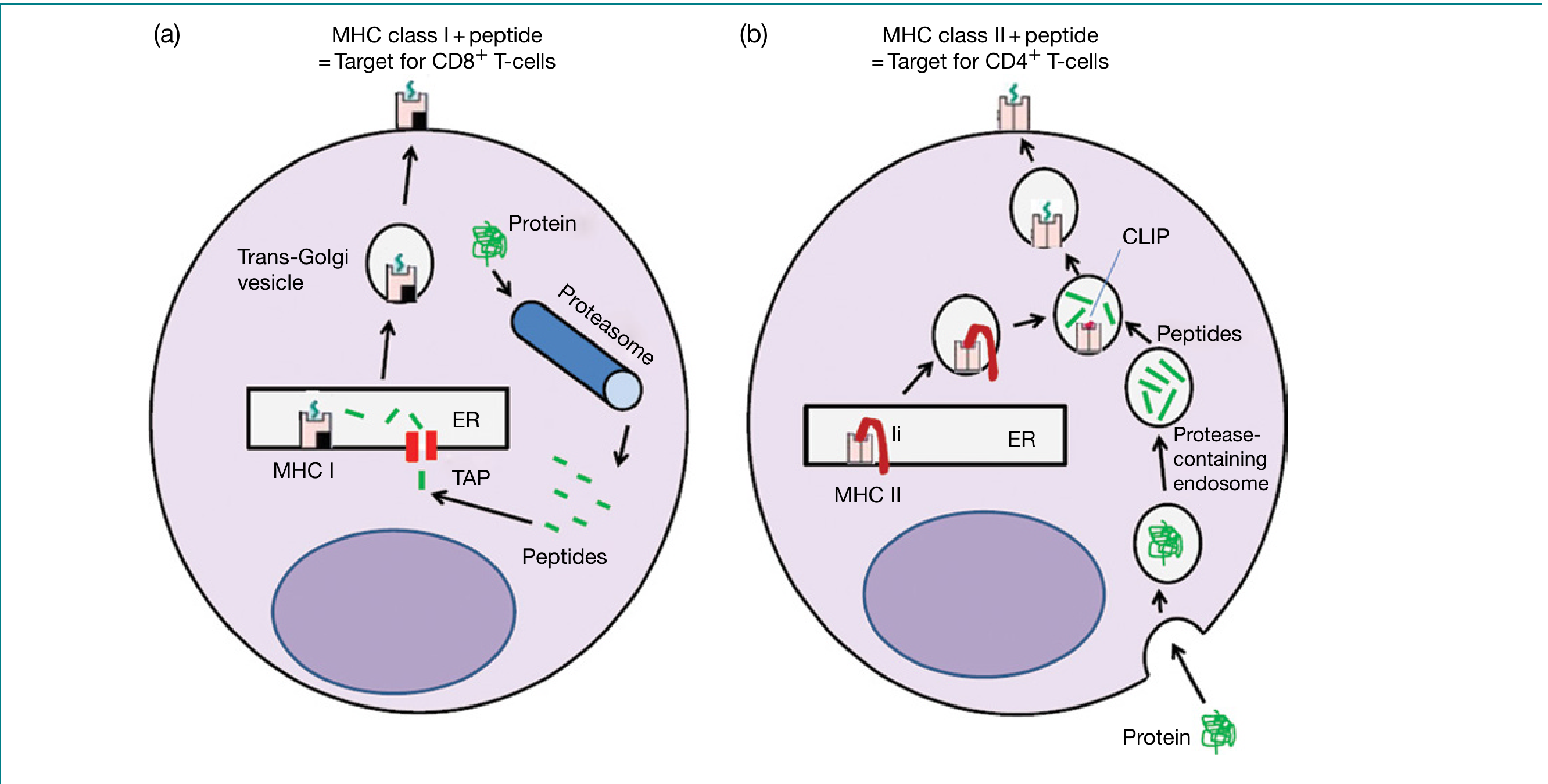
The two pathways are shown below (from Roitt's Essential Immunology):
And the detailed steps (from Cellular and Molecular Immunology):
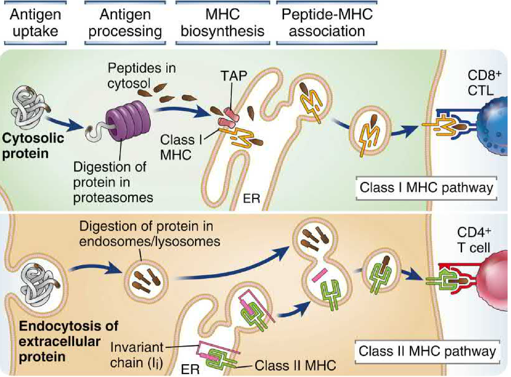
4a. Class I MHC Pathway (Endogenous / Cytosolic Antigens)
This pathway presents intracellular (cytosolic) proteins to CD8+ T cells.
Step 1 - Ubiquitination and proteasomal degradation
Cytosolic proteins targeted for degradation are tagged with polyubiquitin chains. The 26S proteasome (a barrel-shaped structure of 28 subunits in four stacked rings) degrades them into peptides. Viral proteins, tumor neoantigens, and misfolded ER proteins (via ER-associated degradation, ERAD) are all processed this way. IFN-γ replaces three standard proteasomal subunits (β1, β2, β5) with immunoproteasome subunits (LMP2, MECL-1, LMP7) that generate peptides with better C-terminal anchor residues for class I binding.
Step 2 - TAP transport into the ER
Peptides (typically 8-16 amino acids) are transported from the cytosol into the ER lumen by the TAP1/TAP2 heterodimer (Transporter Associated with antigen Processing) - ATP-dependent peptide pumps encoded within the MHC locus itself. Peptides that are too long can be trimmed by ER aminopeptidases (ERAP1, ERAP2 in humans; ERAAP in mice).
Step 3 - Peptide loading complex and MHC assembly
Inside the ER, nascent class I heavy chains assemble with β2m and are stabilized by the peptide loading complex (PLC), which includes: TAP1/2, tapasin (bridges TAP to class I), calreticulin (chaperone), and ERp57 (disulfide isomerase). An optimal peptide of 8-10 residues stabilizes the class I molecule and allows it to be released from the PLC.
Step 4 - Transport to cell surface
The peptide-MHC I complex travels through the Golgi to the plasma membrane, where it is displayed for inspection by CD8+ T cells.
4b. Class II MHC Pathway (Exogenous / Endosomal Antigens)
This pathway presents extracellular proteins internalized by endocytosis to CD4+ T cells.
Step 1 - Antigen uptake
Extracellular proteins are internalized by phagocytosis, macropinocytosis, or receptor-mediated endocytosis into early endosomes.
Step 2 - Endosomal processing
As early endosomes acidify and mature into late endosomes/lysosomes, proteases (cathepsins B, D, H, L, S, and asparagine endopeptidase) digest the internalized proteins into peptides 10-30 residues long. The enzyme GILT (IFN-γ-induced lysosomal thiol reductase) reduces disulfide bonds in engulfed proteins to facilitate unfolding and digestion.
Step 3 - Invariant chain and class II biosynthesis
In the ER, newly synthesized class II αβ heterodimers assemble with the invariant chain (Ii). Ii serves multiple roles:
- Acts as a dedicated chaperone ensuring correct folding
- Occupies the peptide-binding groove (via its CLIP segment - Class II-associated Invariant chain Peptide), blocking premature peptide loading in the ER
- Trimerizes to form a nonameric complex (3 αβ dimers + 3 Ii chains)
- Contains targeting motifs that direct the complex to the endocytic pathway
Step 4 - CLIP removal and HLA-DM editing
The class II-Ii complex is transported to the Golgi, then to MHC class II-enriched compartments (MIICs), where Ii is progressively degraded by cathepsins S/L and asparagine endopeptidase, leaving only the CLIP fragment in the groove. The non-classical MHC molecule HLA-DM (H-2M in mice) catalyzes the exchange of CLIP for high-affinity antigenic peptides. HLA-DO (H-2O) can inhibit HLA-DM activity, modulating the repertoire of presented peptides.
Step 5 - Surface display
Stable peptide-MHC II complexes travel to the plasma membrane. Surface class II levels are regulated by the ubiquitin E3 ligase MARCH-1, which targets class II for degradation; during infection, MARCH-1 is suppressed to increase surface antigen display.
5. Comparative Table: Class I vs. Class II Pathways
| Feature | Class I MHC | Class II MHC |
|---|---|---|
| T cell type activated | CD8+ CTLs | CD4+ helper T cells |
| Source of antigen | Cytosolic proteins (viral, tumor, ERAD) | Endosomal/lysosomal (extracellular antigens) |
| Site of degradation | Proteasome (cytosol) | Late endosomes/lysosomes |
| Proteolytic enzymes | Proteasomal subunits (β1, β2, β5; LMP2, LMP7, MECL-1) | Cathepsins B, D, H, L, S; AEP |
| Peptide transport | TAP1/TAP2 into ER | Ii chain guides class II to endosomes |
| Peptide size | 8-11 amino acids | 12-16 amino acids (up to 30+) |
| Chaperones / accessories | Tapasin, calreticulin, ERp57, calnexin | Invariant chain (Ii), HLA-DM/DO |
| APC expression | All nucleated cells | DCs, macrophages, B cells, thymic epithelium |
| Increased by | IFN-α, IFN-β, IFN-γ | IFN-γ (via CIITA) |
| Outcome | Killing of infected / tumor cells | Macrophage activation, B cell help, antibody production |
6. Cross-Presentation
A critical exception to the "two-pathway" rule is cross-presentation (cross-priming): specialized type 1 conventional dendritic cells (cDC1) can capture exogenous antigens (e.g., from virus-infected cells or tumor cells) and load them onto class I MHC molecules, thereby activating naive CD8+ T cells.
Mechanistically, antigens from phagocytosed cells or debris are taken into vesicles that fuse with ER membrane. By incompletely understood mechanisms, proteins translocate from vesicles into the cytosol, enter the standard proteasome-TAP-class I pathway, and are presented on class I. This allows the immune system to prime antitumor and antiviral CTL responses even when the antigen is not produced within a DC itself.
7. Expression and Regulation
- Class I expression: Constitutive on all nucleated cells; upregulated by IFN-α/β (during early innate antiviral response) and IFN-γ (during adaptive responses). This creates a positive feedback loop: viral infection triggers innate IFN production, which upregulates class I on all cells, making viral antigens more visible to CD8+ T cells.
- Class II expression: Constitutive on professional APCs only; transcriptionally activated by IFN-γ through the master regulator CIITA (class II transactivator). Many pathogens have evolved mechanisms to downregulate class II expression (e.g., herpes viruses blocking CIITA, Mycobacterium inhibiting phagolysosome maturation).
8. Physiological Significance
The segregation of antigen presentation into two pathways has profound functional logic:
- Cytosolic antigens (viruses, intracellular bacteria like Listeria, tumor neoantigens) are presented by class I MHC to CD8+ CTLs, which kill the infected/transformed cell - the only way to eliminate a pathogen producing protein from within a host cell.
- Extracellular antigens (bacteria, toxins, parasites, vaccine antigens) are presented by class II MHC to CD4+ T helper cells, which help macrophages kill phagocytosed microbes and help B cells make antibodies - the appropriate defenses against extracellular threats.
This division ensures that the antigen receptor of a T cell (which cannot by itself distinguish where an antigen comes from) is linked to the correct effector function by the MHC molecule type that co-presents the antigen.
MHC molecules also determine immunodominance: of the many peptides generated by proteolysis of a complex antigen, only those that bind most avidly to the individual's MHC alleles will be efficiently presented. These become the immunodominant epitopes, which has direct applications in vaccine design - synthetic peptides containing such epitopes can serve as candidate vaccines.
9. Non-Classical MHC and CD1
Beyond classical class I and II, there are:
- Non-classical class I molecules (HLA-E, HLA-F, HLA-G): limited polymorphism, specialized roles (e.g., HLA-E presents signal peptides to NK receptors; HLA-G mediates maternal-fetal tolerance)
- CD1 molecules: MHC-like but present lipid antigens (e.g., mycobacterial lipoarabinomannan) to NKT cells and certain T cells
- MR1: presents vitamin B metabolites (riboflavin precursors) to mucosal-associated invariant T (MAIT) cells
Sources:
- Cellular and Molecular Immunology (Abbas, Lichtman, Pillai), Chapter 6
- Roitt's Essential Immunology, Chapter 5
- Janeway's Immunobiology 10e, Chapter 6
dendritic cells
Finding Sources
Finding Sources
Reading File
Finding Sources
Reading File
Finding Sources
Reading File
Finding Sources
Reading File
Loading Image
Reading File
Finding Sources
Reading File
Loading Image
I now have comprehensive material to write a thorough answer on dendritic cells.
Dendritic Cells
Dendritic cells (DCs) are the primary sentinel and professional antigen-presenting cells of the immune system. Named for their long cytoplasmic projections (dendrites), they are uniquely positioned to bridge innate and adaptive immunity - detecting pathogens in tissues and then activating naive T cells in lymphoid organs. Of all APCs (dendritic cells, macrophages, B cells), DCs are the only ones capable of activating naive T lymphocytes.
1. Overview and Position in Immunity
DCs occupy a strategic position at barrier surfaces (skin, intestine, lung) and throughout solid organs. In the steady state they continuously sample the extracellular environment. When they detect danger signals (PAMPs, DAMPs), they transform from sentinels into potent T cell activators. This dual role - sensing then presenting - makes them the essential link between innate detection and adaptive specificity.
"DCs are sentinel cells that sense pathogens as part of the innate immune system and activate naive T-cell responses to initiate adaptive immunity." - Janeway's Immunobiology 10e
2. DC Subsets
There are two broad classes of DCs, with fundamentally different roles:
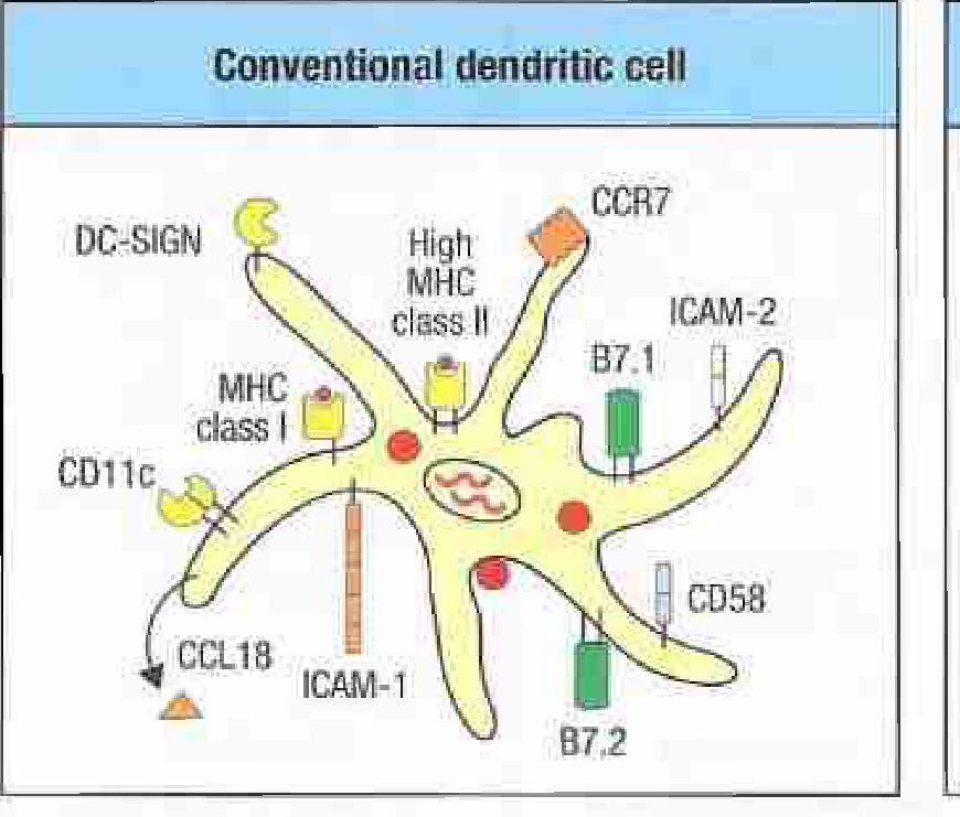
A. Conventional Dendritic Cells (cDCs)
The principal APCs for naive T cell activation. They efficiently ingest and degrade protein antigens, presenting peptides on both MHC class I and class II. Conventionals are subdivided into two major subsets:
| Feature | cDC1 | cDC2 |
|---|---|---|
| Key surface markers | XCR1, BDCA-3 (human), CLEC9A, CD8α (lymphoid) | CD11b, BDCA-1, Dectin-1, SIRPα |
| Nonlymphoid tissue markers | CD103, CD24 | CD11b, CD24, SIRPα |
| Primary function | Cross-presentation to CD8+ T cells; antitumor surveillance | Activation of CD4+ T cells; Th2/Th17 responses |
| Key transcription factor | IRF8 | IRF4 |
| Pathogens targeted | Intracellular (viruses, Listeria) | Extracellular (bacteria, helminths, fungi) |
| Location emphasis | Present in all tissues; abundant in gut (CD103+) | Dominant in gut lamina propria; spleen marginal zone |
cDC1 cells are the main executors of cross-presentation - they capture antigens from virus-infected or tumor cells and load them onto MHC class I to prime naive CD8+ CTLs. This is critical for antitumor immunity and antiviral responses in which the pathogen does not infect DCs directly.
cDC2 cells are the dominant activators of CD4+ helper T cells, directing Th1, Th2, and Th17 polarization depending on the pathogen context and cytokine environment.
B. Plasmacytoid Dendritic Cells (pDCs)
pDCs are morphologically distinct (resembling plasma cells) and are not primary APCs for naive T cells. Their dominant role is the rapid, massive production of type I interferons (IFN-α/β) in response to viral nucleic acids sensed via TLR7 (ssRNA) and TLR9 (CpG DNA). A single pDC can produce 100-1000 times more type I IFN than any other cell type, making them central to early antiviral innate defense. They also produce IFN-α in systemic lupus erythematosus in response to self nucleic acid-immune complexes, contributing to disease pathogenesis.
| Feature | Conventional DCs (cDC) | Plasmacytoid DCs (pDC) |
|---|---|---|
| Primary role | Antigen presentation to naive T cells | Type I IFN production |
| Key PRRs | TLR1-6, CLRs, NLRs, RLRs | TLR7, TLR9 |
| Morphology | Dendritic processes | Plasma cell-like (round) |
| MHC expression | High (especially on maturation) | Low |
| Co-stimulation | High B7.1/B7.2 on maturation | Low |
| Key marker | CD11c+ (high) | BDCA-2, BST2 (CD317), CD123 |
3. DC Life Cycle: Immature to Mature
The process of DC maturation is the key to their function and runs through four stages:
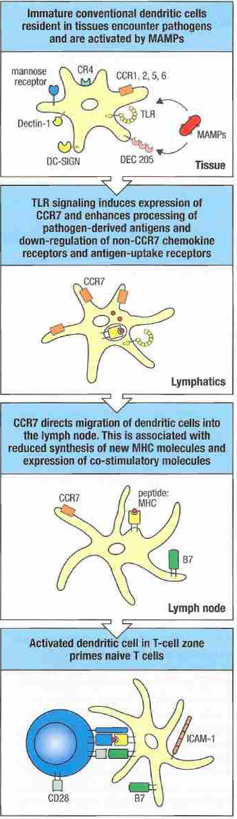
Stage 1 - Immature DCs in Tissues
Tissue-resident immature DCs are highly specialized for antigen capture. They express:
- Pattern recognition receptors: numerous TLRs, C-type lectin receptors (CLRs) like DC-SIGN, Dectin-1, mannose receptor (MRC1/CD206), DEC-205; NLRs; RLRs
- Endocytic receptors for efficient macropinocytosis and phagocytosis
- CCR1, CCR2, CCR5, CCR6: chemokine receptors that retain them in peripheral tissues
At this stage, MHC class II and co-stimulatory molecules (CD80/B7.1, CD86/B7.2) are expressed at low levels on the surface - most class II is in intracellular endosomal compartments.
Stage 2 - Activation by PAMPs/DAMPs
When TLRs and other PRRs detect microbial products, DCs undergo activation/maturation:
- Antigen uptake is dampened and antigen processing and presentation are enhanced - newly synthesized peptide:MHC complexes flood the surface
- MHC class I and II expression increases dramatically
- Co-stimulatory molecules (CD80/B7.1, CD86/B7.2, CD40) are upregulated
- Inflammatory cytokines are secreted: IL-12, IL-18, TNF, IL-6 (the cytokine milieu determines CD4+ T helper cell polarization)
- Chemokines are produced: RANTES (CCL5), MIP-1α (CCL3), IP-10 (CXCL10)
Stage 3 - Migration to Lymphoid Tissue (CCR7)
A critical event during maturation is upregulation of CCR7, which binds CCL19 and CCL21 produced by stromal cells in the T cell zones of lymph nodes. CCR7 expression simultaneously:
- Directs DCs from inflamed tissue through afferent lymphatics to the draining lymph node
- Causes downregulation of tissue-retention receptors (CCR1/3/5 for CCL3/4/5)
Once in the T cell zone (paracortex) of the lymph node, mature DCs slow their synthesis of new MHC molecules (to "freeze" the antigen snapshot from the infection site) and focus on T cell interaction.
Stage 4 - Naive T Cell Priming
Mature DCs in the T cell zone present peptide:MHC complexes to recirculating naive T cells. Effective T cell activation requires three signals:
| Signal | Molecule | Function |
|---|---|---|
| Signal 1 | Peptide:MHC + TCR | Antigen specificity |
| Signal 2 | B7.1 (CD80) / B7.2 (CD86) + CD28 | Co-stimulation; prevents anergy |
| Signal 3 | Cytokines (IL-12, IL-4, TGF-β + IL-6, etc.) | Determines effector T cell subset |
DCs also express ICAM-1 (binds LFA-1 on T cells), ICAM-2, and CD58 (binds CD2), which strengthen the immunological synapse. Without Signal 2 (co-stimulation), T cells encountering antigen become anergic (non-responsive) rather than activated - this is the basis of peripheral tolerance maintained by immature DCs presenting self-peptides.
4. DC Control of Helper T Cell Polarization
The cytokine environment established by the DC (Signal 3) instructs naive CD4+ T cells on which effector phenotype to adopt:
| DC-derived cytokines | T helper subset | Function |
|---|---|---|
| IL-12, IL-18 | Th1 | IFN-γ production; macrophage activation; intracellular pathogens |
| IL-4 (often from other cells) | Th2 | IL-4/IL-5/IL-13; helminth defense; allergy |
| IL-6 + TGF-β | Th17 | IL-17; extracellular bacteria and fungi; neutrophil recruitment |
| IL-10 + TGF-β | T regulatory (Treg) | Immune suppression; tolerance |
The nature of the pathogen (bacterial vs. viral vs. parasitic) and the PRRs it triggers determine which cytokine program the DC activates.
5. Tolerogenic DCs
Not all DC activation leads to immunity. Immature DCs and DCs conditioned by certain signals can become tolerogenic:
- In the steady state, lymphoid tissue-resident immature DCs display self-peptide:MHC complexes but lack co-stimulatory molecules - T cells that contact them undergo deletion or anergy (peripheral tolerance)
- DCs matured in the presence of TSLP, IL-10, or prostaglandin E2 upregulate co-stimulatory molecules but do not produce inflammatory cytokines - this programs Treg induction
- Activation of DCs without CD4+ T cell help, with suboptimal innate stimuli, or in the tumor microenvironment can all yield tolerogenic DCs
- Tolerogenic DCs in tumors actively suppress anti-tumor immunity and are a target of cancer immunotherapy
6. DC Locations and Tissue-Specific Variants
DCs are ubiquitous but have specialized names/phenotypes in certain tissues:
| Location | DC variant | Notes |
|---|---|---|
| Skin (epidermis) | Langerhans cells | Express langerin (CD207), Birbeck granules; seed epidermis from bone marrow; long-lived; important for contact hypersensitivity |
| Skin (dermis) | Dermal DCs | CD11b+; migrate to draining lymph nodes |
| Gut lamina propria | Intestinal DCs | Express CCR9, sample lumen via transepithelial dendrites; key for oral tolerance and IgA responses |
| Lymph nodes | Lymphoid-resident DCs | cDC1 (CD8α+) and cDC2 (CD4+); handle blood-borne antigens |
| Spleen | Splenic DCs | Handle blood-borne antigens; cDC1 in marginal zone, cDC2 in red pulp |
| Lung | Alveolar DCs | Front-line defense against inhaled pathogens and allergens |
| Blood | Circulating DC precursors | Immature precursors seeding tissues |
7. Role in Disease
Autoimmunity: Dysregulated DC activation or failure of tolerogenic DC function contributes to autoimmune diseases. In SLE, pDCs produce pathological amounts of IFN-α in response to self nucleic acid:anti-nuclear antibody complexes internalized via Fc receptors. In rheumatoid arthritis, synovial DCs promote Th1/Th17 responses.
Allergy: DCs conditioned by the epithelial cytokines TSLP, IL-25, and IL-33 drive Th2 polarization, contributing to asthma and atopic dermatitis.
Cancer: Tumor-infiltrating DCs are often immature or tolerogenic due to factors secreted by tumors (VEGF, IL-10, IDO). cDC1 cells are required for successful cross-priming of anti-tumor CD8+ T cells; their abundance in the tumor microenvironment correlates with better prognosis and response to checkpoint immunotherapy.
Vaccination: DCs are the target of most vaccine adjuvants. Adjuvants (alum, MF59, TLR agonists) work by activating DCs, providing the co-stimulatory and cytokine signals needed to break tolerance to the vaccine antigen.
Infectious disease: Many pathogens have evolved mechanisms to subvert DC function - HIV infects DCs via DC-SIGN and exploits their migration to deliver virus to lymph nodes; Mycobacterium tuberculosis prevents phagolysosome maturation in DCs; herpes viruses block class I antigen presentation.
8. Comparison of Professional APCs
| Property | Dendritic Cells | Macrophages | B Cells |
|---|---|---|---|
| Antigen uptake | Macropinocytosis, phagocytosis, receptor-mediated | Phagocytosis (best) | Antigen-specific BCR |
| MHC class II | Low (immature) → High (mature) | Inducible (IFN-γ) | Constitutive; increases on activation |
| Co-stimulation | Constitutive on mature DCs | Inducible | Inducible |
| Primary T cell activated | Naive T cells | Effector/memory T cells | Tfh cells (for B cell help) |
| Location | Ubiquitous (barrier surfaces, organs, lymphoid) | Lymphoid tissue, connective tissue, body cavities | Lymphoid tissue, blood |
| Unique specialization | Naive T cell priming; cross-presentation | Phagocytosis/killing of microbes | Antibody production; antigen presentation to Tfh |
Sources:
- Janeway's Immunobiology 10e, Chapter 9 (sections 9-6, 9-7)
- Cellular and Molecular Immunology (Abbas, Lichtman, Pillai), Chapters 4 & 6
- Firestein & Kelley's Textbook of Rheumatology, Chapter on DC maturation and activation
This is a shared conversation. Sign in to Orris to start your own chat.