
Anatomy of oral cavity ENT long answer question
Reading File
The oral cavity is bounded by lips anteriorly, cheeks laterally, hard/soft palate superiorly, floor of mouth inferiorly, and the oropharyngeal isthmus posteriorly. It contains the tongue, teeth, gums, and opens into the pharynx. Let me search the medical library for comprehensive content.
Searching sources...
Finding Sources
Finding Sources
Reading File
Reading File
Good - Gray's Anatomy has detailed content. Let me read more from it plus search for specific subsections on tongue, palate, salivary glands, and floor of mouth.
Reading File
Finding Sources
Reading File
Reading File
Reading File
Reading File
Reading File
Reading File
Reading File
Reading File
I now have comprehensive content from two authoritative ENT/anatomy sources. Let me fetch a key anatomical diagram image to include in the answer.
Loading Image
Loading Image
I now have all the content needed to write a comprehensive ENT long answer on oral cavity anatomy. Let me compile the full answer.
Anatomy of the Oral Cavity
(Sources: Gray's Anatomy for Students; K.J. Lee's Essential Otolaryngology)
I. INTRODUCTION AND OVERVIEW
The oral cavity is the first part of the digestive tract and serves as the primary inlet for food. It also participates in speech, breathing, and salivation. It is divided by the dental arches (teeth + alveolar bone) into two regions:
- Oral vestibule - Horseshoe-shaped space between the dental arches and the inner surfaces of the cheeks and lips. The oral fissure (mouth opening) opens into it.
- Oral cavity proper - The main space enclosed within the dental arches.
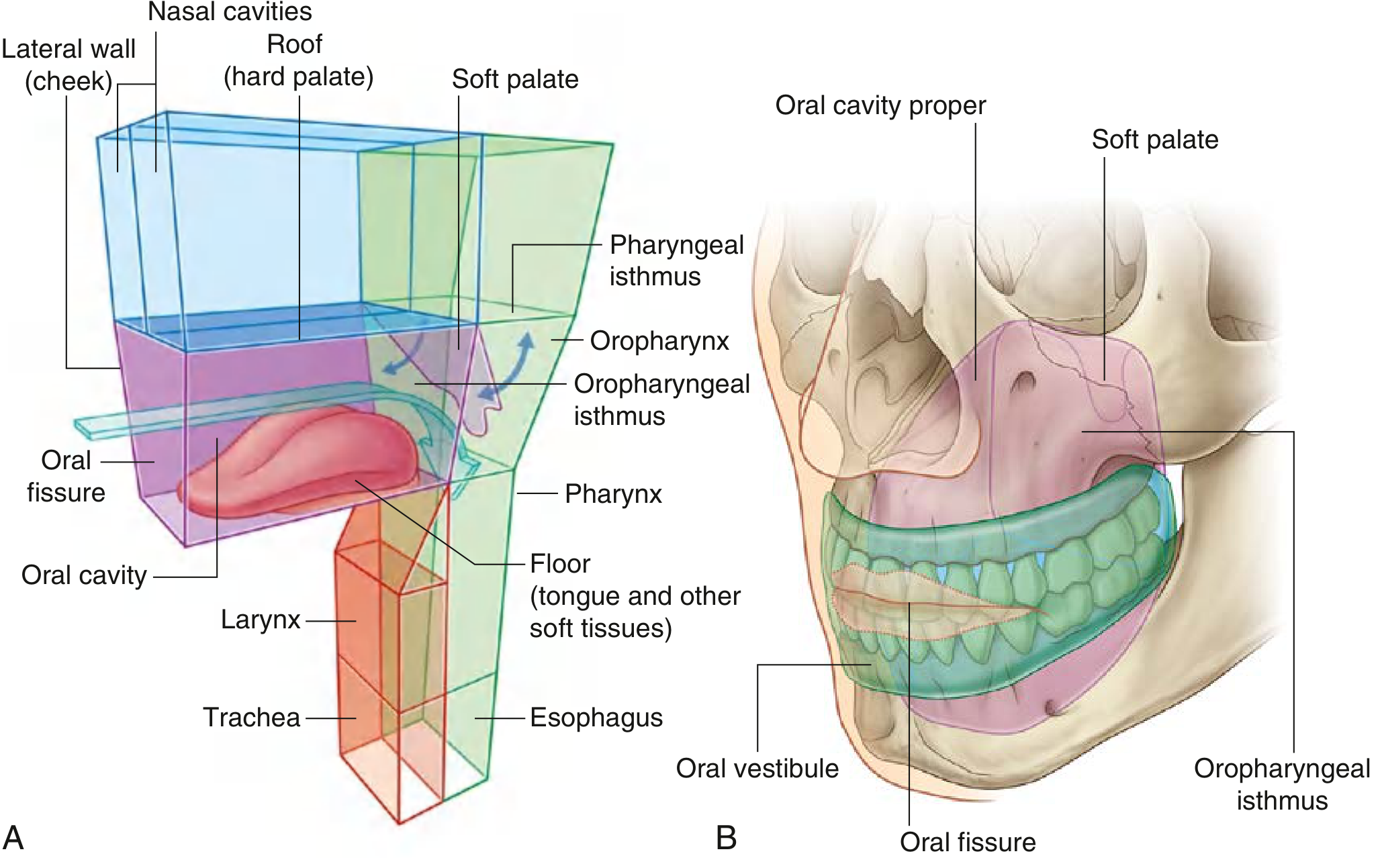
II. BOUNDARIES
| Wall | Structure |
|---|---|
| Roof | Hard palate (anterior 2/3) + Soft palate (posterior 1/3) |
| Floor | Mylohyoid muscle diaphragm, geniohyoid muscles, tongue |
| Lateral walls | Cheeks (buccinator muscle) |
| Anterior opening | Oral fissure (lips) |
| Posterior opening | Oropharyngeal isthmus - communicates with oropharynx |
III. ROOF OF THE ORAL CAVITY - THE PALATE
A. Hard Palate (Anterior 2/3)
- Bony framework: Palatine process of maxilla (anterior) + Horizontal plates of palatine bones (posterior)
- Covering: Stratified squamous epithelium firmly attached to underlying bone
- Foramina:
- Incisive foramen - midline anteriorly; transmits nasopalatine nerve and branch of sphenopalatine artery
- Greater palatine foramen - palpable 1 cm medial to 2nd molar; transmits greater palatine nerve and descending palatine vessels
- Lesser palatine foramen - posterior to greater palatine foramen; transmits lesser palatine nerve and artery to soft palate
- Sensory innervation: Nasopalatine nerve + greater palatine nerve (both from CN V2)
- Blood supply: Maxillary artery → descending palatine artery → greater palatine artery
- Lymphatics: Upper cervical lymphatics or lateral retropharyngeal nodes
B. Soft Palate (Posterior 1/3)
The soft palate attaches to the posterior margin of the hard palate and acts as a "flutter valve":
- Elevation: Closes the pharyngeal isthmus, sealing nasopharynx from oropharynx (prevents food reflux into nasopharynx during swallowing)
- Depression: Closes the oropharyngeal isthmus, sealing oral cavity from oropharynx (allows simultaneous chewing and breathing)
Muscles of the soft palate:
| Muscle | Function | Innervation |
|---|---|---|
| Levator veli palatini | Elevates soft palate, contact with posterior pharyngeal wall | Vagus [X] |
| Tensor veli palatini | Tenses soft palate, opens Eustachian tube | Mandibular nerve [V3] |
| Palatoglossus (anterior pillar) | Approximates palate to tongue, narrows oropharyngeal opening | Vagus [X] |
| Palatopharyngeus (posterior pillar) | Raises larynx and pharynx, closes oropharyngeal aperture | Vagus [X] |
| Musculus uvulae | Shortens uvula | Vagus [X] |
Note: Tensor veli palatini is the only soft palate muscle NOT supplied by the vagus nerve - it is supplied by CN V3.
IV. LATERAL WALLS - THE CHEEKS
- Each cheek consists of: skin (external) → buccinator muscle → oral mucosa (internal)
- Buccinator muscle is the key muscle of the cheeks:
- Origin: Pterygomandibular raphe, alveolar process of maxilla and mandible
- Insertion: Blends with orbicularis oris at the modiolus (fibromuscular nodule at the angle of the mouth)
- Function: Holds cheeks against alveolar arches, keeps food between teeth during chewing
- Innervation (motor): Buccal branch of facial nerve [VII]
- Innervation (sensory): Buccal branch of mandibular nerve [V3]
- The pterygomandibular raphe connects the buccinator anteriorly to the superior pharyngeal constrictor posteriorly, providing structural continuity between the oral and pharyngeal cavities.
V. FLOOR OF THE ORAL CAVITY
Three main structures form the floor:
-
Mylohyoid muscle (paired) - forms a muscular diaphragm filling the U-shaped gap between the two sides of the mandibular body
- Origin: Mylohyoid line of mandible
- Insertion: Body of hyoid bone and a median raphe
- Innervation: Mylohyoid nerve (branch of inferior alveolar nerve, CN V3)
- Function: Elevates floor of mouth and tongue during swallowing; depresses mandible
-
Geniohyoid muscles (paired, cord-like) - run above the mylohyoid, from mental spine of mandible to hyoid bone
- Innervation: C1 fibers traveling with hypoglossal nerve
-
Tongue - overlies the geniohyoid muscles; the largest structure in the oral cavity
Also present: sublingual salivary glands and oral portion of the submandibular glands with their ducts.
VI. THE TONGUE
A. Division
The tongue is divided by the sulcus terminalis (a V-shaped groove) into:
- Anterior 2/3 (oral tongue) - lies within the oral cavity proper
- Posterior 1/3 (tongue base/pharyngeal surface) - lies in the oropharynx
The foramen cecum is a pit at the apex of the sulcus terminalis - the embryological origin point from which the thyroid gland descended (important: source of lingual thyroid and thyroglossal duct cysts).
B. Surface Features
Papillae (cover the anterior 2/3 of dorsal tongue):
| Papilla | Shape | Location | Taste Function |
|---|---|---|---|
| Filiform | Threadlike | Entire anterior 2/3 | None (mechanoreceptors - temperature, texture, pain via CN V) |
| Fungiform | Mushroom-shaped | Anterior 2 cm, diffusely | Yes (taste buds on superior surface) |
| Foliate | Leaf-like folds | Lateral tongue | Yes (taste buds on lateral surface) |
| Circumvallate (vallate) | Large, walled | V-shaped row at junction of anterior and posterior tongue | Yes (taste buds on lateral surfaces) |
Undersurface of tongue: No papillae; has a median mucosal fold (frenulum linguae) that tethers tongue to the floor of mouth. On each side of the frenulum lies a lingual vein, and lateral to this is a fimbriated fold.
Lingual tonsil: Lymphoid tissue on the posterior (pharyngeal) surface of the tongue. No papillae here.
Frenulum: Fold of mucous membrane attaching the inferior anterior tongue to the floor of mouth. Wharton's ducts (submandibular gland ducts) open on either side. Congenital shortness = ankyloglossia (tongue-tie).
C. Muscles of the Tongue
The tongue is completely divided by a median fibrous septum into symmetric left and right halves.
Extrinsic muscles (move the tongue as a whole):
| Muscle | Origin | Insertion | Function | Nerve |
|---|---|---|---|---|
| Genioglossus | Mental spine (superior) of mandible | Body of hyoid + entire length of tongue | Protrudes tongue; depresses center | CN XII |
| Hyoglossus | Body and greater horn of hyoid | Lateral surface of tongue | Depresses and retracts tongue | CN XII |
| Styloglossus | Styloid process of temporal bone | Tip and side of tongue | Retracts and elevates tongue | CN XII |
| Palatoglossus | Palatine aponeurosis of soft palate | Side and dorsum of tongue | Elevates posterior tongue, closes oropharyngeal isthmus | CN X (vagus) |
Intrinsic muscles (alter tongue shape):
| Muscle | Function | Nerve |
|---|---|---|
| Superior longitudinal | Shortens tongue; curls apex and sides upward | CN XII |
| Inferior longitudinal | Shortens tongue; uncurls apex, turns it downward | CN XII |
| Transverse | Narrows and elongates tongue | CN XII |
| Vertical | Flattens and widens tongue | CN XII |
Key Rule: All tongue muscles are supplied by CN XII (hypoglossal), EXCEPT palatoglossus which is supplied by CN X (vagus nerve via the pharyngeal plexus).
D. Blood Supply of the Tongue
- Arterial: Lingual artery (2nd branch of external carotid artery)
- Venous: Lingual vein + vena comitans of the hypoglossal nerve (ranine vein)
E. Lymphatic Drainage of the Tongue
- Tip: Submental nodes (level I)
- Central anterior tongue: Bilateral submandibular and deep cervical nodes
- Lateral anterior tongue: Ipsilateral nodes (levels I-III), may skip to level IV
- Posterior tongue (base): Bilateral deep cervical (jugulodigastric) nodes - clinically important as carcinoma of tongue base metastasizes bilaterally
F. Nerve Supply - Sensory and Motor
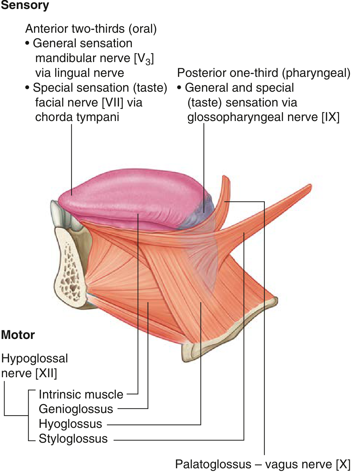
General sensory (touch, pain, temperature):
- Anterior 2/3: Lingual nerve (branch of CN V3 - mandibular nerve)
- Posterior 1/3: Glossopharyngeal nerve (CN IX)
Special sensory (taste):
- Anterior 2/3: Chorda tympani (branch of CN VII - facial nerve), which joins the lingual nerve; fibers travel to geniculate ganglion → nucleus solitarius
- Posterior 1/3: Glossopharyngeal nerve (CN IX) → inferior petrosal ganglion → nucleus solitarius
- Epiglottic taste: Superior laryngeal nerve → Vagus (CN X) → nucleus solitarius
Motor:
- All tongue muscles: Hypoglossal nerve (CN XII)
- Exception - Palatoglossus: Vagus nerve (CN X)
VII. SALIVARY GLANDS AND DUCTS
Major Salivary Glands
| Gland | Duct | Opening |
|---|---|---|
| Parotid | Stensen's duct | Lateral to 2nd upper molar on buccal mucosa |
| Submandibular | Wharton's duct | Floor of mouth on either side of lingual frenulum |
| Sublingual | Ducts of Rivinus (multiple) | Multiple openings into floor of mouth or into submandibular duct |
Saliva production:
- At rest: ~2/3 produced by the submandibular gland
- When stimulated: >1/2 produced by the parotid gland
- Total daily production: 1000-1500 mL/day (~1 mL/min)
- pH: slightly acidic (6-7)
Functions of saliva: lubrication and protection, buffering and clearance, maintenance of tooth integrity, antibacterial activity, taste and digestion.
VIII. SKELETAL FRAMEWORK
Key bones contributing to the oral cavity:
- Maxillae (paired): Palatine process forms anterior 2/3 of hard palate; alveolar process carries upper teeth
- Palatine bones (paired): Horizontal plates form posterior 1/3 of hard palate
- Mandible: Bears lower teeth; body forms the lateral and anterior walls of the floor
- Mylohyoid line on inner surface is the origin of the mylohyoid muscle
- Mandibular foramen on medial ramus transmits inferior alveolar nerve and vessels
- Retromolar triangle and pterygomandibular raphe attachment are located behind last molar
- Hyoid bone: U-shaped bone connecting the floor of oral cavity to the pharynx and larynx below
- Sphenoid: Contributes pterygoid hamulus (for tensor veli palatini and pterygomandibular raphe attachment)
IX. NERVE SUPPLY SUMMARY
| Structure | General Sensation | Taste | Motor |
|---|---|---|---|
| Anterior 2/3 tongue | Lingual nerve (CN V3) | Chorda tympani (CN VII) | CN XII |
| Posterior 1/3 tongue | CN IX | CN IX | CN XII (except palatoglossus - CN X) |
| Hard palate | Nasopalatine + greater palatine (CN V2) | - | - |
| Soft palate | Lesser palatine nerve (CN V2) | - | CN X (except tensor veli palatini - CN V3) |
| Cheeks | Buccal branch (CN V3) | - | Facial nerve (CN VII) |
| Upper teeth and gingiva | CN V2 | - | - |
| Lower teeth and gingiva | Inferior alveolar nerve (CN V3) | - | - |
| Floor of mouth | Lingual nerve (CN V3) | - | CN V3 (mylohyoid) |
X. BLOOD SUPPLY SUMMARY
- Upper oral cavity (palate, upper gingiva): Branches of maxillary artery (descending palatine, sphenopalatine)
- Lower oral cavity and tongue: Lingual artery and facial artery
- Cheeks and lips: Facial artery (superior and inferior labial branches)
XI. LYMPHATIC DRAINAGE SUMMARY
- Upper lip and cheek: Parotid/pre-auricular and submandibular nodes
- Lower lip: Submental (midline) and submandibular (lateral) nodes
- Hard palate: Upper cervical and retropharyngeal nodes
- Tongue tip: Submental nodes
- Tongue body/base: Submandibular → deep cervical (jugulodigastric) nodes; base drains bilaterally
XII. FUNCTIONS OF THE ORAL CAVITY
- Digestion: Initial mechanical processing of food (mastication); initiation of enzymatic digestion by salivary amylase
- Speech: Tongue, lips, teeth, and palate shape sounds produced by the larynx
- Breathing: Alternative airway when nasal passage is obstructed; used clinically for endotracheal intubation
- Swallowing: Oropharyngeal isthmus can be opened/closed by soft palate and tongue movements
CLINICAL PEARLS (ENT Examination Context)
- Tongue deviation on protrusion: Toward the side of CN XII lesion (ipsilateral genioglossus is paralyzed; the intact contralateral side pushes tongue toward the affected side)
- Lingual thyroid / thyroglossal duct cyst: Both arise from incomplete descent of thyroid from the foramen cecum
- Ankyloglossia: Short frenulum restricting tongue movement; may affect speech and breastfeeding
- Submandibular duct stones (sialolithiasis): Wharton's duct is longer and more tortuous than other ducts; most common site for salivary calculi
- Parotid duct (Stensen's duct) pierces the buccinator opposite the upper 2nd molar - a landmark during cheek surgery
- Ludwig's angina: Rapidly spreading cellulitis of the submandibular space and floor of mouth - can elevate the tongue and cause airway compromise
- Unilateral soft palate palsy: Uvula deviates away from the lesion (intact levator pulls uvula to normal side)
Sources: Gray's Anatomy for Students (9780323934237); K.J. Lee's Essential Otolaryngology (9781260122237)
This is a shared conversation. Sign in to Orris to start your own chat.