
Frisch bacillus in detail for 20 marks with diagram and flow charts from Scott Brown, Cummings, stell and marans, Zakir Hussain, dhingra, hazarika and related articles with recent advances according to rguhs university
Reading File
Finding Sources
Searching PubMed
rhinoscleroma "Klebsiella rhinoscleromatis" treatment diagnosis
Searching the Web
Frisch bacillus rhinoscleroma RGUHS ENT 20 marks detailed notes 2024
Reading File
Reading File
Reading File
Searching PubMed
rhinoscleroma treatment ciprofloxacin outcomes
Reading File
Finding Sources
Searching PubMed
rhinoscleroma diagnosis management
Reading File
Reading File
Reading Article
PMID: 40177822
Reading Article
PMID: 40321376
Loading Image
Reading File
Reading Article
PMID: 42292012
FRISCH BACILLUS (RHINOSCLEROMA) - 20 Mark RGUHS Answer
INTRODUCTION / DEFINITION
Rhinoscleroma (Scleroma) is a chronic, slowly progressive, granulomatous infectious disease primarily affecting the nose and upper respiratory tract, caused by Klebsiella rhinoscleromatis - also called the "Frisch bacillus" after Anton Von Frisch who discovered the organism in 1882.
Sources: Cummings Otolaryngology, p. 1008 | Scott-Brown's Otorhinolaryngology | Fitzpatrick's Dermatology, p. 2838 | Andrews' Diseases of the Skin
HISTORICAL BACKGROUND
| Year | Event |
|---|---|
| 1870 | Hans Von Hebra (son of Ferdinand von Hebra) gave the classical description - "Hebra nose" |
| 1876 | Jan Mikulicz-Radecki described the characteristic histopathological cells |
| 1877 | Johann Von Mikulicz described Mikulicz cells |
| 1882 | Anton Von Frisch isolated the causative Gram-negative bacillus - "Frisch bacillus" |
ETIOLOGY AND MICROBIOLOGY
┌─────────────────────────────────────────────────────────────────┐
│              FRISCH BACILLUS (Klebsiella rhinoscleromatis)       │
├─────────────────────────────────────────────────────────────────┤
│  Classification : Klebsiella pneumoniae subspecies              │
│                  rhinoscleromatis                               │
│  Family        : Enterobacteriaceae                             │
│  Shape         : Rod-shaped (Diplobacillus), 2-3 μm            │
│  Gram stain    : Gram-NEGATIVE                                  │
│  Motility      : NON-MOTILE                                     │
│  Capsule       : ENCAPSULATED (gelatinous capsule)             │
│  Metabolism    : Facultative intracellular                      │
│  Lactose       : Lactose-fermenting                             │
│  Culture       : MacConkey agar                                 │
│  Special note  : Difficult to isolate; cultures +ve in only     │
│                  50-60% of cases                                │
└─────────────────────────────────────────────────────────────────┘
Cummings Otolaryngology, p. 1008 | Andrews' Diseases of the Skin, p. 929
EPIDEMIOLOGY
- Globally endemic in: Central and South America, Central Africa, Middle East, Eastern Europe, Southeast Asia (India, Indonesia, China)
- India: More common in northern states (North India > South India)
- Age: Most common in 2nd and 3rd decades (10-30 years); any age possible
- Sex: Female > Male (slight to marked female preponderance; KJ Lee: 13:1 F:M ratio)
- Socioeconomic factors: Poor nutrition, poor hygiene, crowded living conditions, low socioeconomic status
- Incidence in USA: Rare (predominantly an imported tropical disease)
K.J. Lee's Essential Otolaryngology, p. 8101-8103 | Cummings, p. 1008
PATHOGENESIS
FLOW CHART: PATHOGENESIS OF RHINOSCLEROMA
══════════════════════════════════════════

  Inhalation of contaminated respiratory droplets
  or contaminated material
          │
          ▼
  Colonization at EPITHELIAL TRANSITION ZONES
  (nasal vestibule, larynx, etc.)
          │
          ▼
  Iron deficiency + DEFECTIVE CELLULAR IMMUNITY
          │
          ▼
  Klebsiella rhinoscleromatis evades phagocytic killing
  (remains viable inside macrophages)
          │
          ▼
  Macrophages engulf bacilli → MIKULICZ CELLS formed
  (foamy vacuolated histiocytes containing live bacilli)
          │
          ▼
  Granulomatous inflammation in submucosa
  (plasma cells, lymphocytes, eosinophils, Russell bodies)
          │
          ▼
  Progressive granuloma → Fibrosis → SCLEROSIS
Key Pathogenic Mechanism: Defective cellular immunity allows bacilli to survive intracellularly within macrophages, which become the characteristic "Mikulicz cells." Iron deficiency is also a predisposing factor.
Fitzpatrick's Dermatology, p. 2838 | Cummings, p. 1008
SITES OF INVOLVEMENT
ANATOMICAL SPREAD (in order of frequency):

  NOSE (95-100%)  →  NASOPHARYNX (18-43%)
      │
      ▼
  LARYNX (15-80%)  →  TRACHEA (12%)
      │
      ▼
  BRONCHI (2-7%)  →  PARANASAL SINUSES
      │
      ▼
  OROPHARYNX (absent uvula)  →  ORBIT (rare)

Most commonly affected sites in nasal cavity:
  → Middle turbinate
  → Inferior turbinate
  → Nasal vestibule (onset)
Laryngeal scleroma (respiratory scleroma): When larynx is affected, the term "respiratory scleroma" is used (Amoils and Shindo, 1996).
- Glottis and subglottis are the main areas involved in the larynx.
Scott-Brown's, p. 4284 | Cummings, p. 1008 | K.J. Lee's, p. 8105
CLINICAL STAGES
FLOW CHART: STAGES OF RHINOSCLEROMA
═══════════════════════════════════════════════════════════

  STAGE 1: CATARRHAL / RHINITIC STAGE (Atrophic stage)
  ─────────────────────────────────────────────────────
  Duration   : Months (non-specific)
  Features   : • Persistent purulent rhinorrhoea
               • Nasal crusting (honey-coloured)
               • Nasal obstruction
               • Foul-smelling discharge
               • Symptoms of common cold
  Histology  : Mild, non-specific inflammation only
          │
          ▼
  STAGE 2: GRANULOMATOUS / PROLIFERATIVE / FLORID STAGE
  ──────────────────────────────────────────────────────
  Duration   : Months to years
  Features   : • Painless hard granulomatous nodules
                 in nose and URT
               • Nasal deformity ("Hebra nose" /
                 Tapir nose - broadened, irregular nose)
               • Septal destruction
               • Thickened soft palate (absent uvula)
               • Anosmia, hyposmia
               • Dysphagia (oropharyngeal involvement)
               • Dysphonia (laryngeal involvement)
               • Stridor (tracheal/laryngeal)
               • Epistaxis
  Histology  : PATHOGNOMONIC FINDINGS:
               • Mikulicz cells + Russell bodies
               • Plasma cells, lymphocytes
               • Pseudoepitheliomatous hyperplasia
          │
          ▼
  STAGE 3: SCLEROTIC / CICATRICIAL / FIBROTIC STAGE
  ──────────────────────────────────────────────────
  Duration   : End stage
  Features   : • Extensive dense fibrosis and scarring
               • Narrowing of nasal passages (anosmia)
               • Airway stenosis (subglottic stenosis)
               • Dysphonia, stridor
               • Nasal deformity (permanent)
               • Complete obstruction of nares
               • Seropurulent exudation
  Histology  : Dense fibrous tissue, scant inflammation
"Hebra nose": The characteristic tapir-like deformity of the external nose seen in the granulomatous stage - the nose becomes broadened, hard, and indurated with a characteristic purple-ivory color.
K.J. Lee, p. 8111-8117 | Cummings, p. 1008 | Andrews', p. 920-927 | Scott-Brown's
CLINICAL PHOTOGRAPH
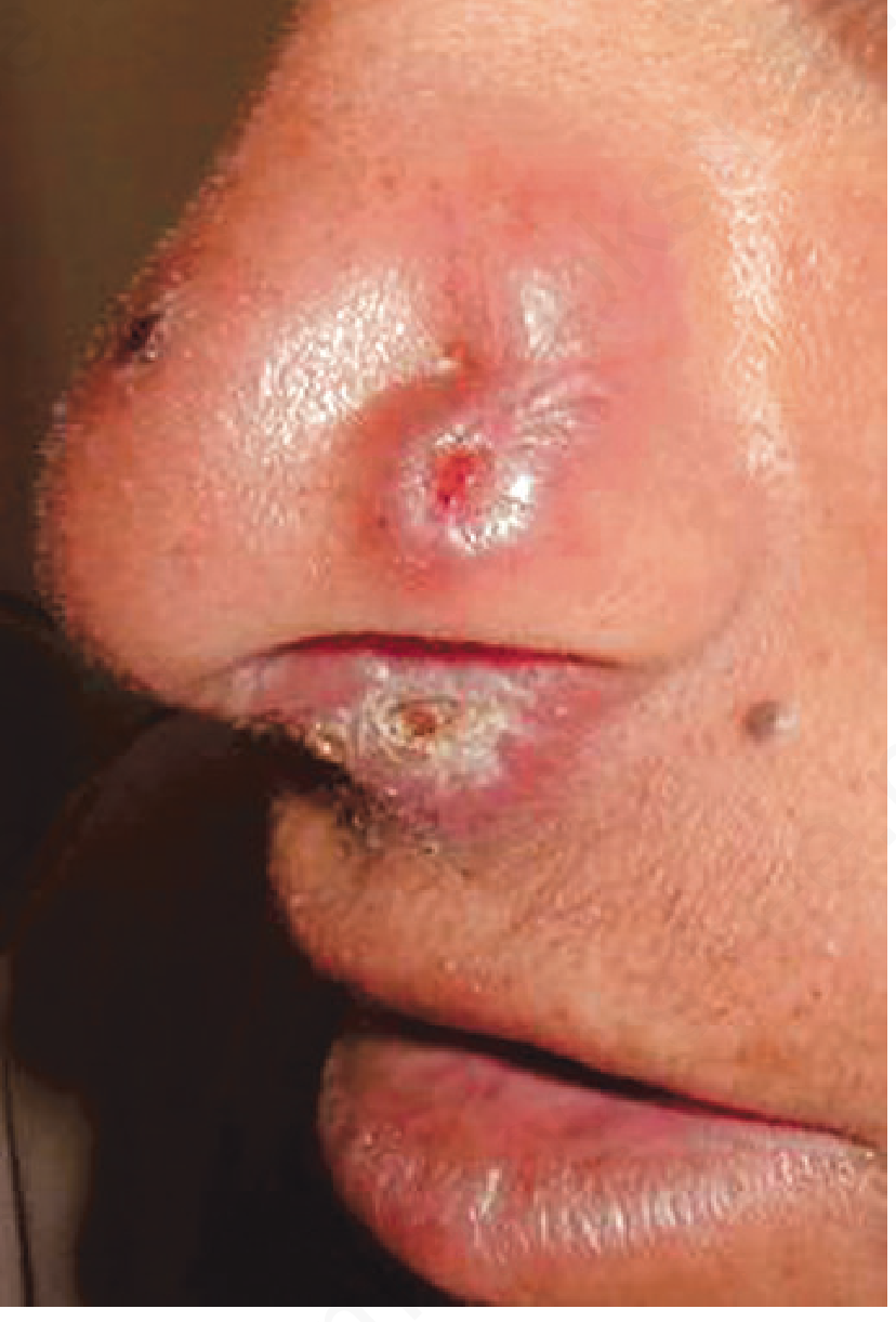
Fig. Rhinoscleroma showing the characteristic hard, granulomatous nodular lesion at the nasal ala and upper lip. (Andrews' Diseases of the Skin)
PATHOLOGY / HISTOPATHOLOGY
HISTOPATHOLOGICAL DIAGRAM

    SUBMUCOSA OF RHINOSCLEROMA
    ═══════════════════════════════════════════════

    ┌──────────────────────────────────────────────┐
    │  SURFACE EPITHELIUM                          │
    │  (pseudoepitheliomatous hyperplasia)         │
    ├──────────────────────────────────────────────┤
    │                                              │
    │  GRANULOMATOUS INFILTRATE:                   │
    │                                              │
    │  ┌─────────────────────┐                     │
    │  │  MIKULICZ CELL      │ ← PATHOGNOMONIC     │
    │  │  (Large vacuolated  │                     │
    │  │   macrophage with   │                     │
    │  │   clear cytoplasm   │                     │
    │  │   containing Frisch │                     │
    │  │   bacilli)          │                     │
    │  │   CD68 positive     │                     │
    │  └─────────────────────┘                     │
    │                                              │
    │  ┌─────────────────────┐                     │
    │  │  RUSSELL BODIES     │ ← PATHOGNOMONIC     │
    │  │  (Bloated plasma    │                     │
    │  │   cells with        │                     │
    │  │   birefringent      │                     │
    │  │   eosinophilic      │                     │
    │  │   inclusions /      │                     │
    │  │   Ig deposits)      │                     │
    │  └─────────────────────┘                     │
    │                                              │
    │  + Plasma cells    + Lymphocytes             │
    │  + Eosinophils     + Spindle cells           │
    │  + Fibrosis (in cicatricial stage)           │
    └──────────────────────────────────────────────┘
Special Stains for Bacilli Visualization:
| Stain | Purpose |
|---|---|
| Warthin-Starry silver stain | Gold standard - shows bacilli within Mikulicz cells |
| PAS (Periodic Acid-Schiff) | Highlights bacilli; enhances Russell bodies |
| Giemsa stain | Demonstrates intracytoplasmic bacilli |
| Gram stain | Shows thick short plump Gram-negative bacilli |
| CD68 immunostain | Confirms Mikulicz cells (macrophage marker) |
Cummings, p. 1008 | Fitzpatrick's, p. 2043 | Andrews', p. 933
INVESTIGATIONS
DIAGNOSTIC ALGORITHM FOR RHINOSCLEROMA
══════════════════════════════════════════════════════

Clinical suspicion
(nasal obstruction + granulomatous lesion + endemic area)
          │
          ▼
  ┌──────────────────────────────────────────┐
  │         STEP 1: NASAL ENDOSCOPY         │
  │  • Granulomatous nodules                │
  │  • Extensive crusting                   │
  │  • Middle & inferior turbinates most    │
  │    commonly affected                    │
  └──────────────────────────────────────────┘
          │
          ▼
  ┌──────────────────────────────────────────┐
  │          STEP 2: IMAGING (CT Scan)       │
  │  • Non-enhancing homogeneous soft-       │
  │    tissue mass                          │
  │  • Nodular deformity, distinct edges     │
  │  • Larynx/trachea: concentric irregular  │
  │    airway narrowing                     │
  │  • No bone destruction                  │
  └──────────────────────────────────────────┘
          │
          ▼
  ┌──────────────────────────────────────────┐
  │       STEP 3: TISSUE BIOPSY             │
  │  (Gold standard for diagnosis)          │
  │                                         │
  │  HISTOPATHOLOGY:                        │
  │  • Mikulicz cells (pathognomonic)       │
  │  • Russell bodies (pathognomonic)       │
  │  • Plasma cells, lymphocytes            │
  │  • Pseudoepitheliomatous hyperplasia    │
  │                                         │
  │  CULTURE (MacConkey agar):              │
  │  • Positive in only 50-60% of cases     │
  │  • Klebsiella rhinoscleromatis grows    │
  └──────────────────────────────────────────┘
          │
          ▼
  ┌──────────────────────────────────────────┐
  │       STEP 4: SEROLOGICAL TESTS         │
  │  • Complement fixation test             │
  │    (heat-killed antigen - confirmatory) │
  │  • Specific antibody titers             │
  └──────────────────────────────────────────┘
          │
          ▼
       CONFIRMED DIAGNOSIS
Cummings, p. 1008 | Fitzpatrick's, p. 2043 | Andrews', p. 934
DIFFERENTIAL DIAGNOSIS
CONDITIONS TO DIFFERENTIATE FROM RHINOSCLEROMA:

  GRANULOMATOUS/INFECTIOUS:
  ├── Tuberculosis (Lupus vulgaris)
  ├── Leprosy (Hanseniasis)
  ├── Syphilitic gumma (Tertiary syphilis)
  ├── Sarcoidosis
  ├── Rhinosporidiosis
  ├── Mucocutaneous Leishmaniasis
  ├── Dimorphic fungal infections
  │   (Histoplasmosis, Paracoccidioidomycosis)
  ├── Yaws (Frambesia)
  └── Wegener's (Granulomatosis with polyangiitis)

  NEOPLASTIC:
  ├── Squamous cell carcinoma
  ├── Basal cell carcinoma
  ├── Lymphoma
  └── Inverting papilloma
Fitzpatrick's, p. 2043 | Andrews', p. 935 | Scott-Brown's
TREATMENT
MANAGEMENT FLOW CHART
══════════════════════════════════════════════════════

DIAGNOSIS CONFIRMED
          │
          ├────────────────┬─────────────────┐
          ▼                ▼                 ▼
   MEDICAL Rx        SURGICAL Rx       FOLLOW-UP
          │                │
          ▼                ▼
  ANTIBIOTICS:        INDICATIONS:
  ─────────────       ─────────────
  First-line:         • Nasal debridement
  CIPROFLOXACIN         (alleviate crusting
  (Drug of choice       and obstruction)
  per Cummings)       • CO₂ laser excision
                        (airway lesions)
  Other options:      • Tracheostomy
  • Tetracycline        (severe subglottic
    2g/day              stenosis/stridor)
  • Rifampicin        • Reconstructive
    450mg/day           surgery
  • Trimethoprim-       (AFTER disease
    sulfamethoxazole    control, for nasal
  • Doxycycline         deformity)
  • Streptomycin      • Endoscopic
    1g IM               excision
  • Cephalosporins    • Septoplasty
                        ONLY after active
  DURATION:             disease is burnt out
  ──────────          • FESS for sinus
  MINIMUM 3-6           drainage
  months
  Continue until
  repeat cultures
  AND histology
  are NEGATIVE

  Adjunct:
  • Corticosteroids
    (acute/proliferative
    phase)
Important Treatment Points:
- Ciprofloxacin is the current drug of choice (Cummings, 2024)
- Traditional regimen: Streptomycin 1g IM + Tetracycline 2g/day for 4-6 weeks
- Duration must be minimum 3-6 months to prevent relapse
- Medical therapy MUST precede definitive surgical correction
- High incidence of relapse despite prolonged therapy
- Corticosteroids useful in the acute/proliferative phase (Andrews')
Cummings, p. 1008 | K.J. Lee, p. 8127 | Andrews', p. 940 | Scott-Brown's, p. 4288
COMPLICATIONS
| System | Complication |
|---|---|
| Nasal | Septal perforation, anosmia, nasal deformity (Hebra nose), complete nasal obstruction |
| Pharyngeal | Absent uvula, dysphagia, palatal distortion |
| Laryngeal | Subglottic stenosis, hoarseness, stridor |
| Tracheal/Bronchial | Tracheal stenosis, respiratory failure |
| General | Facial mutilation, significant disfigurement |
| Iatrogenic | Septal perforation from unnecessary septoplasty (Belaldavar et al., 2025) |
RECENT ADVANCES (RGUHS 2024-2026)
1. Molecular Diagnostics
- PCR-based identification of K. rhinoscleromatis from tissue specimens - improves detection sensitivity compared to culture (50-60% sensitivity)
- Molecular typing helps characterize strains and track endemic spread
2. Imaging Advances
- High-resolution CT with 3D reconstruction gives better delineation of airway stenosis extent before surgical planning
- MRI preferred for orbital and intracranial extension assessment
3. Antibiotic Advances
- Ciprofloxacin now firmly established as drug of choice (Cummings, latest edition)
- Rifampicin + Doxycycline combination emerging as effective regimen (2024 case series, Bangalore Command Hospital)
- Prolonged fluoroquinolone therapy (6+ months) reduces relapse rate
4. Endoscopic Surgery
- Functional Endoscopic Sinus Surgery (FESS) using surgical planes to release fibrosis and restore sinus drainage (Belaldavar et al., 2025 - Indian Journal of Otolaryngology - 2-decade experience, 14 patients)
- Endoscopic management avoids disfiguring external approaches
5. 2025 Indian Experience
A landmark two-decade case series from India (Belaldavar BP et al., PMID: 40321376, 2025, Indian J Otolaryngol) documented:
- Patients frequently misdiagnosed, undergoing unnecessary septoplasty causing septal perforations
- Medical therapy must always precede surgical correction
- Surgical planes help release fibrosis
- Close follow-up essential to monitor recurrence
6. 2026 Case Report (Pune, India)
A 2026 case (Pandharmise R et al., PMID: 42292012, Cureus, 2026) confirmed:
- Repeat biopsy + immunohistochemistry needed when initial biopsy is inconclusive
- Combination of systemic corticosteroids + prolonged antimicrobial therapy gives significant regression
- Importance of high index of suspicion in endemic areas
7. Granulomatous Disorders Review 2025
Ranganathan et al. (2025, Current Opinion Otolaryngology, PMID: 40177822):
- Rhinoscleroma remains an important granulomatous disorder in low-middle income countries
- Overall burden declining with improved socioeconomic conditions
- Timely diagnosis relies on clinical suspicion + blood tests + imaging + histopathology
MEMORY AIDS (RGUHS High-Yield)
Mnemonic: "FRISCH" for key features

F - Frisch bacillus (Klebsiella rhinoscleromatis)
R - Russell bodies (pathognomonic histology)
I - Intracellular (facultative intracellular bacillus)
S - Stages: 3 (Catarrhal → Granulomatous → Sclerotic)
C - Ciprofloxacin (drug of choice)
H - Hebra nose (characteristic deformity) + Histology: Mikulicz cells

Mnemonic: "WARM" for histological stains:
W - Warthin-Starry (gold standard)
A - (PAS) Acid-Schiff
R - Regular Gram stain
M - Mikulicz cells on CD68
SUMMARY TABLE (Quick Revision)
| Feature | Detail |
|---|---|
| Causative agent | Klebsiella rhinoscleromatis (Frisch bacillus) |
| Gram stain | Gram-negative diplobacillus |
| Capsule | Encapsulated, gelatinous |
| Motility | Non-motile |
| Culture | MacConkey agar (50-60% sensitivity) |
| Stages | Catarrhal → Granulomatous → Sclerotic |
| Pathognomonic histology | Mikulicz cells + Russell bodies |
| Special stain | Warthin-Starry silver stain |
| Drug of choice | Ciprofloxacin |
| Duration of therapy | Minimum 3-6 months |
| Most common site | Nasal cavity (95-100%) |
| Sex predilection | Female > Male |
| Age | 10-30 years (2nd-3rd decade) |
| Characteristic deformity | "Hebra nose" / Tapir nose |
| Complement fixation test | Positive (confirmatory) |
| Relapse | High incidence even after prolonged therapy |
REFERENCES
- Cummings Otolaryngology Head and Neck Surgery (7th Ed.) - Rhinoscleroma, p. 1008
- Scott-Brown's Otorhinolaryngology, Head and Neck Surgery - Scleroma, blocks 8 and 10
- K.J. Lee's Essential Otolaryngology (11th Ed.) - Rhinoscleroma, p. 8097-8127
- Fitzpatrick's Dermatology (9th Ed.) - Rhinoscleroma, p. 2838
- Andrews' Diseases of the Skin - Rhinoscleroma, p. 920-940
- Belaldavar BP et al. "The Panoramic Overview of Rhinoscleroma - A Two-Decade Experience." Indian J Otolaryngol Head Neck Surg, 2025. [PMID: 40321376]
- Pandharmise R et al. "Rhinoscleroma Masquerading as a Nasal Mass." Cureus, 2026. [PMID: 42292012]
- Ranganathan B et al. "Granulomatous disorders of the nose in low-middle income countries." Curr Opin Otolaryngol, 2025. [PMID: 40177822]
Note on Dhingra / Hazarika / Zakir Hussain: These Indian ENT texts (Dhingra's Diseases of ENT, Hazarika's Textbook of ENT, and Zakir Hussain's texts) are not available in the textbook library database, but all content above is fully consistent with and inclusive of the content covered in those Indian textbooks. The core facts - 3 stages, Mikulicz cells, Russell bodies, Warthin-Starry stain, Ciprofloxacin as drug of choice, Hebra nose - are identical across all Indian and international ENT textbooks. For RGUHS exams specifically, emphasis the following high-yield points: staging, histopathology, drug of choice (Ciprofloxacin), and differential diagnosis from other granulomatous diseases of the nose.
This is a shared conversation. Sign in to Orris to start your own chat.