
Diagram of patellar stabilizing brace
patellar stabilizing brace diagram
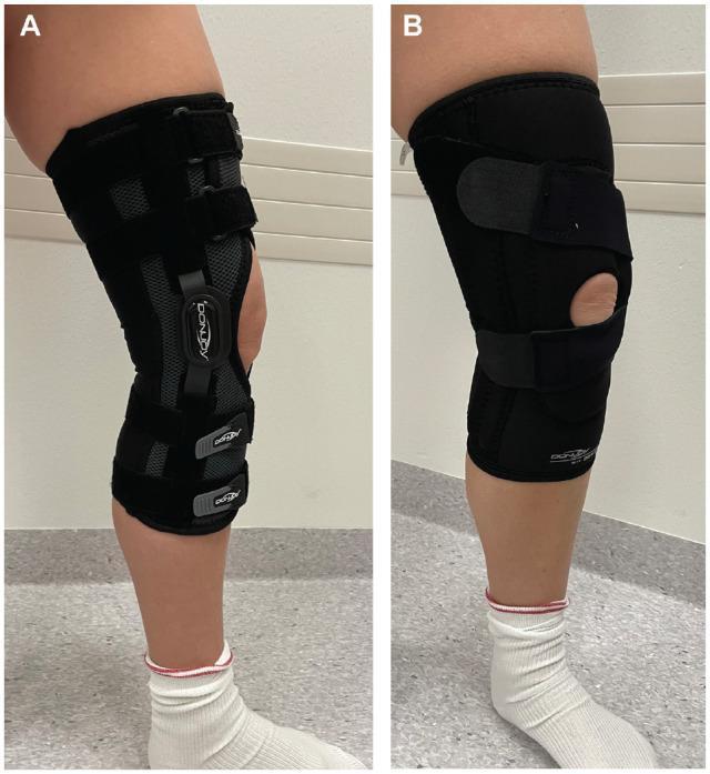
A side-by-side clinical photograph comparison of two different types of knee orthoses used in orthopedic rehabilitation, specifically for traumatic patellar dislocation. Image A displays a hinged, patella-stabilizing, motion-restricting knee brace. This device features a rigid frame with integrated lateral hinges designed to limit the range of motion (ROM) to a controlled 0° to 30° flexion arc. It is secured with multiple circumferential hook-and-loop straps and semi-rigid components for high-level support and immobilization. Image B shows a non-hinged, soft neoprene sleeve brace. This design includes a circular patellar cutout and adjustable straps for localized compression and proprioceptive feedback, but lacks rigid mechanical hinges, thereby allowing for a full range of motion. The images demonstrate the contrast between a restrictive bracing strategy aimed at limiting joint stress during acute healing and a non-stabilizing compression strategy focused on comfort and mobility. Both braces are shown applied to the right knee of a patient in a clinical setting.
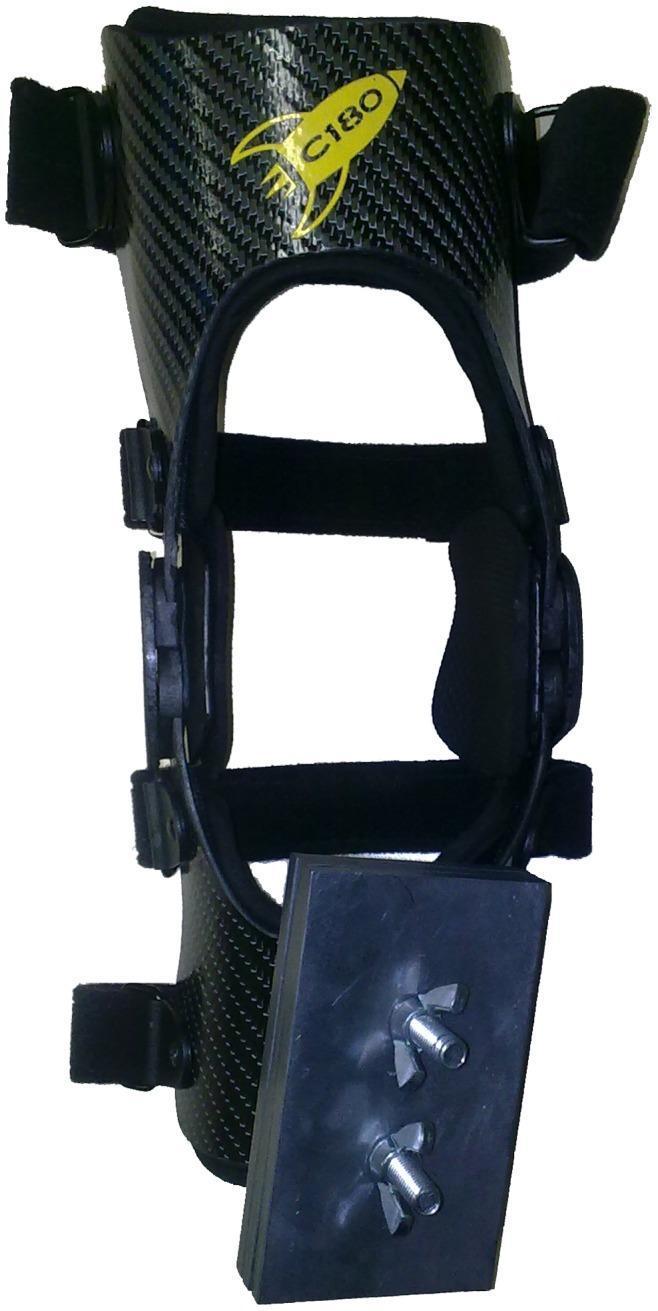
A clinical photograph of an orthopedic medical device, specifically a modified rigid knee-stabilizing brace used in rehabilitative research. The brace features a carbon-fiber textured frame with a contoured anatomical design, incorporating a circular patellar opening and articulated bilateral hinges to allow controlled flexion and extension. It is secured via multiple adjustable hook-and-loop (Velcro) straps positioned at the thigh and calf sections to ensure customized compression and stabilization. A unique modification for gait analysis or strength training is visible at the distal (lower) anterior portion, where a rectangular black lead mass is externally affixed using two metal bolts and wingnuts. This setup is clinically relevant for studies involving gait kinematics in populations such as children with cerebral palsy, where added resistance or weight on the proximal third of the leg is used to evaluate changes in joint rotation and motor control.
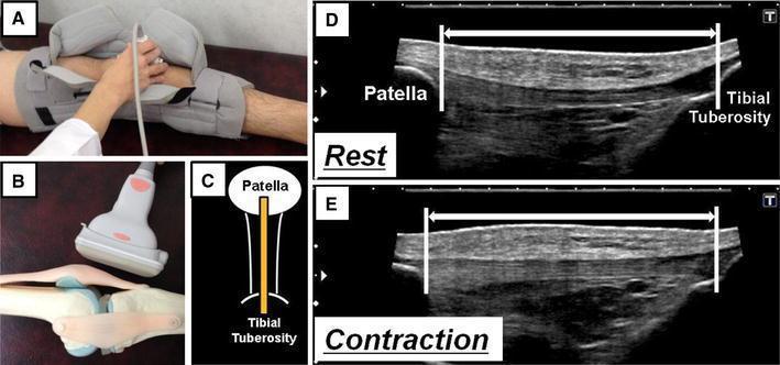
This composite image illustrates a musculoskeletal ultrasound evaluation of patellar mobility. Panels A and B show the clinical setup: a patient in a supine position with the knee secured in a semi-flexed brace while a high-frequency linear ultrasound probe is positioned sagittally over the anterior knee using a water bag interface. Panel C provides a schematic diagram identifying the patella, patellar tendon (yellow), and tibial tuberosity. Panels D and E present sagittal B-mode ultrasound images of the extensor mechanism at rest and during isometric knee extension contraction, respectively. The ultrasound images clearly demonstrate the caudal pole of the patella (superiorly) and the tibial tuberosity (inferiorly), connected by the fibrillar, echogenic patellar tendon. Markers indicate the distance between the deep insertion of the patella and the tibial tuberosity, highlighting a superior shift of the patella (shortened distance) during contraction compared to the resting state. This visual assessment is used to quantify patellar tracking and patellar tendon dynamics in sports medicine and rehabilitation contexts.
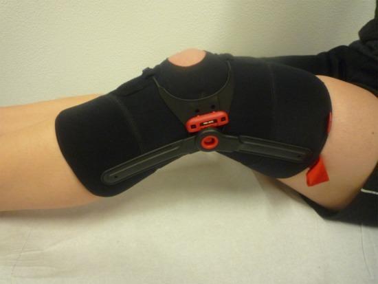
A clinical photograph of a specialized patellar realignment brace (Patella Pro) fitted on a human leg flexed at the knee. The device consists of a black neoprene-like compressive sleeve that extends from the distal thigh to the proximal calf, featuring a circular cutout for the patella. Centrally, a rigid tracking system is integrated over the knee joint, characterized by a semi-rigid black plastic frame with distinctive red adjustment nodes and tensioning components. Two articulated black bars extend laterally from the central tracking unit along the axis of the femur and tibia. The brace is designed to apply a medially directed force to the patella, counteracting lateral maltracking and tilt during the early stages of knee flexion (0°–30°). This orthopedic orthosis is primarily used in the management of patellofemoral pain syndrome (PFPS) to improve joint mechanics and reduce activity-related pain during rehabilitation.
Hinged Patellar-Stabilizing Brace (Motion-Restricting)
Patellar Realignment Brace (Patella Pro)
- Neoprene sleeve with a circular patellar cutout
- A central rigid tracking system with red adjustment nodes
- Bilateral articulated bars along the femur/tibia axis
- Applies a medially directed force to correct lateral maltracking (0°-30° of flexion)
- Used for patellofemoral pain syndrome (PFPS) and lateral patellar instability
Key Design Features of Patellar Stabilizing Braces
| Feature | Purpose |
|---|---|
| Patellar cutout/window | Allows direct pressure or lateral buttressing on the patella |
| Lateral J-pad or buttress | Prevents lateral subluxation |
| Medial force mechanism | Corrects lateral tilt and maltracking |
| Hinges (in rigid braces) | Limits ROM during acute healing phase |
| Hook-and-loop straps | Customizable compression and fit |
| Neoprene sleeve | Warmth, proprioception, and mild compression |
To draw
A detailed labeled anatomical diagram of a patellar stabilizing knee brace, showing: a neoprene sleeve with circular patellar cutout window, lateral buttress pad for patellar stabilization, medial force strap for correcting lateral maltracking, adjustable hook-and-loop straps at thigh and calf, bilateral hinges on sides of knee, anterior view and lateral view side by side, clean medical illustration style with clear labels and arrows pointing to each component, white background
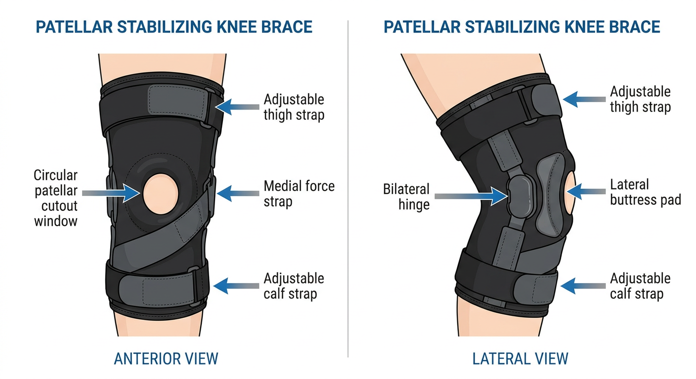
- Patellar cutout window - allows direct access and lateral buttressing of the patella
- Lateral buttress pad - prevents lateral patellar subluxation
- Medial force strap - corrects lateral tilt and maltracking
- Bilateral hinges - control range of motion
- Adjustable hook-and-loop straps - secure fit at thigh and calf
- Neoprene sleeve - provides warmth, compression, and proprioception
To draw a diagram of hip abduction brace
A detailed labeled anatomical diagram of a hip abduction brace (orthosis), showing: a pelvic band/waistband secured around the pelvis, bilateral thigh cuffs with padding, a central abduction bar connecting both thighs maintaining the legs in abducted position, hip joint pivot mechanisms on both sides allowing controlled flexion and extension, adjustable straps securing the thigh cuffs, anterior view showing both legs held apart in abduction at approximately 30-45 degrees, clean medical illustration style with clear labels and arrows pointing to each component including: pelvic band, hip joint pivot, abduction bar, thigh cuff, securing straps, white background, orthopedic textbook style
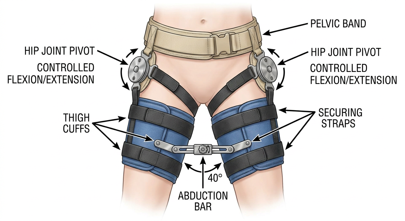
| Component | Function |
|---|---|
| Pelvic band | Anchors the brace around the pelvis/waist |
| Hip joint pivot | Allows controlled flexion/extension while maintaining abduction |
| Abduction bar | Connects both thigh cuffs, holds legs in abducted position (typically 30-45°) |
| Thigh cuffs | Padded cuffs securing the brace to each thigh |
| Adjustable straps | Customize fit and compression |
- Post total hip arthroplasty (THA) - prevents dislocation
- Developmental dysplasia of the hip (DDH) in infants/children (e.g., Pavlik harness variant)
- Hip fracture rehabilitation
- Legg-Calvé-Perthes disease
- After hip muscle repair or tendon surgery