
in articular cartilage injury, why it appears dark in mri
Articular cartilage appears dark on MRI primarily because it has a very low water content relative to other tissues, and its tightly packed collagen matrix restricts water mobility - leading to short T2 relaxation times and low signal on standard sequences.
Searching sources...
Reading File
Finding Sources
Finding Sources
Reading File
Finding Sources
Finding Sources
Reading File
Finding Sources
Now I have excellent content from the textbook. Let me also fetch the key figure showing the three-layer cartilage pattern.
Loading Image
Finding Sources
I now have all the information needed for a thorough answer. Here is the complete explanation:
Why Articular Cartilage (and Its Injury) Appears Dark on MRI
Normal Cartilage Has a Trilaminar Appearance
On routine T2-weighted MRI, normal articular cartilage is NOT simply dark - it shows a three-layer pattern:
| Layer | MRI Signal | Reason |
|---|---|---|
| Deep layer (adjacent to subchondral bone) | Low signal (dark) | Collagen fibers oriented perpendicular to the field; calcified layer with little free water |
| Middle/radial zone | Intermediate signal | Mixed collagen orientation, more free water |
| Superficial zone (lamina splendans) | Low signal (dark) | Collagen fibers oriented parallel to the surface; tightly packed, restricted water |
This regional variation is "largely due to T2 variations caused by the orientation of the collagen fibrils relative to the magnetic field" - Imaging Anatomy: Bones, Joints, Vessels & Nerves, Vol. 3
The Core Physics: Why Collagen Orientation = Dark Signal
MRI signal intensity on T2W images depends on how freely water molecules move (tumble) in tissue:
- Free water tumbles fast → long T2 → bright signal
- Bound water (water tightly associated with macromolecules like collagen) tumbles slowly → short T2 → dark signal
Articular cartilage contains:
- Type II collagen in tightly packed, highly organized arrays
- Water makes up 70-75% of cartilage bulk, but much of it is bound to the collagen-proteoglycan matrix, not free
- The tightly bound water has a very short T2 relaxation time, producing low (dark) signal
The Magic Angle Effect
When collagen fibers run at 55° to the main magnetic field (B0), a phenomenon called the magic angle effect occurs - the dipolar interactions between water protons cancel out and signal increases (becomes bright). This is why the middle transitional zone, where collagen runs obliquely, appears brighter than the superficial and deep zones where fibers run parallel or perpendicular to the field.
Why Injured/Damaged Cartilage Appears Dark
In cartilage injury, the signal changes depend on what has happened to the matrix:
| Pathology | MRI Appearance | Mechanism |
|---|---|---|
| Loss of proteoglycans (early degeneration) | Variable - often dark on T1W | Proteoglycans normally bind water; their loss reduces bound water content and disrupts normal signal |
| Collagen disorganization | Loss of normal trilaminar pattern | Normal signal depends on ordered collagen orientation; disruption abolishes the magic-angle gradient |
| Fibrillation / surface irregularity | Low signal defect on T2W | Physical loss of cartilage tissue = signal void |
| Full-thickness defect | Focal dark area with fluid filling | Absent cartilage replaced by low-signal scar or exposed bone |
| Calcified cartilage | Dark on both T1W and T2W | Calcium has no mobile protons; both sequences show signal void |
| Fibrocartilage repair tissue | Low signal | Type I collagen (scar cartilage) has even tighter water binding than normal hyaline cartilage (type II) |
The textbook specifically notes: "Abnormal signal within the damaged articular cartilage is a common finding in chondromalacia, fibrocartilage formation after injury, chondrocalcinosis, and cartilage defects."
Zonal Architecture - Key to Understanding Signal Loss
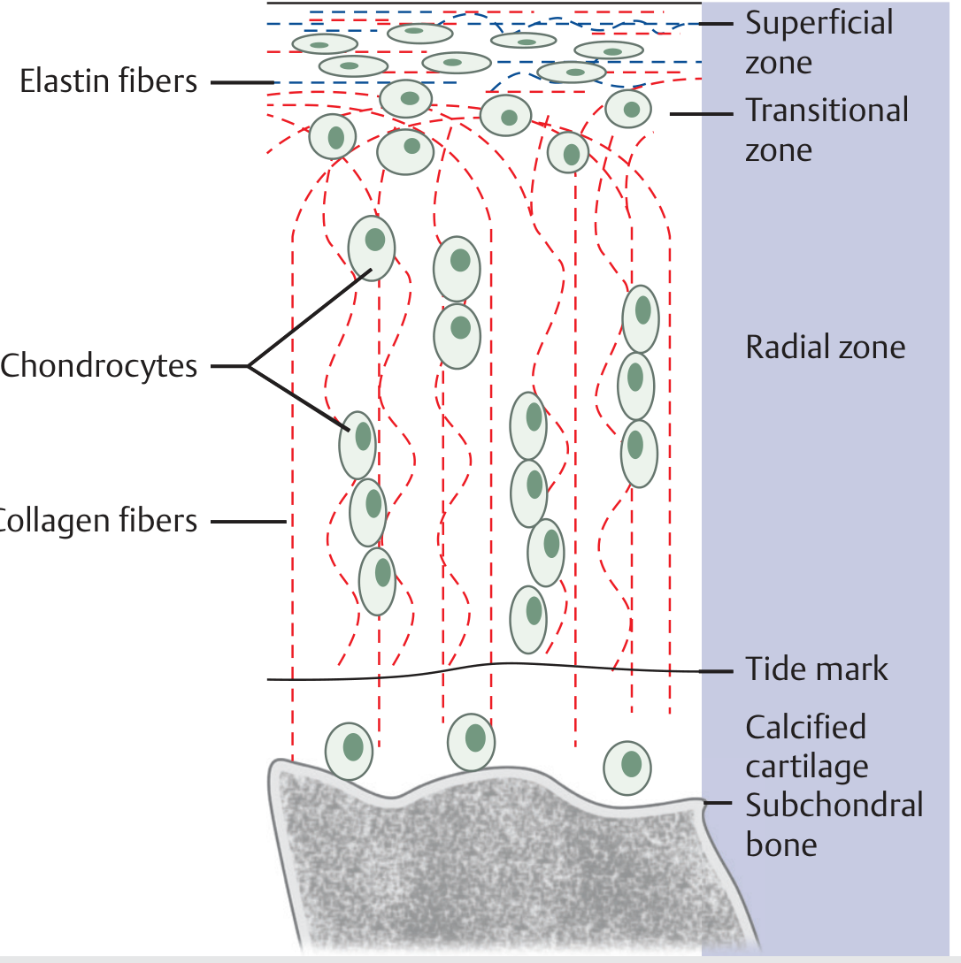
The four zones of articular cartilage. The ordered arrangement of collagen fibers (shown in red) is the structural basis for the MRI signal pattern. Injury disrupts this order.
- Superficial zone: Collagen parallel to surface - normally dark, injury causes surface fissures visible as defects
- Transitional zone: Oblique collagen - normally intermediate signal
- Radial zone: Perpendicular collagen - normally dark; this is the thickest zone and most important for load bearing
- Calcified cartilage + tidemark: Always dark due to calcium and dense matrix
Best MRI Sequences for Cartilage Evaluation
- T2W with fat suppression: Best contrast between cartilage surface and joint fluid; detects defects
- Proton density-weighted (PD): Best for internal cartilage abnormalities and surface defects
- Gradient echo (GRE/SPGR): Increased signal in articular cartilage, better visualization of the three layers
- T1W: Shows hyaline cartilage contents but poor contrast with adjacent joint fluid
The key clinical point: On T2W MRI, loss of the normal trilaminar signal pattern - particularly disruption of the dark-bright-dark layering - is an early sign of cartilage injury even before a frank defect is visible.
Source: Imaging Anatomy Text and Atlas Volume 3, Bones, Joints, Vessels and Nerves
This is a shared conversation. Sign in to Orris to start your own chat.