
Polymorphous light eruption
"polymorphous light eruption"
polymorphic light eruption diagnosis prevention treatment DermNet
polymorphous light eruption clinical photos papular rash sun exposed skin
https://dermnetnz.org/topics/polymorphic-light-eruption
Typical features
- Itchy or burning red bumps, small blisters, or patchy plaques
- Appears a few hours to 1-4 days after strong sun exposure
- Often affects the upper chest, neck, outer arms, and legs. The face is often relatively spared.
- Common in spring or early summer, or after a sunny holiday
- Clears over days to a few weeks if further UV exposure is avoided
- Often improves later in summer as the skin develops tolerance, called hardening
Cause
Management and prevention
- Avoid further sun exposure during an active flare.
- Use broad-spectrum, water-resistant SPF 50+ sunscreen, applied generously before exposure and reapplied every 2 hours and after swimming.
- Wear protective clothing, a broad-brimmed hat, and seek shade.
- Increase early-season sun exposure gradually rather than suddenly.
- Cool emollients or calamine can relieve discomfort. An oral antihistamine may help itch.
- A clinician may prescribe a topical corticosteroid for a significant flare. People with recurrent severe PMLE may be referred for pre-season UVB phototherapy or other specialist treatment.
Important alternatives to rule out
Morphia in derma
morphea
morphea diagnosis treatment DermNet
morphea localized scleroderma clinical photograph plaque
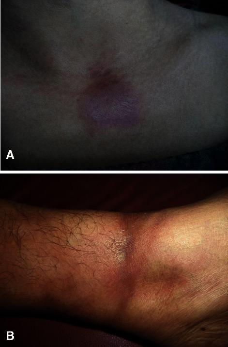
Clinical photograph presenting two distinct morphological variants of morphea (localized scleroderma). Image A displays plaque-type morphea on the upper chest, characterized by a solitary, well-demarcated, oval-shaped plaque with brownish hyperpigmentation and a visibly indurated, slightly waxy texture. Image B illustrates linear morphea on the lower leg, featuring a band-like, hyperpigmented, and indurated lesion that circumferentially involves the limb. Both images demonstrate the characteristic skin thickening and pigmentary changes associated with localized scleroderma. These visuals serve as a comparative educational tool for distinguishing between localized plaque and linear distribution patterns in dermatological diagnosis. The lesions signify an underlying autoimmune-mediated inflammatory process leading to increased collagen deposition in the dermis.
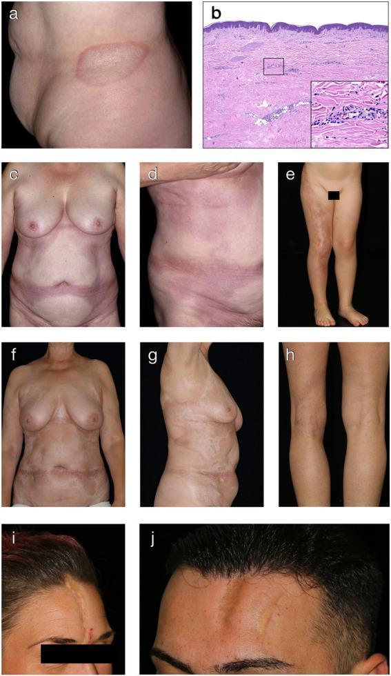
A composite medical image illustrating various clinical and histological presentations of morphea (localized scleroderma). (A) Clinical photograph of plaque-type morphea on the trunk, showing a well-defined oval patch with a sclerotic, ivory-white center and a peripheral erythematous-violaceous 'lilac ring' indicative of active inflammation. (B) H&E stained histopathology (40x, 200x) showing a thinned epidermis, significant dermal sclerosis with thick eosinophilic collagen bundles, and periadnexal lymphocytic infiltrates. (C-D, F-G) Examples of generalized morphea on the torso, progressing from early inflammatory erythematous-violaceous coalescing patches (C-D) to late-stage sclerotic pink-white patches with central induration (F-G). (E) Linear morphea presenting as a band-like atrophic hyperpigmented plaque following Blaschko lines on a limb. (H) Deep morphea showing limb asymmetry due to underlying tissue atrophy. (I-J) Linear morphea en coup de sabre, characterized by depressed, sclerotic paramedian forehead plaques and cicatricial alopecia. This collection serves as a diagnostic guide for recognizing different clinical subtypes and evolutionary stages of morphea.
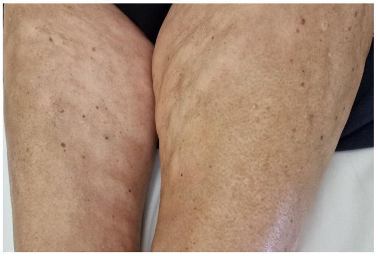
This clinical photograph displays the inner thighs of a patient exhibiting signs of plaque-type morphea, a localized form of scleroderma. The skin demonstrates significant morphological changes, characterized by multiple hypopigmented, ivory-colored plaques. These lesions are primarily round to oval, varying in size, with some appearing as isolated spots while others have coalesced into larger, irregularly shaped patches. The surface texture of the affected skin shows subtle central induration and unevenness, consistent with cutaneous sclerosis. In addition to the large hypopigmented regions, there are scattered small, hyperpigmented macules and nevi throughout the field. The distribution is bilateral and asymmetric. This visual serves as an educational example of localized scleroderma (morphea) in the context of systemic conditions like monoclonal gammopathy, illustrating characteristic central whiteness and loss of normal skin texture within active or chronic plaques.
https://dermnetnz.org/topics/morphoea
Morphea
Clinical appearance and stages
- Early active stage: pink, red, or violaceous patch, often with a peripheral lilac ring
- Sclerotic stage: central ivory-white or yellow-white, shiny, hard, bound-down plaque
- Late stage: brown or lighter pigmentation, thinning/atrophy, possible hair loss and reduced sweating in the area
Main types
- Plaque morphea: one or several oval, localized hardened plaques. Most common adult form.
- Linear morphea: a band-like line of thickened skin, commonly in children; may cross a joint and restrict growth or movement.
- Generalized morphea: many coalescing plaques over several body regions.
- Deep morphea / morphea profunda: extends into subcutaneous tissue, fascia, muscle, or bone.
- En coup de sabre: linear, depressed plaque on the forehead/scalp, sometimes associated with hair loss and eye or neurologic complications.
Is it systemic sclerosis?
Diagnosis
- Skin biopsy
- Blood tests, mainly to assess associated autoimmune features or exclude alternatives
- MRI or ultrasound if deep disease, a joint-crossing lesion, or craniofacial linear morphea is suspected
Treatment
- Limited superficial plaque disease: observation, moisturisers, topical corticosteroid or tacrolimus in selected active lesions, and sometimes phototherapy
- More extensive superficial disease: phototherapy, especially UVA1 where available
- Active linear, generalized, deep, or function-threatening disease: dermatologist-led systemic treatment, commonly methotrexate, sometimes with systemic corticosteroids initially; mycophenolate may be an alternative
- Physiotherapy/occupational therapy: particularly important when a limb or joint is involved, to prevent contracture and preserve motion