
Points to remember of meiosis and mitosis for final examination
Reading File
Now let me search the medical library for meiosis and mitosis content in parallel.
Finding Sources
Searching the Web
meiosis mitosis key points differences exam summary
Now let me read the best textbook sources - Thompson & Thompson Genetics (has both mitosis and meiosis sections) and Histology (detailed mitosis phases), plus Ganong's physiology section.
Reading File
Reading File
Finding Sources
Let me read the Thompson & Thompson meiosis section and the Histology detailed meiosis section in parallel:
Reading File
Reading File
Reading File
Let me also grab the meiosis phases from Thompson & Thompson and the Ganong physiology section:
Reading File
Reading File
Reading File
I now have comprehensive information from the textbooks. Let me also fetch the key comparison diagram:
Loading Image
I now have everything needed for a thorough exam-ready answer. Here is the complete study guide:
Meiosis & Mitosis - Exam Points to Remember
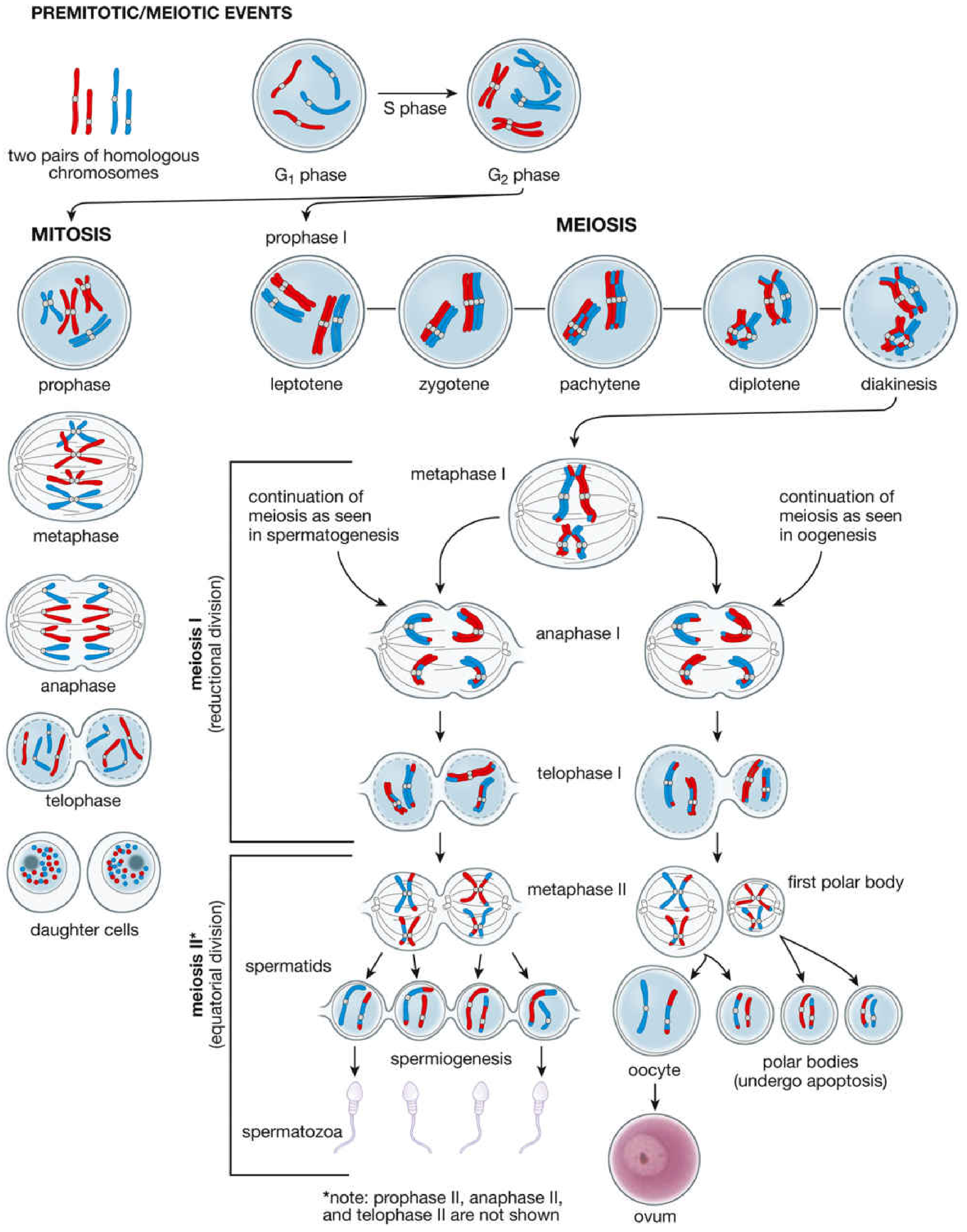
MITOSIS
Definition
Mitosis is a process of chromosome segregation and nuclear division followed by cell division that produces two daughter cells with the same chromosome number and DNA content as the parent cell. - Histology: A Text and Atlas
The Cell Cycle (must know)
| Phase | Key Events |
|---|---|
| G1 (Gap 1) | Cell growth; preparation for DNA synthesis |
| S phase | DNA replication - chromosome number stays 2n, but DNA content doubles (2n → 4d) |
| G2 (Gap 2) | Cell growth continues; chromosomes begin condensing |
| M phase | Mitosis + cytokinesis |
| G0 | Quiescent/arrested state (some cells exit cycle here) |
Key point: At the start of S phase, chromosome number = 2n, DNA = 2d. At the end of S phase, chromosome number = 2n, DNA = 4d. - Histology: A Text and Atlas
Five Stages of Mitosis (PPMAT)
- Prophase - Chromosomes condense and become visible; each consists of two sister chromatids held by cohesins; mitotic spindle begins to form from centrioles; nucleolus disappears
- Prometaphase - Nuclear envelope dissolves into small vesicles (resembles sER); kinetochore protein complexes appear on each chromatid; microtubules attach to kinetochores
- Metaphase - Chromosomes are maximally condensed; align at the equatorial plane (metaphase plate); spindle has 3 microtubule types: astral, polar, and kinetochore microtubules
- Anaphase - Centromeres split; sister chromatids separate and migrate to opposite poles; each chromatid now becomes an independent daughter chromosome
- Telophase - Chromosomes decondense; nuclear envelope re-forms around each daughter nucleus; followed by cytokinesis (cleavage of cytoplasm)
The term mitosis describes karyokinesis (nuclear division); cytokinesis = cytoplasmic division. Together they complete cell division. - Histology: A Text and Atlas
Key Points - Mitosis
- Produces 2 daughter cells, genetically identical to parent
- Occurs in somatic (body) cells - for growth, tissue renewal, wound repair, embryonic development
- DNA content: 4d → 2d in each daughter cell
- Chromosomes can be best analyzed at metaphase or prometaphase (maximally condensed)
- Mitotic nondisjunction in somatic cells → chromosomal mosaicism (e.g., some cases of Down syndrome) and is a step in tumor formation - Thompson & Thompson Genetics
MEIOSIS
Definition
Meiosis involves two sequential nuclear divisions followed by cell divisions that produce 4 haploid (1n) gametes from a single diploid (2n) cell, each with half the chromosomes and half the DNA of somatic cells. - Histology: A Text and Atlas
Overall Structure
- One S phase (DNA replication: 2n → 4d) followed by two divisions
- No S phase between meiosis I and meiosis II
- Meiosis I = Reductional division (2n → 1n; 4d → 2d)
- Meiosis II = Equatorial division (1n stays 1n; 2d → 1d)
MEIOSIS I (Reductional Division)
Prophase I - THE MOST COMPLEX STAGE (has 5 sub-stages: LZPDD)
| Sub-stage | Key Event |
|---|---|
| Leptotene | Chromosomes begin to condense |
| Zygotene | Homologous chromosomes begin to align (synapsis begins); paired homologues = bivalents |
| Pachytene | Synapsis complete; crossing over (genetic recombination) takes place; synaptonemal complex present |
| Diplotene | Synaptonemal complex breaks down; crossover points visible as chiasmata |
| Diakinesis | Chromosomes maximally condensed; further preparation for division |
The synaptonemal complex is a ribbonlike proteinaceous structure essential to recombination. It holds paired homologues (bivalents) together during synapsis. - Thompson & Thompson Genetics
Metaphase I
- Nuclear membrane disappears; spindle forms
- Bivalents (homologous pairs) align at equatorial plane with centromeres oriented toward opposite poles
- This is different from mitosis (where individual chromosomes align, not pairs)
Anaphase I
- Homologous chromosomes (not sister chromatids) separate and move to opposite poles - this is disjunction
- Chromosome number halved: 2n → 1n
- The 23 pairs assort independently → 2²³ (>8 million) possible combinations
- Each chromosome is now a mix of paternal and maternal segments due to crossing over
Telophase I + Cytokinesis
- Two haploid cells formed, each with 1n chromosomes but 2d DNA (still two chromatids per chromosome)
MEIOSIS II (Equatorial Division)
- Resembles mitosis but no preceding S phase
- Chromosome number does not change (stays 1n)
- DNA content reduced from 2d → 1d
- Sister chromatids separate in anaphase II
- Result: 4 haploid cells with 1n chromosomes and 1d DNA
MALE vs FEMALE MEIOSIS
| Feature | Spermatogenesis (Male) | Oogenesis (Female) |
|---|---|---|
| Products | 4 spermatids - all equal, structurally identical | 1 ovum + 3 polar bodies |
| Cytoplasm | Equally distributed | Unequal - ovum gets most cytoplasm |
| Polar bodies | Not formed | Polar bodies receive little cytoplasm and degenerate |
| Functional gametes | All 4 spermatids become spermatozoa | Only 1 ovum is functional |
In humans, the first polar body does not divide (differs from some other species). - Histology: A Text and Atlas
GENETIC VARIATION FROM MEIOSIS
Two mechanisms ensure each gamete is genetically unique:
- Crossing over (recombination) - homologous segments of DNA are exchanged between non-sister chromatids during pachytene; creates novel combinations within each chromosome
- Independent assortment - random orientation of bivalents at metaphase I gives 2²³ (>8 million) chromosome combinations
A typical large human chromosome contains 3-5 segments alternately of paternal and maternal origin after crossing over. - Thompson & Thompson Genetics
MEDICAL RELEVANCE (High-yield exam points)
| Condition | Mechanism |
|---|---|
| Trisomy (e.g., Down syndrome - Trisomy 21) | Meiotic nondisjunction - most commonly in oogenesis; most common mutational mechanism in humans |
| Chromosomal mosaicism | Mitotic nondisjunction early after fertilization |
| Aneuploidy in tumors | Mitotic nondisjunction in somatic cells; abnormal chromosome segregation in rapidly dividing tissues (e.g., colon cancer) |
Meiotic nondisjunction, particularly in oogenesis, is responsible for chromosomally abnormal fetuses in at least several percent of all recognized pregnancies. - Thompson & Thompson Genetics
COMPARISON TABLE - MITOSIS vs MEIOSIS
| Feature | Mitosis | Meiosis |
|---|---|---|
| Where it occurs | Somatic cells | Germ cells (gonads) |
| Purpose | Growth, repair, replacement | Gamete production |
| Number of divisions | 1 | 2 |
| Daughter cells | 2 | 4 |
| Chromosome number | 2n → 2n (diploid) | 2n → 1n (haploid) |
| DNA content | 4d → 2d | 4d → 1d |
| Genetic identity | Identical to parent | Unique (crossing over + independent assortment) |
| Synapsis / crossing over | NO | YES (Prophase I) |
| Homologue pairing | NO | YES |
| Anaphase event | Sister chromatids separate | Homologues separate (Meiosis I); then chromatids separate (Meiosis II) |
| Prophase complexity | Simple | Complex (5 sub-stages) |
| Duration | Relatively short | Much longer (especially in females - oocytes arrested in prophase I for years) |
QUICK MNEMONICS
- Mitosis stages: PPMAT - Prophase, Prometaphase, Metaphase, Anaphase, Telophase
- Prophase I sub-stages: LZPDD - Leptotene, Zygotene, Pachytene, Diplotene, Diakinesis
- Crossing over happens at Pachytene (P for Pachytene = P for recombination/Pairing done)
- Chiasmata are visible at Diplotene (the physical cross-shaped structures where exchange occurred)
- Meiosis I = Reduction (number drops); Meiosis II = Equatorial (like mini-mitosis, splits chromatids)
Sources: Thompson & Thompson Genetics and Genomics in Medicine, 9th ed. | Histology: A Text and Atlas with Correlated Cell and Molecular Biology | Ganong's Review of Medical Physiology, 26th ed.
This is a shared conversation. Sign in to Orris to start your own chat.