
Give single rooted teeth photo
Reading File
Searching sources...
Finding Sources
Finding Sources
Finding Sources
Finding Sources
Reading File
Reading File
Now let me find the actual images of teeth from the textbooks:
Finding Sources
Reading File
Reading File
Found the images. Let me fetch both:
Loading Image
Loading Image
Here are the single-rooted teeth images from the medical textbooks:
Single-Rooted Teeth
Single-rooted teeth include: Incisors (central and lateral) and Canines. These are the teeth with one root each.
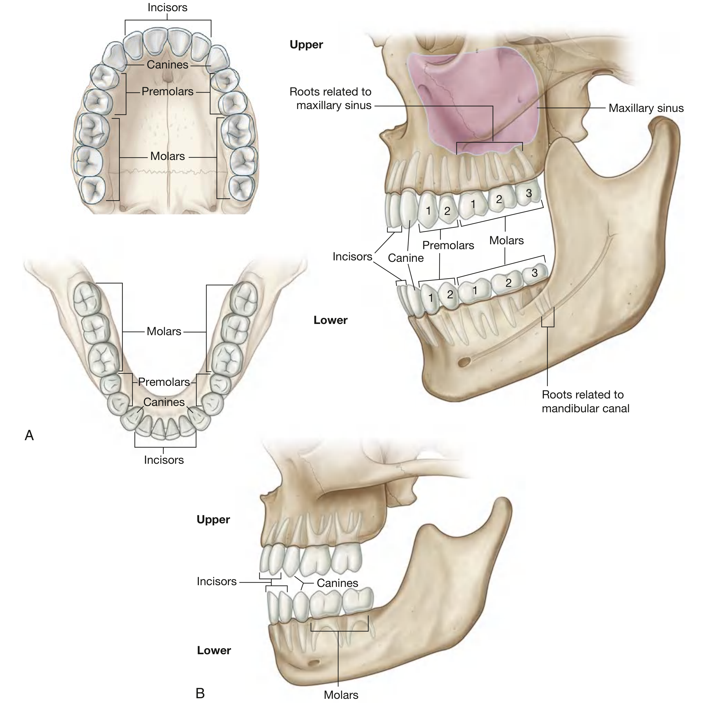
Fig. 8.281 - Adult upper and lower permanent teeth (A) and deciduous teeth (B). Note the incisors and canines anteriorly, which are single-rooted. - Gray's Anatomy for Students
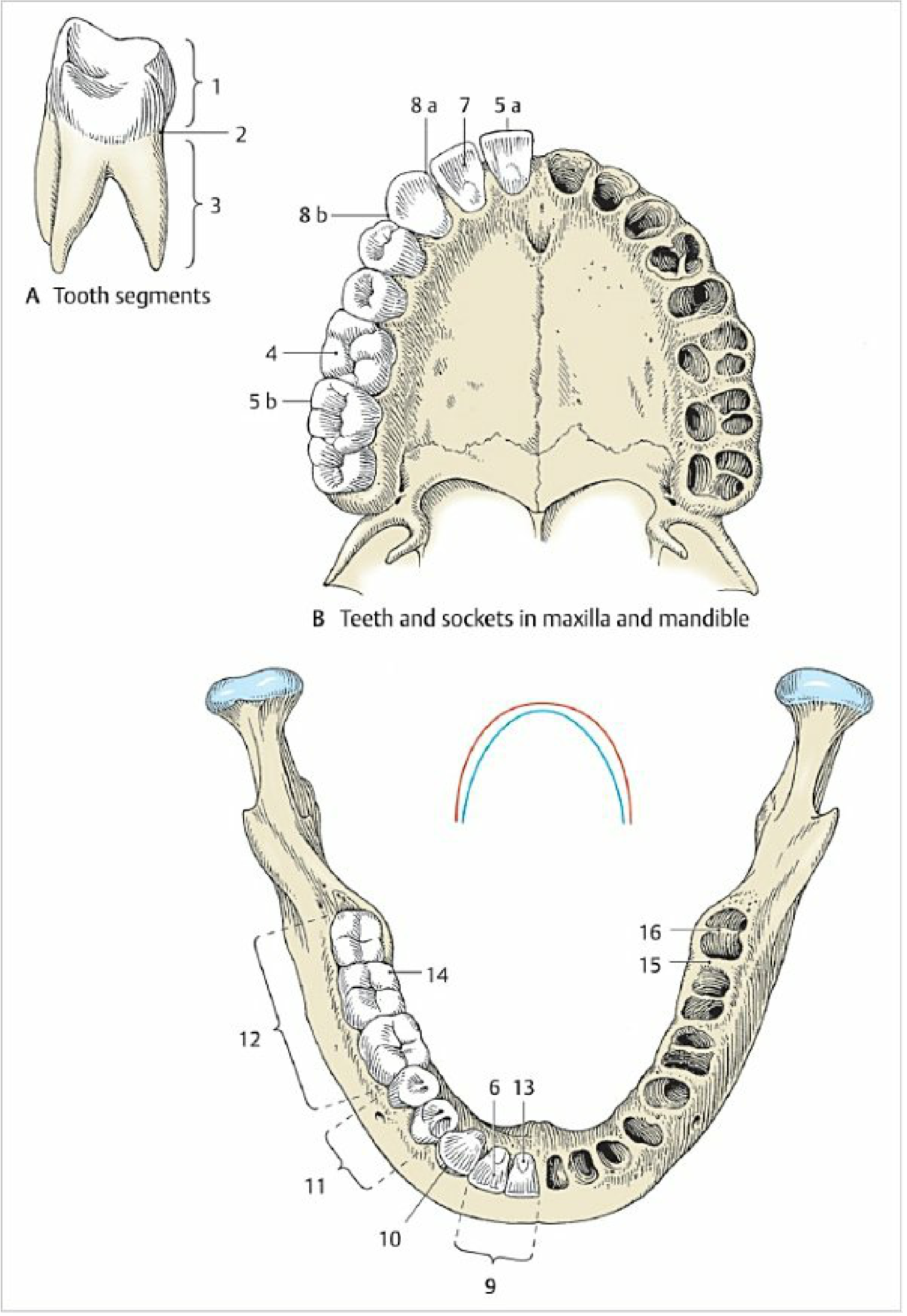
Fig. 4.9 - Teeth and sockets in the maxilla and mandible. The single-rooted incisor and canine sockets (anterior) are clearly distinct from multi-rooted molar sockets. - Color Atlas of Human Anatomy, Vol. 2
Summary of Single-Rooted Teeth
| Tooth | Root Count | Function |
|---|---|---|
| Central Incisor | 1 (long, conical) | Cutting |
| Lateral Incisor | 1 (long, conical) | Cutting |
| Canine | 1 (longest root in mouth) | Tearing/Grasping |
| 1st Premolar (lower) | Usually 1 | Grinding |
| 2nd Premolar | Usually 1 | Grinding |
Note: Upper first premolars may occasionally have two roots. The lower premolars are typically single-rooted. Molars are multi-rooted (3 roots upper, 2 roots lower).
Sources: Gray's Anatomy for Students; Color Atlas of Human Anatomy, Internal Organs Vol. 2
Photos
Finding Sources
Searching the Web
single rooted teeth clinical photo incisor canine anatomy
Finding Sources
Loading Image
Loading Image
Loading Image
Loading Image
Here are multiple photos/diagrams of single-rooted teeth from medical textbooks and anatomy references:
1. Longitudinal Section of a Human Canine Tooth (single-rooted)
The classic single-rooted tooth cross-section showing crown, neck, and one root with all internal structures:

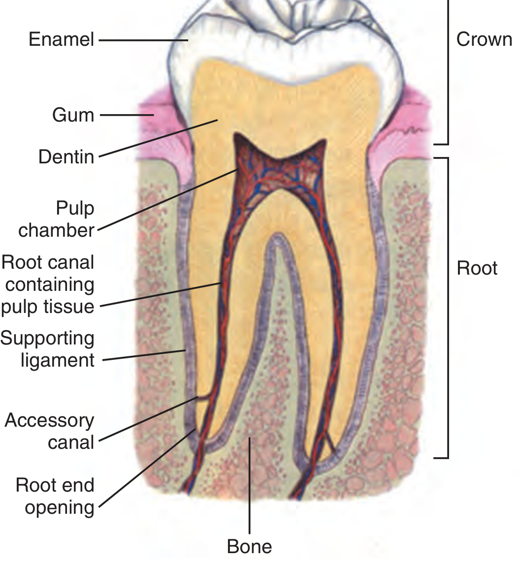
2. Dental Anatomic Unit - Single-Rooted Tooth Internal Structure
From Roberts & Hedges' Clinical Procedures in Emergency Medicine - shows enamel, dentin, pulp, root canal, and supporting ligament of a single-rooted tooth:
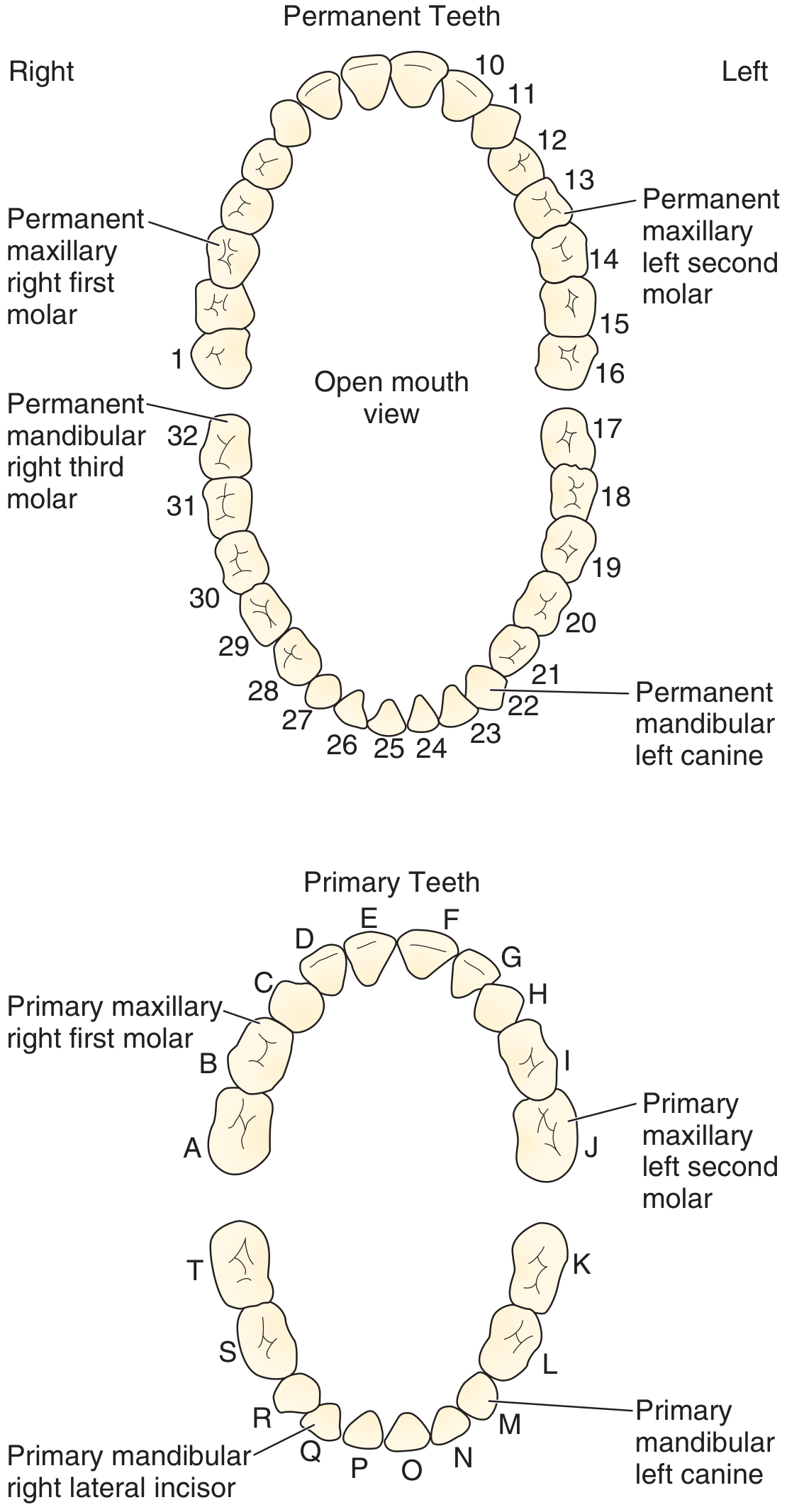
3. Permanent & Primary Teeth Identification Chart
Incisors (front teeth, #6-11 upper, #22-27 lower) and canines are all single-rooted:
Key Points on Single-Rooted Teeth
| Tooth | Notes |
|---|---|
| Central Incisor | 1 root, chisel-shaped crown, cuts food |
| Lateral Incisor | 1 root, similar to central but smaller |
| Canine | 1 root - the longest root in the mouth, pointed cusp |
| Lower Premolars | Usually 1 root (upper 1st premolar may have 2) |
Sources: Roberts & Hedges' Clinical Procedures in Emergency Medicine; Gray's Anatomy for Students; Color Atlas of Human Anatomy Vol. 2
This is a shared conversation. Sign in to Orris to start your own chat.