
Explain me about types of autorefractometers subjective and objective with photos of bith and them and priciple of working and mechanism of it uses advantage and disadvantages of both
Reading File
Now let me search the medical library and web simultaneously for comprehensive content on autorefractometers.
Finding Sources
Searching the Web
types of autorefractometers subjective objective principle working mechanism advantages disadvantages
Finding Sources
Reading Web Page
https://www.ncbi.nlm.nih.gov/books/NBK580520
Searching the Web
subjective autorefractor Badal optometer photo objective autorefractor clinical image Scheiner principle
Reading Web Page
https://uomus.edu.iq/img/lectures21/MUCLecture_2024_11193175…
Searching the Web
objective autorefractor machine photo Nidek Topcon AND subjective autorefractor phoropter photo clinical use ophthalmology
Loading Image
Loading Image
Loading Image
Loading Image
Searching the Web
Badal optometer subjective autorefractor image phoropter subjective refraction device photo ophthalmology
Loading Image
Loading Image
Loading Image
Now I have comprehensive information and images to produce a thorough, well-illustrated answer.
Autorefractometers: Subjective and Objective - Complete Guide
An autorefractometer (or optometer) is an automated electronic instrument that measures the refractive error of the eye - the sphere, cylinder, and axis - to determine the correct spectacle or contact lens prescription. Two broad categories exist: subjective and objective autorefractometers.
1. Historical Background
- Early subjective optometers (1895-1920): Required the patient to manually adjust optics until the target appeared clearest. Examples: Badal optometer, Young's optometer. They fell out of favor due to instrument accommodation (the eye relaxes focus toward the instrument instead of infinity).
- Early objective optometers: Required the examiner to judge when the retinal image appeared sharpest. Less accurate than retinoscopy.
- Modern autorefractors (post-1960): Electronic, computer-controlled, use infrared light, CCD cameras, and microprocessors. No patient input required.
2. Subjective Autorefractometers
What They Are
Subjective autorefractors require active patient cooperation - the patient adjusts the instrument or responds to questions until they achieve the sharpest, most comfortable image. They are essentially automated phoropters or optometers that guide the patient through the refraction process.
Examples
- Badal Optometer (historical, first developed ~1880s by Father Badal)
- Young's Optometer
- Modern digital phoropters (e.g., Nidek RT-5100, Marco TRS-5100)
- Portable subjective autorefractors (e.g., NeOptix handheld devices)
Photo - Modern Subjective Refraction (Phoropter)
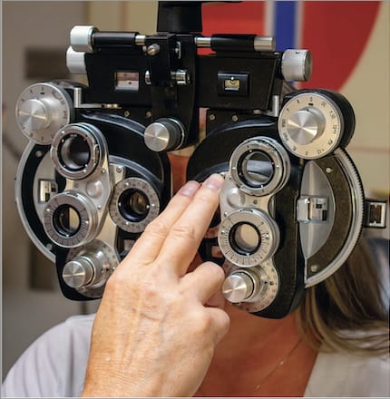
A phoropter (the classic subjective refraction instrument) - the patient indicates which lens setting gives the clearest vision.
Portable Subjective Autorefractor (Modern)

A modern handheld subjective optometer design - patient adjusts focus until image is sharp.
Principle of Working
The Badal optometer principle underlies most subjective devices:
- A high-powered lens (the Badal lens) is placed at its own focal length from the eye's far point
- Moving the target through this lens changes the vergence reaching the eye linearly - 1 mm of movement = 1 diopter change, regardless of the patient's refractive error
- The patient reports when the target appears sharpest - at that point, the instrument lens position is converted into a diopter reading
- Astigmatism is measured by rotating a target (e.g., fan or cross chart) and adjusting cylinder until all lines appear equally sharp
Mechanism
- Patient places their eye at the eyepiece
- A visible fixation target (letters, symbols) is presented at a virtual optical infinity via the Badal lens
- Patient adjusts focusing knobs (or responds to prompts on digital phoropters) until the target is sharpest
- The instrument records lens position and calculates sphere, cylinder, axis
- Multiple meridians are tested (typically sphere, then astigmatic correction)
Advantages of Subjective Autorefractors
| Advantage | Detail |
|---|---|
| Gold standard accuracy | Incorporates patient's own visual experience and neural processing |
| Better final prescription | Result reflects what the patient actually sees best, not just optical power |
| Accounts for higher-order aberrations | Patient selects best perceived image, not just best optical focus |
| No accommodation artifact | Modern digital phoropters can fog the eye before testing |
| Works post-refractive surgery | More reliable than objective in eyes with irregular optics |
| Presbyopia correction | Patient can titrate near addition directly |
| Patient confidence | Patient feels involved in the process |
Disadvantages of Subjective Autorefractors
| Disadvantage | Detail |
|---|---|
| Requires cooperation | Cannot be used in non-communicative patients, very young children, cognitive impairment |
| Instrument accommodation | Eye may relax focus toward the device rather than optical infinity, causing myopic over-minus |
| Time-consuming | Considerably longer than objective refraction |
| Examiner skill dependent | Requires trained optometrist/ophthalmologist to guide the test |
| Subjective variability | Patient responses may fluctuate; same patient may give different answers at different visits |
| Not usable under cycloplegia alone | Cycloplegic drops eliminate accommodation but patient must still be able to respond |
3. Objective Autorefractometers
What They Are
Objective autorefractors measure refractive error without any input from the patient. They project infrared light into the eye, analyze the reflected wavefront from the retina, and compute sphere, cylinder, and axis automatically. These are the most commonly used autorefractors today.
Examples
- Nidek ARK-1e (Scheiner-based)
- Topcon KR-1, KR-800, KR-800S
- Huvitz HRK-8000A
- Reichert RA-600
- Canon RK-F2
- Welch Allyn SureSight (handheld)
- Plusoptix A12 (pediatric, open-field)
Photo - Objective Autorefractor (Nidek ARK-1e)
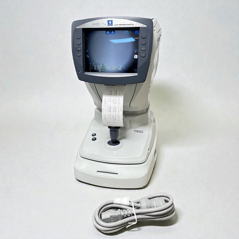
Nidek ARK-1e: one of the most widely used objective autorefractor-keratometers in clinical practice. The printed output shows sphere, cylinder, and axis values for both eyes.
Photo - Nidek Auto Refractor (another model)

Principles of Working
Modern objective autorefractors operate on one of three core principles:
A. Scheiner's Double Pinhole Principle (most widely used today)
- First described by Christoph Scheiner in 1619
- A dual-pinhole aperture is placed in front of the eye
- Parallel infrared rays from a source enter through two separate pinholes
| Eye condition | What happens | What is seen |
|---|---|---|
| Emmetropic | Both rays converge on the retina | Single point |
| Myopic | Rays cross before the retina | Two crossed (uncrossed diplopia) spots |
| Hyperopic | Rays have not yet converged at retina | Two uncrossed (crossed diplopia) spots |
- The autorefractor positions its Badal lens until the two pinhole images merge (null point = emmetropia or full correction)
- This is repeated across at least 3 meridians to calculate sphere, cylinder, and axis
- Infrared LEDs substitute for the pinholes in modern devices
B. Retinoscopic (Optometric) Principle
- Based on the same principle as clinical retinoscopy
- An infrared slit or spot is projected into the eye and swept across the pupil
- The direction of motion of the fundus reflex is analyzed:
- "With" motion = hyperopia
- "Against" motion = myopia
- No motion = emmetropia (neutralization point)
- The device adjusts its Badal lens until neutralization is achieved
- The lens position at neutralization = refractive error of that meridian
C. Best-Focus (Image Quality Analysis) Principle
- An infrared target ring (mire) is projected onto the retina
- The instrument continuously adjusts the focal plane via a Badal optometer
- A CCD camera captures images of the ring from the fundus
- The system detects when the ring image is sharpest (highest contrast, best-defined edges)
- That lens position = best focal correction
- Used in systems like the early Dioptron (Coopervision, 1970s)
D. Knife-Edge Principle
- A knife-edge (fine blade edge) is placed in the measurement beam
- Detects asymmetry in the light distribution across the pupil
- The Badal lens is adjusted until the light distribution becomes uniform (null)
- Similar to the Foucault knife-edge test in telescope mirror testing
E. Ray Deflection (Wavefront) Principle
- Used in modern wavefront aberrometers
- A narrow laser beam is scanned across the pupil in an array
- Exit angles of reflected rays are measured
- A wavefront map is reconstructed showing refractive error across the entire pupillary area
Core Hardware Components
Every objective autorefractor has these essential elements:
-
Infrared light source - near-infrared (NIR) at 780-950 nm wavelength
- NIR is used because: invisible to patient (no accommodation stimulus, no photophobia, no pupil constriction), strong fundus reflectance, and good transmission through ocular media
- Main NIR disadvantage: more scattered from fundus compared to visible light
-
Badal optometer (fogging lens system) - moves along an optical rail
- Linear relationship between lens position and diopter correction
- Constant target magnification regardless of position
-
Fixation target - a visible colored image (e.g., hot-air balloon, house) viewed at apparent optical infinity to relax accommodation
-
Beam splitter / semi-silvered mirror - separates the outgoing illumination path from the incoming reflected path
-
CCD camera or photodetector array - captures the retinal reflex
-
Microprocessor - analyzes the photodetector signals, drives the Badal lens to neutralization, calculates sphere/cylinder/axis, and prints results
-
Chin rest + forehead rest - for head positioning and alignment
-
Joystick control - for the examiner to align the instrument with the patient's pupil
How a Measurement is Performed (Step by Step)
- Patient places chin on chin rest, forehead on forehead rest
- Examiner aligns the instrument with the patient's right eye using the joystick (crosshair centered on pupil)
- Patient fixates on the internal target (usually a picture or colored light)
- The instrument auto-focuses: projects NIR light into the eye
- Reflected light from the retina is captured by the CCD
- The processor analyzes the reflection and adjusts the Badal lens
- At the null point, the sphere power is recorded
- Process repeats for at least 3 meridians (e.g., 0°, 60°, 120°)
- Sphere, cylinder, and axis are calculated using the sine-squared function across meridians
- Usually 3-5 automatic measurements are averaged
- A printed ticket is produced showing: R/L sphere, cylinder, axis, and keratometry (if combined ARK)
- A correction of approximately -0.50 to -0.75 D is added automatically to compensate for chromatic aberration (infrared vs. visible light)
Advantages of Objective Autorefractors
| Advantage | Detail |
|---|---|
| No patient cooperation needed | Ideal for children, elderly, cognitively impaired, non-verbal patients |
| Fast | Complete measurement in 3-10 seconds |
| Repeatable, objective data | Not influenced by patient responses or examiner judgment |
| Starting point for subjective refraction | Provides a reliable baseline; reduces total chair time by 30-50% |
| Prints results | No risk of transcription error; results stored/printed immediately |
| Cycloplegia | Works perfectly under cycloplegic drops (cyclopentolate, atropine) for pediatric refraction |
| Simultaneous keratometry | Most modern ARKs measure corneal curvature (K readings) at the same time |
| High volume | Can screen hundreds of patients in mass vision screening programs |
| Pupil dilation not required | Measures through an undilated pupil in normal conditions |
Disadvantages of Objective Autorefractors
| Disadvantage | Detail |
|---|---|
| Accommodation artifact | Even with fogging, some patients over-accommodate, making result too myopic ("myopic shift") |
| Cannot replace subjective refraction | Always used as a starting point only - final prescription must be refined subjectively |
| Unreliable post-refractive surgery | Eyes after LASIK, PRK, or RK have irregular corneas that confuse the ring/pinhole system |
| Irregular astigmatism | Keratoconus, corneal scars - reflex is distorted; readings are inaccurate |
| Dense media opacities | Dense cataracts, corneal opacities block the infrared signal |
| Small pupils | Miosis (<2.5 mm) prevents adequate sampling of the pupillary aperture |
| Fixation instability | Nystagmus or poor fixation gives variable results |
| Bulk and cost | Table-mounted units are large and expensive; not portable for field use |
| Measures only lower-order aberrations | Standard ARs cannot measure higher-order aberrations (coma, spherical aberration) - need aberrometer for that |
4. Side-by-Side Comparison: Subjective vs. Objective
| Feature | Subjective Autorefractor | Objective Autorefractor |
|---|---|---|
| Patient input | Required | Not required |
| Speed | Slow (5-15 min) | Fast (seconds) |
| Accuracy as final Rx | Gold standard | Starting point only |
| Use in children | Difficult | Excellent (with cycloplegia) |
| Accommodation control | Fog/cycloplegia needed | Fog/cycloplegia needed |
| Post-LASIK reliability | Good | Poor |
| Keratoconus | Reliable (patient judges) | Unreliable |
| Operator skill | High | Low |
| Equipment cost | Moderate-high | Moderate |
| Portability | Some handheld models | Mostly table-mounted |
| Prints result | Digital phoropters can | Yes, standard |
| Typical clinical role | Final prescription | Screening + starting Rx |
5. Contraindications
Both types are relatively safe, but measurement is unreliable or impossible in:
- Mentally disabled or uncooperative patients (subjective only)
- Patients with postural problems preventing chin-rest use
- Gross vision loss (light perception or less)
- Acute traumatic eye injury
- Artificial intraocular lens implants (mild inaccuracy in objective ARs)
- Atrophic bulbi
- Very small children (< 1 year)
6. Clinical Significance
- Autorefraction is used as a starting point for subjective refraction, not as a standalone prescription
- Readings should always be refined with a subjective refraction (Snellen chart, duochrome test, JCC for cylinder)
- In cycloplegic refraction (children under 12, suspected latent hyperopia), objective autorefraction under cycloplegia is the standard of care
- Post-refractive surgery cases: subjective over-refraction is always preferred
- Wavefront aberrometry (a specialized form of objective autorefraction) provides the full higher-order aberration profile for custom laser vision correction
Sources:
- StatPearls: Autorefractors (NCBI Bookshelf NBK580520)
- Frank's Hospital Workshop: Automated Refraction - Design and Applications
- University of Mosul Lecture 9: Autorefractometer (Karrar Eidan)
- University of Arizona Visual Optics Lab: Optometer / Badal Principle
- Nidek ARK-1e Clinical Documentation
This is a shared conversation. Sign in to Orris to start your own chat.