
Foam cell histopathology d/d
Reading File
Foam cells are lipid-laden macrophages with a characteristic vacuolated "foamy" cytoplasm seen on H&E staining. The differential is broad - let me pull authoritative textbook content.
Searching sources...
Finding Sources
Finding Sources
Good results. Now let me read the key sections for comprehensive content, and fetch relevant histology images.
Reading File
Reading File
Finding Sources
Finding Sources
Reading File
Loading Image
Excellent. Now let me get more content on specific foam cell conditions - xanthogranuloma, Virchow cells in leprosy, malakoplakia, atherosclerosis, and storage diseases.
Finding Sources
Finding Sources
Finding Sources
I now have comprehensive information. Let me also fetch a key foam cell image from atherosclerosis context and get Robbins content on lepromatous leprosy.
Loading Image
Reading File
I now have all the material needed for a comprehensive, well-structured answer. Let me compile it.
Foam Cell Histopathology - Differential Diagnosis
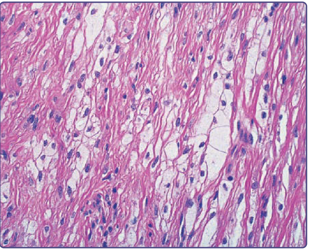
Foam cells are lipid-laden macrophages (histiocytes) with a characteristic pale, vacuolated, "foamy" cytoplasm on H&E due to dissolved neutral lipids. They are not a single diagnosis - they appear across a broad range of metabolic, infectious, neoplastic, and reactive conditions.
Histological Appearance
- Large macrophages with abundant, pale vacuolated cytoplasm (lipid washed out by formalin fixation leaves clear spaces)
- Small, eccentric or central nucleus
- On H&E: optically clear or finely granular cytoplasm
- Special stains: Oil Red O / Sudan Black on frozen sections confirms lipid
- CD68 positive (macrophage marker); adipophilin IHC helps identify lipid accumulation
Differential Diagnosis by System/Category
1. XANTHOMAS (Skin/Soft Tissue)
The prototype foam cell lesion. Macrophages engulf lipid via scavenger receptors after excess lipoprotein deposition in tissues.
| Type | Key Histologic Features |
|---|---|
| Eruptive xanthoma | Foam cells in reticular dermis; mixed neutrophil + lymphocyte infiltrate early; extracellular lipid clefts |
| Tuberous xanthoma | Large foam cell aggregates in dermis; fibrosis; few inflammatory cells; cholesterol esters visible under polarized light |
| Tendinous xanthoma | Similar to tuberous but even larger foam cells |
| Plane xanthoma / Xanthelasma | Superficial dermal foam cells; non-inflammatory; minimal fibrosis; thin epidermis (eyelid clue) |
| Verruciform xanthoma | Foam cells restricted to dermal papillae; overlying epithelial hyperplasia/acanthosis |
Note: Formalin fixation removes lipid leaving artifactual clefts. Adipophilin/CD68 IHC confirms foam cell identity. - Dermatology 2-Volume Set 5e
2. NON-LANGERHANS CELL HISTIOCYTOSES
| Condition | Distinguishing Features |
|---|---|
| Juvenile Xanthogranuloma (JXG) | Dermal histiocytes + Touton giant cells (wreath of nuclei with peripheral foamy cytoplasm); eosinophils; no lipid disorder required; CD68+/S100-/CD1a- |
| Xanthoma Disseminatum | Foam cells + Touton cells on mucous membranes; associated DI |
| Papular Xanthoma | More foam cells, fewer giant cells than JXG |
| Multicentric Reticulohistiocytosis | Large histiocytes with "ground-glass" eosinophilic cytoplasm (NOT foam cells) - key distinguishing point |
| Erdheim-Chester Disease | Foamy CD68+/CD1a- histiocytes with fibrosis; long bone involvement |
3. ATHEROSCLEROSIS (Vascular)
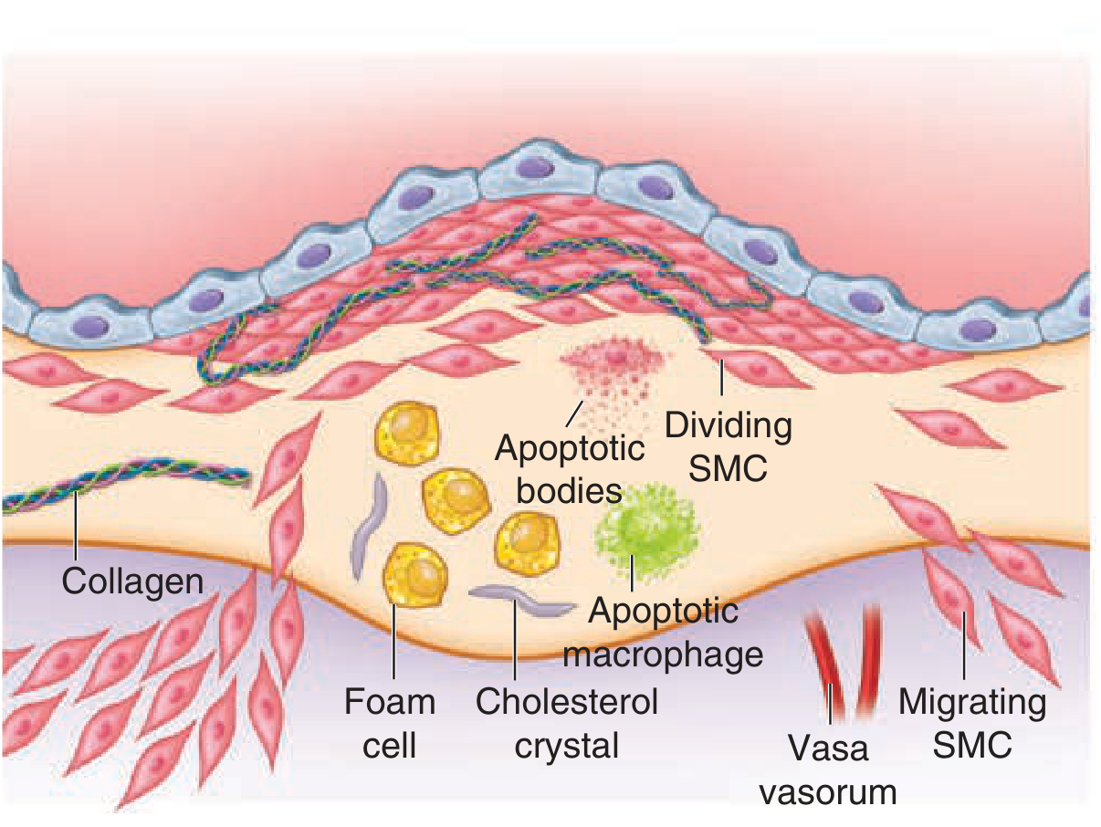
- Fatty streak: Earliest atherosclerotic lesion - subendothelial accumulation of foam cells (macrophage-derived + smooth muscle cell-derived)
- Macrophages ingest oxidized LDL via scavenger receptors → foam cell formation
- Progress to fibrous plaques with foam cell-rich lipid core, cholesterol clefts, necrotic debris, and fibrous cap
- Fuster and Hurst's The Heart, 15th Edition
4. LEPROSY (Lepromatous Type)
- Virchow cells (Lepra cells): Lipid-laden macrophages stuffed with Mycobacterium leprae organisms ("globi")
- H&E: Foamy macrophages in dermis; grenz zone (Unna's grenz zone - clear subepidermal zone)
- AFB stain (Fite-Faraco): Reveals masses of acid-fast bacilli within the foamy cytoplasm
- Lepromatous = multibacillary; no granulomas (poor T-cell response)
- Involves skin, peripheral nerves, testes, anterior eye
- Robbins, Cotran & Kumar Pathologic Basis of Disease; Andrews' Diseases of the Skin
Key distinguishing feature from xanthomas: AFB positivity, grenz zone, no lipid disorder
5. LIPID STORAGE DISEASES
| Disease | Organ/Site | Cell Appearance |
|---|---|---|
| Niemann-Pick Disease (A/B) | Lung, liver, spleen, bone marrow | Large foamy macrophages; sphingomyelin on EM - lamellar "myelin figures" |
| Niemann-Pick Type C | Lung (BAL foam cells), liver | Similar foamy infiltrates; pulmonary fibrosis in long-standing disease |
| Gaucher Disease | Spleen, liver, BM | Gaucher cells - "crumpled tissue paper" cytoplasm (glucocerebroside); NOT true foam cells but often in D/D |
| Cholesteryl Ester Storage Disease / Wolman Disease | Intestinal mucosa, liver | Foam cells infiltrating intestinal villi; mutation in LIPA gene |
- Fishman's Pulmonary Diseases; Sleisenger and Fordtran's GI & Liver Disease
6. XANTHOGRANULOMATOUS INFLAMMATION (Infectious/Reactive)
| Condition | Site | Features |
|---|---|---|
| Xanthogranulomatous Pyelonephritis | Kidney | Foamy macrophages in renal parenchyma; usually E. coli/Proteus; staghorn calculi; CD68+ |
| Malakoplakia | Bladder, kidney, GI tract | Foamy von Hansemann cells with Michaelis-Gutmann bodies (targetoid calcospherites, PAS/von Kossa+); macrophage dysfunction with gram-neg infections |
| Megalocytic Interstitial Nephritis | Kidney | Similar macrophage dysfunction pattern |
- National Kidneys Foundation Primer on Kidney Diseases, 8e
7. TUMORS / NEOPLASTIC CONDITIONS
| Tumor | Foam Cell Context |
|---|---|
| Clear Cell Carcinoma (kidney, endometrium) | Clear cells due to glycogen/lipid - NOT true foam cells; PAS+, CD10+/EMA+ |
| Adrenocortical Carcinoma | Lipid-rich clear cells |
| Hibernoma | Brown fat tumor - multivacuolated lipoblasts |
| Liposarcoma (Well-differentiated) | Lipoblasts with scalloped nuclei |
| Dermatofibroma | May contain incidental xanthomatous (foamy) cells; storiform pattern |
| Giant Cell Tumor of Tendon Sheath | Foamy histiocytes + hemosiderin + giant cells - DDx for tendinous xanthoma |
8. MISCELLANEOUS
| Condition | Notes |
|---|---|
| Whipple's Disease | Small intestinal villi stuffed with PAS+ macrophages (contain T. whipplei); foamy appearance; villi expanded - "foam cells" in villi |
| Langerhans Cell Histiocytosis | CD1a+/S100+/langerin+ (Birbeck granules on EM); NOT true foam cells but in clinical D/D |
| Rosai-Dorfman Disease | Emperipolesis (lymphocytes within histiocyte cytoplasm); S100+/CD68+ |
| GVHD | Dermal xanthomatous cells in biopsies may indicate hepatic GVHD |
| Pulmonary Alveolar Proteinosis | Foamy alveolar macrophages; PAS+ granular material |
| Post-obstructive pneumonia | Endogenous lipoid pneumonia - foamy alveolar macrophages |
| HIV + IgA gammopathy | Papular neutrophilic xanthoma - foam cells intermingled with neutrophils + nuclear dust |
Key Discriminating Stains
| Stain | Purpose |
|---|---|
| Oil Red O / Sudan Black (frozen) | Confirms neutral lipid |
| CD68 (PGM1/KP1) | Macrophage marker |
| Adipophilin IHC | Lipid droplet protein; aids foam cell ID on paraffin |
| AFB (Fite-Faraco) | Leprosy (Virchow cells) |
| PAS | Whipple's macrophages; Gaucher cells |
| CD1a / Langerin | Langerhans cells (distinguish from foam cells) |
| Michaelis-Gutmann (PAS, von Kossa) | Malakoplakia |
| S100 | Rosai-Dorfman (positive); xanthoma foam cells (negative) |
Quick Summary Table
| Category | Condition | Key Distinguishing Feature |
|---|---|---|
| Metabolic | Xanthomas | Lipid disorder; adipophilin+; no organisms |
| Histiocytosis | JXG | Touton giant cells; children; no lipid disorder |
| Vascular | Atherosclerosis | Intimal location; cholesterol clefts |
| Infectious | Lepromatous leprosy | AFB+ Virchow cells; grenz zone |
| Storage | Niemann-Pick | EM lamellar figures; sphingomyelinase deficiency |
| Infectious-reactive | Xanthogranulomatous pyelonephritis | Kidney; gram-neg infection |
| Infectious-reactive | Malakoplakia | Michaelis-Gutmann bodies |
| GI infection | Whipple's disease | PAS+ macrophages in small bowel villi |
Sources: Dermatology 2-Volume Set 5e; Fitzpatrick's Dermatology Vol 1-2; Robbins, Cotran & Kumar Pathologic Basis of Disease; Andrews' Diseases of the Skin; National Kidneys Foundation Primer on Kidney Diseases, 8e; Fuster and Hurst's The Heart, 15th Edition; Sleisenger and Fordtran's GI & Liver Disease; Harrison's Principles of Internal Medicine 22E
Compiled all in one page
Asking for Preferences
What format should the one-page document be in?
Reading File
Running Command
mkdir -p /home/daytona/workspace/foam-cell-dd && ls /home/daytona/workspace/foam-cell-dd/
Writing File
~/foam-cell-dd/generate_pdf.py
from reportlab.lib.pagesizes import A4
from reportlab.lib import colors
from reportlab.lib.units import mm, cm
from reportlab.platypus import (
SimpleDocTemplate, Table, TableStyle, Paragraph, Spacer,
HRFlowable, KeepTogether
)
from reportlab.lib.styles import getSampleStyleSheet, ParagraphStyle
from reportlab.lib.enums import TA_CENTER, TA_LEFT, TA_JUSTIFY
from reportlab.platypus import BaseDocTemplate, PageTemplate, Frame
OUTPUT = "/home/daytona/workspace/foam-cell-dd/Foam_Cell_Histopathology_DD.pdf"
# ── Colour palette ────────────────────────────────────────────────────────────
DARK_BLUE = colors.HexColor("#1a2e4a")
MED_BLUE = colors.HexColor("#2563a8")
LIGHT_BLUE = colors.HexColor("#dbeafe")
ACCENT = colors.HexColor("#e07b39")
ROW_ALT = colors.HexColor("#f0f6ff")
ROW_WHITE = colors.white
HEADER_BG = colors.HexColor("#1a2e4a")
SUBHDR_BG = colors.HexColor("#2563a8")
GREEN_BG = colors.HexColor("#d1fae5")
YELLOW_BG = colors.HexColor("#fef9c3")
PINK_BG = colors.HexColor("#fce7f3")
PURPLE_BG = colors.HexColor("#ede9fe")
ORANGE_BG = colors.HexColor("#ffedd5")
TEAL_BG = colors.HexColor("#ccfbf1")
# ── Styles ────────────────────────────────────────────────────────────────────
styles = getSampleStyleSheet()
def make_style(name, parent='Normal', **kw):
return ParagraphStyle(name, parent=styles[parent], **kw)
title_style = make_style('TitleS', fontSize=16, textColor=colors.white,
alignment=TA_CENTER, fontName='Helvetica-Bold',
spaceAfter=2)
subtitle_style = make_style('SubTitle', fontSize=9, textColor=colors.HexColor("#bfdbfe"),
alignment=TA_CENTER, fontName='Helvetica')
sect_style = make_style('SectHdr', fontSize=8, textColor=colors.white,
fontName='Helvetica-Bold', alignment=TA_LEFT)
cell_style = make_style('CellS', fontSize=7, textColor=DARK_BLUE,
fontName='Helvetica', leading=9)
cell_bold = make_style('CellB', fontSize=7, textColor=DARK_BLUE,
fontName='Helvetica-Bold', leading=9)
cell_hdr = make_style('ColHdr', fontSize=7, textColor=colors.white,
fontName='Helvetica-Bold', leading=9, alignment=TA_CENTER)
note_style = make_style('NoteS', fontSize=6.5, textColor=colors.HexColor("#374151"),
fontName='Helvetica-Oblique', alignment=TA_CENTER)
key_style = make_style('KeyS', fontSize=7, textColor=DARK_BLUE,
fontName='Helvetica-Bold', leading=9)
footer_style = make_style('FootS', fontSize=6, textColor=colors.grey,
fontName='Helvetica', alignment=TA_CENTER)
def P(text, style=cell_style): return Paragraph(text, style)
def B(text): return Paragraph(text, cell_bold)
def H(text): return Paragraph(text, cell_hdr)
# ── Table helper ──────────────────────────────────────────────────────────────
def section_header(title, color=SUBHDR_BG, cols=4):
t = Table([[Paragraph(title, sect_style)]], colWidths=['100%'])
t.setStyle(TableStyle([
('BACKGROUND', (0,0), (-1,-1), color),
('TOPPADDING', (0,0), (-1,-1), 3),
('BOTTOMPADDING', (0,0), (-1,-1), 3),
('LEFTPADDING', (0,0), (-1,-1), 6),
]))
return t
def make_table(headers, rows, col_widths, row_colors=None):
data = [headers] + rows
t = Table(data, colWidths=col_widths, repeatRows=1)
style_cmds = [
('BACKGROUND', (0,0), (-1,0), MED_BLUE),
('GRID', (0,0), (-1,-1), 0.3, colors.HexColor("#d1d5db")),
('VALIGN', (0,0), (-1,-1), 'TOP'),
('TOPPADDING', (0,0), (-1,-1), 2.5),
('BOTTOMPADDING', (0,0), (-1,-1), 2.5),
('LEFTPADDING', (0,0), (-1,-1), 3),
('RIGHTPADDING', (0,0), (-1,-1), 3),
('ROWBACKGROUNDS', (0,1), (-1,-1), [ROW_WHITE, ROW_ALT]),
]
if row_colors:
for ri, rc in row_colors:
style_cmds.append(('BACKGROUND', (0, ri+1), (-1, ri+1), rc))
t.setStyle(TableStyle(style_cmds))
return t
# ── Build document ────────────────────────────────────────────────────────────
doc = SimpleDocTemplate(
OUTPUT,
pagesize=A4,
leftMargin=10*mm, rightMargin=10*mm,
topMargin=8*mm, bottomMargin=8*mm,
title="Foam Cell Histopathology - Differential Diagnosis",
author="Orris Medical"
)
W = A4[0] - 20*mm # usable width
story = []
# ═══════════════════════════════════════════════════════════════════
# TITLE BANNER
# ═══════════════════════════════════════════════════════════════════
banner = Table(
[[P("FOAM CELL HISTOPATHOLOGY", title_style)],
[P("Differential Diagnosis — Quick Reference", subtitle_style)]],
colWidths=[W]
)
banner.setStyle(TableStyle([
('BACKGROUND', (0,0), (-1,-1), DARK_BLUE),
('TOPPADDING', (0,0), (-1,-1), 6),
('BOTTOMPADDING', (0,0), (-1,-1), 6),
('LEFTPADDING', (0,0), (-1,-1), 8),
]))
story.append(banner)
story.append(Spacer(1, 3))
# ═══════════════════════════════════════════════════════════════════
# DEFINITION BOX
# ═══════════════════════════════════════════════════════════════════
def_text = (
"<b>Foam cells</b> are lipid-laden macrophages with <b>pale, vacuolated, \"foamy\" cytoplasm</b> on H&E "
"(lipid dissolved by formalin → clear vacuoles). CD68+, Adipophilin+. "
"Oil Red O / Sudan Black on <b>frozen sections</b> confirms neutral lipid. "
"They occur across metabolic, infectious, neoplastic and reactive conditions."
)
def_box = Table([[P(def_text, make_style('Def', fontSize=7.2, textColor=DARK_BLUE,
fontName='Helvetica', leading=10))]],
colWidths=[W])
def_box.setStyle(TableStyle([
('BACKGROUND', (0,0), (-1,-1), LIGHT_BLUE),
('TOPPADDING', (0,0), (-1,-1), 5),
('BOTTOMPADDING', (0,0), (-1,-1), 5),
('LEFTPADDING', (0,0), (-1,-1), 8),
('RIGHTPADDING', (0,0), (-1,-1), 8),
('BOX', (0,0), (-1,-1), 0.8, MED_BLUE),
]))
story.append(def_box)
story.append(Spacer(1, 4))
# ═══════════════════════════════════════════════════════════════════
# MAIN DIFFERENTIAL TABLE
# ═══════════════════════════════════════════════════════════════════
story.append(section_header("▶ MAIN DIFFERENTIAL DIAGNOSIS"))
story.append(Spacer(1, 2))
W1, W2, W3, W4 = 32*mm, 42*mm, 52*mm, 34*mm
cw = [W1, W2, W3, W4]
hdr = [H("Category / Disease"), H("Organ / Site"), H("Key Histologic Features"), H("Distinguishing Clue")]
rows_main = [
# XANTHOMAS
[B("XANTHOMAS"), P(""), P(""), P("")],
[P("Eruptive xanthoma"),
P("Reticular dermis"),
P("Small foam cells; neutrophil + lymphocyte infiltrate (early); extracellular lipid clefts"),
P("Hypertriglyceridaemia; AFB –")],
[P("Tuberous / Tendinous xanthoma"),
P("Skin / tendons"),
P("Large foam cell aggregates; fibrosis; cholesterol esters (polarised light)"),
P("Cholesterol clefts; familial dyslipidaemia")],
[P("Xanthelasma / Plane xanthoma"),
P("Eyelid / skin"),
P("Superficial dermal foam cells; non-inflammatory; minimal fibrosis"),
P("Thin epidermis; eyelid location")],
[P("Verruciform xanthoma"),
P("Oral mucosa / genital"),
P("Foam cells confined to dermal papillae; overlying epithelial hyperplasia"),
P("HPV unrelated; unique papillary pattern")],
# HISTIOCYTOSES
[B("NON-LANGERHANS HISTIOCYTOSES"), P(""), P(""), P("")],
[P("Juvenile Xanthogranuloma (JXG)"),
P("Skin (head/neck)"),
P("Histiocytes + Touton giant cells (wreath nuclei + peripheral foam); eosinophils"),
P("CD68+, S100–, CD1a–; children; no lipid disorder")],
[P("Xanthoma Disseminatum"),
P("Mucosa / skin / CNS"),
P("Foam cells + Touton cells; DI association"),
P("Normolipidaemia; mucous membrane involvement")],
[P("Erdheim-Chester Disease"),
P("Long bones, retroperitoneum"),
P("Foamy CD68+ histiocytes + dense fibrosis; Touton-like giant cells"),
P("BRAF V600E mutation; long bone osteosclerosis")],
# VASCULAR
[B("VASCULAR"), P(""), P(""), P("")],
[P("Atherosclerosis (fatty streak)"),
P("Intima (aorta, coronaries)"),
P("Subendothelial foam cells (macrophage + SMC-derived); cholesterol clefts; necrotic core in advanced plaques"),
P("Intimal location; no grenz zone; evolves to fibrous plaque")],
# INFECTIOUS
[B("INFECTIOUS"), P(""), P(""), P("")],
[P("Lepromatous Leprosy\n(Virchow / Lepra cells)"),
P("Dermis, nerves, testes"),
P("Lipid-laden macrophages filled with M. leprae globi; Grenz zone (clear sub-epidermal band); no true granulomas"),
P("AFB+ (Fite-Faraco); globi; leonine facies")],
[P("Whipple's Disease"),
P("Small intestinal villi"),
P("Villi expanded by PAS+ macrophages (T. whipplei); foamy cytoplasm"),
P("PAS+++ (diastase-resistant); PCR T. whipplei")],
# STORAGE
[B("LIPID STORAGE DISEASES"), P(""), P(""), P("")],
[P("Niemann-Pick (A/B)"),
P("Lung, liver, spleen, BM"),
P("Large foamy macrophages; sphingomyelin; lamellar 'myelin figures' on EM"),
P("Sphingomyelinase deficiency; EM diagnostic")],
[P("Niemann-Pick Type C"),
P("Lung, liver"),
P("Foamy macrophages; pulmonary fibrosis in long-standing disease; BAL foam cells"),
P("NPC1/NPC2 gene; cholesterol transport defect")],
[P("Cholesteryl Ester Storage / Wolman Disease"),
P("Intestinal mucosa, liver"),
P("Foam cells infiltrating intestinal villi; adrenal calcification (Wolman)"),
P("LIPA gene mutation; adrenal calcification on imaging")],
# XANTHOGRANULOMATOUS INFECTION
[B("XANTHOGRANULOMATOUS INFLAMMATION"), P(""), P(""), P("")],
[P("Xanthogranulomatous Pyelonephritis"),
P("Kidney"),
P("Foamy CD68+ macrophages replacing renal parenchyma; staghorn calculi; necrosis"),
P("E. coli / Proteus; staghorn calculus; CD68+")],
[P("Malakoplakia"),
P("Bladder, kidney, GI"),
P("Von Hansemann cells (foamy macrophages) + Michaelis-Gutmann bodies (targetoid calcospherites)"),
P("MG bodies: PAS+, von Kossa+, iron stain+")],
# TUMOURS
[B("TUMOURS / NEOPLASTIC"), P(""), P(""), P("")],
[P("Clear Cell RCC"),
P("Kidney"),
P("Clear cells (glycogen/lipid); delicate 'chicken-wire' vasculature; no true foam cell aggregates"),
P("PAS+, CD10+, EMA+, PAX8+; NOT CD68+")],
[P("Well-Diff. Liposarcoma"),
P("Retroperitoneum, thigh"),
P("Lipoblasts with scalloped hyperchromatic nuclei; MDM2+"),
P("MDM2/CDK4 amplification; not pure foam cells")],
[P("Giant Cell Tumour Tendon Sheath"),
P("Tendon / joint"),
P("Foamy histiocytes + hemosiderin + multinucleated giant cells + fibrous stroma"),
P("CSF1 rearrangement; DDx tendinous xanthoma")],
# MISC
[B("MISCELLANEOUS"), P(""), P(""), P("")],
[P("Lipoid Pneumonia (endogenous)"),
P("Lung / alveoli"),
P("Foamy alveolar macrophages; oil-like material in alveolar spaces"),
P("Post-obstruction or aspiration; Oil Red O+")],
[P("Pulmonary Alveolar Proteinosis"),
P("Lung"),
P("Foamy macrophages + granular PAS+ alveolar material"),
P("PAS+ proteinaceous material; GM-CSF antibodies")],
[P("GVHD (cutaneous)"),
P("Skin"),
P("Dermal xanthomatous cells in biopsies"),
P("Context: post-HSCT; may indicate hepatic GVHD")],
]
row_colors_main = []
cat_rows = [0, 5, 10, 12, 15, 19, 22, 26] # 0-indexed among data rows
cat_bg = [colors.HexColor("#1e3a5f"), colors.HexColor("#1e3a5f"), colors.HexColor("#1e3a5f"),
colors.HexColor("#1e3a5f"), colors.HexColor("#1e3a5f"), colors.HexColor("#1e3a5f"),
colors.HexColor("#1e3a5f"), colors.HexColor("#1e3a5f")]
t_main = Table([hdr] + rows_main, colWidths=cw, repeatRows=1)
style_cmds = [
('BACKGROUND', (0,0), (-1,0), MED_BLUE),
('GRID', (0,0), (-1,-1), 0.3, colors.HexColor("#d1d5db")),
('VALIGN', (0,0), (-1,-1), 'TOP'),
('TOPPADDING', (0,0), (-1,-1), 2),
('BOTTOMPADDING', (0,0), (-1,-1), 2),
('LEFTPADDING', (0,0), (-1,-1), 3),
('RIGHTPADDING', (0,0), (-1,-1), 3),
]
# alternating row backgrounds for data rows
for i in range(len(rows_main)):
ri = i + 1
if i in cat_rows:
style_cmds.append(('BACKGROUND', (0, ri), (-1, ri), DARK_BLUE))
style_cmds.append(('SPAN', (0, ri), (-1, ri)))
else:
bg = ROW_ALT if i % 2 == 0 else ROW_WHITE
style_cmds.append(('BACKGROUND', (0, ri), (-1, ri), bg))
t_main.setStyle(TableStyle(style_cmds))
story.append(t_main)
story.append(Spacer(1, 4))
# ═══════════════════════════════════════════════════════════════════
# BOTTOM ROW: Stains | Key Distinguishing Points
# ═══════════════════════════════════════════════════════════════════
story.append(section_header("▶ KEY DISCRIMINATING STAINS"))
story.append(Spacer(1, 2))
stain_cw = [28*mm, 60*mm, 72*mm]
stain_hdr = [H("Stain"), H("Purpose"), H("Positive In")]
stain_rows = [
[B("Oil Red O / Sudan Black"),
P("Confirms neutral lipid (frozen sections only)"),
P("Xanthomas, lipoid pneumonia, lipid storage diseases")],
[B("CD68 (KP1 / PGM1)"),
P("Macrophage lineage marker"),
P("All foam cell lesions; strongly + in xanthogranulomatous inflammation")],
[B("Adipophilin IHC"),
P("Lipid droplet protein; works on paraffin"),
P("Xanthomas, foam cells in standard sections")],
[B("AFB – Fite-Faraco"),
P("Acid-fast mycobacteria"),
P("Lepromatous leprosy (Virchow cells); globi within macrophages")],
[B("PAS (diastase-resistant)"),
P("Intracellular organisms / glycoprotein"),
P("Whipple's disease (PAS++++); NOT positive in xanthomas")],
[B("Michaelis-Gutmann bodies\n(PAS, von Kossa, Iron)"),
P("Targetoid calcospherites in macrophages"),
P("Malakoplakia only")],
[B("CD1a / Langerin / S100"),
P("Langerhans cell markers"),
P("LCH (CD1a+, S100+); absent in all foam cell xanthomas")],
[B("CSF1R / BRAF V600E"),
P("Molecular markers"),
P("Giant cell tumour (CSF1R); Erdheim-Chester (BRAF V600E)")],
]
t_stain = make_table(stain_hdr, stain_rows, stain_cw)
story.append(t_stain)
story.append(Spacer(1, 4))
# ═══════════════════════════════════════════════════════════════════
# TOUTON GIANT CELL NOTE
# ═══════════════════════════════════════════════════════════════════
touton_note = Table([[
P("<b>Touton Giant Cell</b> — Pathognomonic foamy giant cell: central wreath of nuclei "
"surrounded by <i>eosinophilic</i> cytoplasm (inner) and <i>foamy/xanthomatous</i> cytoplasm (outer). "
"Seen in: <b>JXG</b> (most characteristic), xanthoma disseminatum, xanthomas, necrobiotic xanthogranuloma. "
"<b>NOT</b> seen in: lepromatous leprosy, atherosclerosis, malakoplakia, or storage diseases.",
make_style('ToutonS', fontSize=7, textColor=colors.HexColor("#1e3a5f"),
fontName='Helvetica', leading=9))
]], colWidths=[W])
touton_note.setStyle(TableStyle([
('BACKGROUND', (0,0), (-1,-1), YELLOW_BG),
('BOX', (0,0), (-1,-1), 0.8, colors.HexColor("#ca8a04")),
('TOPPADDING', (0,0), (-1,-1), 5),
('BOTTOMPADDING', (0,0), (-1,-1), 5),
('LEFTPADDING', (0,0), (-1,-1), 8),
('RIGHTPADDING', (0,0), (-1,-1), 8),
]))
story.append(touton_note)
story.append(Spacer(1, 3))
# ═══════════════════════════════════════════════════════════════════
# QUICK MNEMONICS
# ═══════════════════════════════════════════════════════════════════
mn_cw = [W/3, W/3, W/3]
mn_hdr = [H("AFB+ Foam Cells"), H("PAS+ Foam Cells"), H("Touton Giant Cells")]
mn_rows = [
[P("Lepromatous leprosy\n(Virchow / Lepra cells)\n• Fite-Faraco +\n• Grenz zone\n• No granulomas"),
P("Whipple's disease\n(T. whipplei)\n• Villi expanded\n• Diastase-resistant\n• PCR confirms"),
P("JXG (most typical)\nXanthoma disseminatum\nTuberous xanthoma\nNecrobiotic xanthogranuloma")]
]
t_mn = make_table(mn_hdr, mn_rows, mn_cw)
story.append(t_mn)
story.append(Spacer(1, 3))
# ═══════════════════════════════════════════════════════════════════
# FOOTER
# ═══════════════════════════════════════════════════════════════════
footer_txt = (
"Sources: Dermatology 2-Vol Set 5e | Fitzpatrick's Dermatology | Robbins & Cotran Pathologic Basis of Disease | "
"Andrews' Diseases of the Skin | Harrison's Principles of Internal Medicine 22E | "
"Fuster & Hurst's The Heart 15e | Sleisenger & Fordtran's GI & Liver Disease | "
"National Kidney Foundation Primer 8e • Compiled by Orris Medical AI"
)
story.append(Paragraph(footer_txt, footer_style))
# ── Build ─────────────────────────────────────────────────────────────────────
doc.build(story)
print(f"PDF written to: {OUTPUT}")
Running Command
cd /home/daytona/workspace/foam-cell-dd && python generate_pdf.py 2>&1
Running Command
ls -lh /home/daytona/workspace/foam-cell-dd/Foam_Cell_Histopathology_DD.pdf
Your one-page PDF is ready:
Foam Cell Histopathology DD
PDF Document · PDF
What's included on the page:
- Definition box - foam cell basics, stain notes
- Main D/D table (4 columns: Category, Site, Key Histology, Distinguishing Clue) covering:
- Xanthomas (eruptive, tuberous, tendinous, xanthelasma, verruciform)
- Non-Langerhans histiocytoses (JXG, xanthoma disseminatum, Erdheim-Chester)
- Vascular (atherosclerosis/fatty streak)
- Infectious (lepromatous leprosy/Virchow cells, Whipple's disease)
- Lipid storage diseases (Niemann-Pick A/B/C, Wolman/CESD)
- Xanthogranulomatous inflammation (XGP, malakoplakia)
- Tumours (clear cell RCC, liposarcoma, GCTS)
- Miscellaneous (lipoid pneumonia, PAP, GVHD)
- Key discriminating stains table (Oil Red O, CD68, adipophilin, AFB, PAS, MG bodies, CD1a, molecular markers)
- Touton giant cell highlighted note box
- Quick mnemonics - AFB+ / PAS+ / Touton+ foam cell conditions side by side
- Full source citations in footer
This is a shared conversation. Sign in to Orris to start your own chat.