
Definition of myopia, hypermetropia, and astigmatism.
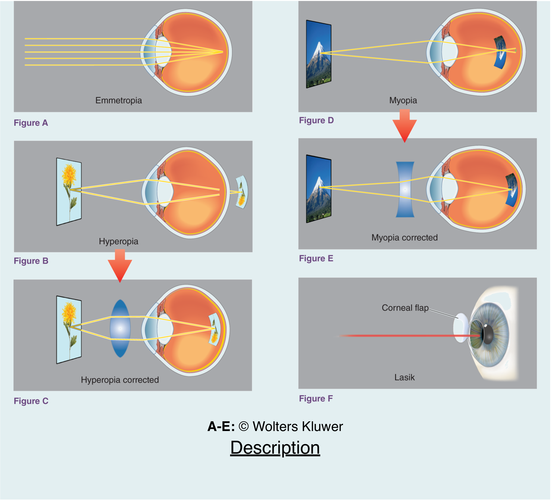
Myopia (nearsightedness) is where distant objects appear blurry because the focal point falls in front of the retina - either due to an elongated eyeball or excessive corneal/lens curvature. Hypermetropia (farsightedness) is the opposite: the focal point falls behind the retina, making near objects blurry, typically due to a shorter eyeball or flatter cornea. Astigmatism occurs when the cornea or lens has an irregular (non-spherical) curvature, causing light to focus at multiple points rather than one, producing blurred or distorted vision at all distances.
Searching sources...
Reading File
Finding Sources
Finding Sources
Now let me read the primary definition sections from Goldman-Cecil and Kanski's:
Reading File
Reading File
Reading File
Finding Sources
Now let me get the Kanski definitions for myopia, hypermetropia, and astigmatism, plus fetch the diagram image:
Finding Sources
Loading Image
Let me now find the dedicated myopia/hypermetropia/astigmatism sections in Kanski's for their formal definitions:
Finding Sources
Finding Sources
I now have solid textbook content. Let me look for the refractive errors chapter that would have the classic definitions, and also check if there's a dedicated optics/refraction chapter in Kanski's:
Reading File
I have sufficient textbook content from Goldman-Cecil Medicine and Kanski's Clinical Ophthalmology to provide a thorough answer. Let me now compile the comprehensive response.
Refractive Errors: Myopia, Hypermetropia, and Astigmatism
These three conditions are all ametropic states - meaning the eye fails to bring parallel rays of light to a single focus on the retina in the resting (unaccommodated) state. They are the most common causes of correctable visual impairment worldwide.
1. Myopia (Nearsightedness)
Definition: A refractive error in which parallel rays of light from a distant object are brought to a focus in front of the retina, rather than on it. The result is blurred distance vision with relatively preserved near vision.
Mechanism / Aetiology:
- Axial myopia (most common): the eyeball is longer than normal. The axial length of a normal eye is 20-24 mm; in myopia it exceeds this range.
- Refractive (curvature) myopia: excessive curvature of the cornea or lens increases refractive power beyond what is needed.
Clinical Features:
- Usually discovered in childhood when children cannot see the blackboard at school.
- Progresses until approximately age 20-25 years.
- Pathologic (degenerative) myopia: a partly heritable condition with progressive posterior scleral weakening, increasing axial length (sometimes >40 mm), and formation of a posterior staphyloma. Complications include retinal holes, retinal detachment, choroidal neovascularization, and intraocular hemorrhage. Associated syndromes include Marfan, Stickler, and Cornelia de Lange.
- Rapidly progressing myopia after age 25 should prompt evaluation for diabetes mellitus or cataract development (nuclear sclerosis in high myopia can paradoxically increase myopic error).
Correction: Concave (diverging, minus) lenses; contact lenses; LASIK (corrects up to 6-8 D depending on corneal thickness); SMILE procedure; phakic IOL implants for high degrees.
2. Hypermetropia / Hyperopia (Farsightedness)
Definition: A refractive error in which parallel rays of light from a distant object converge to a focus behind the retina. The eye is effectively "underpowered" for its length.
Mechanism / Aetiology:
- Axial hypermetropia (most common): the eyeball is shorter than average axial length.
- Refractive hypermetropia: the cornea or lens has insufficient curvature (flatter than normal), reducing total refractive power.
Clinical Features:
- Near vision is typically more affected than distance vision, but in significant degrees both are blurred.
- Young patients can compensate with accommodation (the crystalline lens increases its curvature to add focusing power), masking the defect until their 40s when the lens loses pliability (presbyopia).
- When compensation fails, the first pair of glasses may need to correct for both distance and near (bifocals).
- Short axial length is anatomically associated with a narrow anterior chamber angle, predisposing to narrow-angle and acute angle-closure glaucoma.
- High bilateral hypermetropia is a common cause of ametropic amblyopia in children.
Correction: Convex (converging, plus) lenses; LASIK (up to 4 D); surface ablation; conductive keratoplasty (CK) for low-moderate degrees; clear lens exchange or phakic lens implants for high degrees.
3. Astigmatism
Definition: A refractive error arising from non-spherical (toric) curvature of the cornea and/or lens, so that rays of light in different meridians are focused at different points rather than at a single point. This causes blurred or distorted vision at all distances.
Mechanism / Aetiology:
- Regular astigmatism: the cornea has two principal meridians of maximal and minimal curvature that are perpendicular to each other. This is a variation in anatomy - most people have some degree of it - and it is not inherently pathological.
- With-the-rule: vertical meridian is steepest (most common in young people).
- Against-the-rule: horizontal meridian is steepest (more common in older patients).
- Irregular astigmatism: multiple irregular focal points, usually from corneal disease - scarring due to infection or trauma, pterygium, keratoconus, or post-surgical changes. Irregular astigmatism often cannot be corrected fully with spectacles and requires rigid contact lenses.
Symptoms: Blurry vision and "shadowing" of images, difficulty with fine detail, and eyestrain.
Correction:
- Regular astigmatism: cylindrical (toric) lenses in spectacles or soft toric contact lenses.
- Laser ablation (PRK corrects up to ~3 D, LASIK up to ~5 D of astigmatism).
- Irregular astigmatism: rigid gas-permeable contact lenses; corneal procedures for the underlying cause.
Summary Comparison
| Feature | Myopia | Hypermetropia | Astigmatism |
|---|---|---|---|
| Focal point | In front of retina | Behind retina | Multiple points (different meridians) |
| Axial length | Long (>24 mm) | Short | Usually normal |
| Blurred vision | Distance | Near (and distance) | All distances |
| Correcting lens | Concave (minus) | Convex (plus) | Cylindrical (toric) |
| Key risk | Retinal detachment (high myopia) | Angle-closure glaucoma | Amblyopia (high/anisometropic) |
Sources: Goldman-Cecil Medicine, 27th ed., Ch. 391; Kanski's Clinical Ophthalmology: A Systematic Approach, 10th ed., Ch. 8.
Definition, clinical features, investigations and management of myopia.
Myopia is a refractive error where parallel light rays focus in front of the retina, causing blurred distance vision. It is usually due to an elongated axial length or excessive corneal/lens curvature. It typically presents in childhood, progresses through adolescence, and stabilizes in the mid-20s. Management includes spectacles, contact lenses, and refractive surgery (LASIK, PRK, phakic IOLs).
Searching sources...
Reading File
This is a broad topic - I'll run multiple parallel searches across the library.
Finding Sources
Finding Sources
Good. Now let me read the core sections in parallel - Kanski's degenerative myopia chapter, Wills Eye Manual, and Goldman-Cecil:
Reading File
Reading File
Reading File
Excellent content. Now let me read more of the Kanski degenerative myopia section (diagnosis, investigations, management) and the Wills Eye management section:
Reading File
Reading File
Now let me get the Kanski treatment/management section and the refractive correction options, plus fetch the key fundus image:
Reading File
Loading Image
Let me read the treatment section of Kanski's degenerative myopia, and also get the fundus findings image (tessellated fundus, lacquer cracks, staphyloma):
Reading File
Loading Image
Now I have comprehensive material. Let me compile the full, well-structured answer.
Myopia
1. Definition
Myopia (nearsightedness) is a refractive error in which parallel rays of light entering the eye - from a distant object - are brought to a focus in front of the retina rather than on it, resulting in a blurred retinal image. Near objects remain relatively clear because the divergent rays from a close source naturally focus further back.
Optical classification:
- Axial myopia (most common): the eyeball is longer than normal. The axial length of a normal eye is 20-24 mm; in myopia it exceeds this, and in pathologic myopia can reach 40 mm.
- Refractive (curvature) myopia: the cornea or lens has excessive curvature, increasing refractive power beyond what is needed for the axial length.
Refractive power classification:
| Grade | Dioptres (D) |
|---|---|
| Low myopia | Up to -3.00 D |
| Moderate myopia | -3.00 to -6.00 D |
| High myopia | > -6.00 D |
Aetiology: Myopia results from complex hereditary and environmental factors. Strong evidence exists for a causative association with prolonged near visual activity (reading, computer use). Spending time outdoors in sunlight is protective. High myopia (>-6 D) affects over 2% of adult Western populations and up to 10% of East Asians.
2. Classification
A. Simple (Physiological) Myopia
- The most common form; usually develops during school age.
- Typically stable by age 20-25 years.
- Fully correctable with glasses, contact lenses, or refractive surgery.
- Not associated with structural ocular disease.
B. Pathologic (Degenerative) Myopia
- Characterized by progressive anteroposterior elongation of the scleral envelope with secondary structural ocular changes due to mechanical stretching.
- A significant cause of legal blindness; maculopathy is the most common cause of visual loss in this group.
- Defined by: refractive correction > -6.00 D and/or axial length ≥ 26 mm.
C. Causes of Acquired / Accelerated Myopia
Rapidly progressing myopia after age 25 should prompt evaluation for:
- Diabetes mellitus - reversible osmotic changes in the crystalline lens
- Nuclear cataract - increased refractive index of sclerosing lens nucleus
- Drug-induced - miotic drops (e.g., pilocarpine), sulfa drugs, tetracyclines
- Corneal ectasia - keratoconus or post-refractive surgery
3. Clinical Features
Symptoms
- Blurred distance vision - the cardinal symptom; near vision is relatively preserved
- Squinting to improve image clarity (reducing aperture narrows the circle of least confusion)
- Discovered during childhood school screening (inability to read the blackboard)
- Eyestrain and headaches from sustained near work in some cases
- In pathologic myopia: decreased central vision, often asymptomatic until middle adulthood when complications arise (macular hemorrhage, CNV)
Signs - External / Anterior
- Enlarged globe (buphthalmos-like appearance) in high myopia - pseudoproptosis can occur
- Deep anterior chamber (associated with increased axial length)
- High myopia can cause early-onset nuclear cataract and posterior subcapsular opacity
Signs - Fundus (Pathologic / High Myopia)
These are the hallmark posterior segment findings:
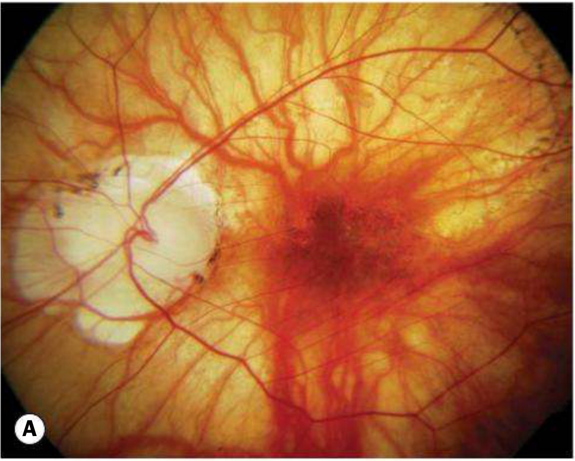
- Tessellated (tigroid) fundus - diffuse attenuation of the RPE with visibility of large choroidal vessels, giving a tiled appearance
- Anomalous optic disc - unusually small, large, or tilted; peripapillary chorioretinal atrophy (commonly as a temporal crescent)
- Focal chorioretinal atrophy - patchy areas where choroidal vessels and even bare sclera are visible
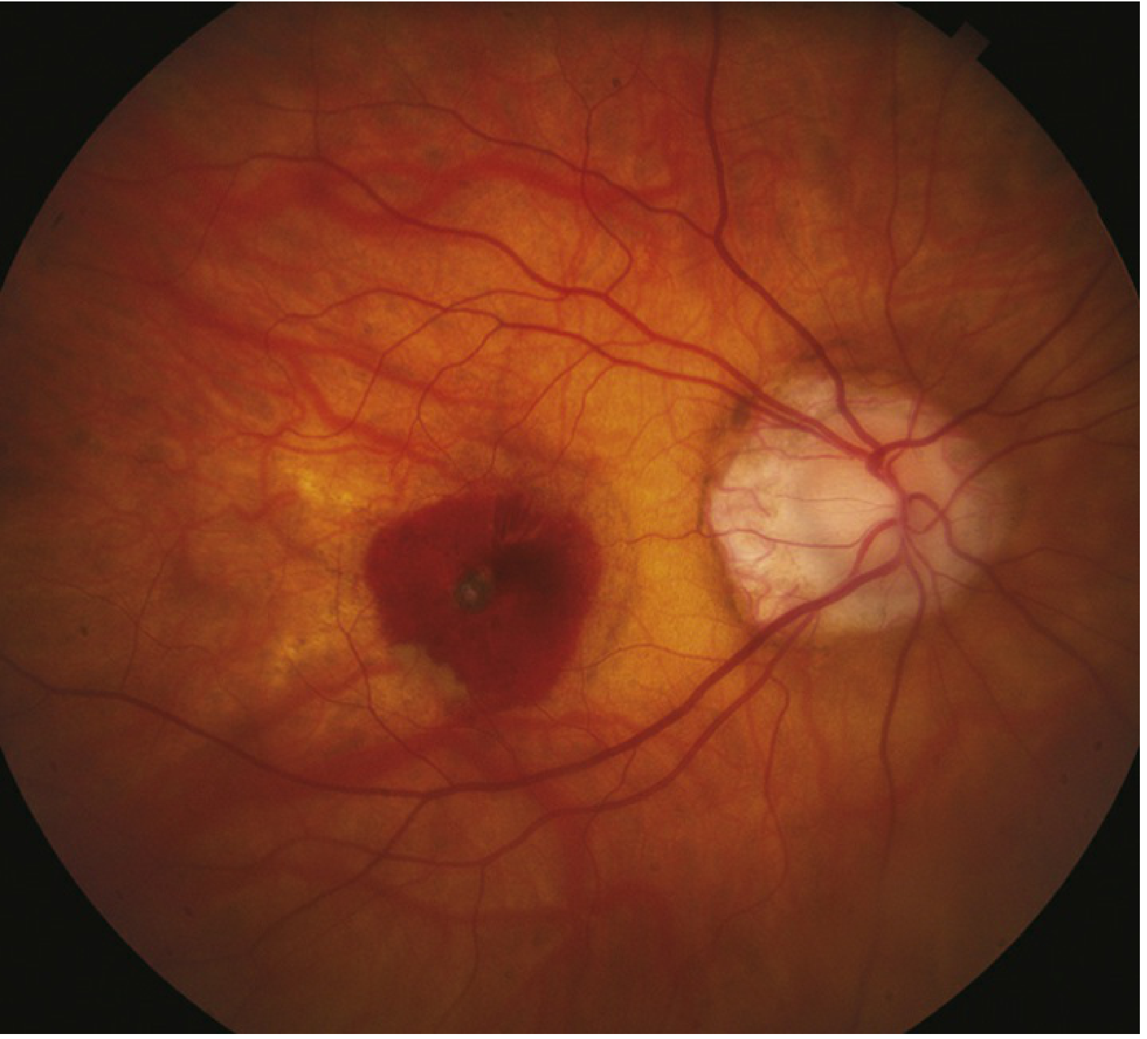
- Lacquer cracks - fine, irregular, yellow lines criss-crossing the posterior pole; ruptures in the RPE-Bruch membrane-choriocapillaris complex (in ~5% of highly myopic eyes); can be complicated by macular neovascularization (MNV)
- Subretinal "coin" hemorrhages - develop from lacquer cracks in the absence of MNV
- Fuchs spot - a pigmented macular scar representing a regressed area of MNV
- Posterior staphyloma - focal ectasia of the posterior sclera (peripapillary or macular), present in approximately one-third of eyes with pathologic myopia; associated with macular hole formation and "dome-shaped macula"
- Lattice degeneration - peripheral retinal thinning with risk of retinal breaks
- Macular neovascularization (MNV) - occurs in ~10% of highly myopic eyes; presents as a grey/green subretinal lesion with subretinal blood or exudate; the prognosis for MNV in young myopic patients is better than in AMD
Systemic Associations of High Myopia
- Marfan syndrome
- Stickler syndrome
- Down syndrome
- Ehlers-Danlos syndrome
- Noonan syndrome
- Pierre-Robin syndrome
- Prematurity
- Cornelia de Lange syndrome
4. Investigations
Refractive Assessment
- Manifest refraction - subjective refraction without cycloplegia; establishes the working correction
- Cycloplegic refraction - after instillation of a cycloplegic agent (e.g., cyclopentolate 1%); removes accommodative tone, giving the true refractive error; especially important in children and young adults
- Autorefraction / retinoscopy - objective measures used to guide subjective refraction
Visual Acuity
- Snellen/LogMAR chart at 6 m (20 ft) for distance VA
- Pinhole test: improvement with pinhole confirms a refractive (not structural) cause of blurred vision
Biometry
- A-scan ultrasound or optical biometry (IOLMaster) to measure axial length - valuable in monitoring myopia progression and for surgical planning (phakic IOL, cataract)
- Keratometry - measures corneal curvature (K-readings); important before refractive surgery
Posterior Segment Investigations (for High/Pathologic Myopia)
- Dilated fundus examination with indirect ophthalmoscopy - mandatory; search for retinal breaks, detachment, lattice degeneration. Scleral depression should be performed with care over a staphyloma
- Slit lamp biomicroscopy with 60-D or 90-D lens - examine the macula, search for MNV (subretinal grey-green lesion, subretinal blood/fluid, exudate)
- Optical Coherence Tomography (OCT) - the key imaging investigation:
- Identifies MNV, foveal retinoschisis (a cause of vision loss in high myopia), macular holes, macular retinal detachment over a staphyloma, and intrachoroidal cavitation
- OCT angiography (OCTA) can detect MNV without dye injection
- Intravenous fluorescein angiography (IVFA) - for suspected MNV when OCT is equivocal
- IOP measurement - by applanation tonometry (note: Schiotz or Tono-pen may underestimate IOP in highly myopic eyes due to altered scleral rigidity)
- Visual fields - in suspected glaucoma; note that myopic visual field defects can mimic early glaucoma; progressive visual field loss in the absence of progressive myopia suggests true glaucoma
5. Management
A. Optical Correction (Non-Surgical)
Spectacles
- Concave (diverging, minus) lenses are the standard correction
- Fully correctable in simple myopia (20/40 or better in 95%+ of patients)
- In pathologic myopia, spectacles are palliative; correction is limited by the severity of the refractive error and posterior segment abnormalities
Contact Lenses
- Soft, rigid gas-permeable (RGP), or orthokeratology lenses
- RGP lenses are preferred for irregular corneas
B. Myopia Control (Slowing Progression in Children)
Modern evidence supports the following in children aged 5-15 years:
| Intervention | Evidence |
|---|---|
| Low-dose atropine 0.01% eyedrops at bedtime | Significantly slows myopia progression; reasonable for children with >1 D/year progression |
| High-add (+2.50 D) centre-distance soft multifocal contact lens | BLINK trial shows dose-dependent slowing in ages 7-11 with <5 D myopia |
| Orthokeratology | Overnight rigid lenses reshape the cornea, reducing progression |
| Increased outdoor time | Sunlight exposure is protective; should be actively encouraged |
C. Refractive Surgery (Surgical Correction)
| Procedure | Range correctable | Notes |
|---|---|---|
| PRK (Surface ablation) | Up to -6 D myopia | No flap; preferred in thin corneas; slower recovery |
| LASIK | Moderate to high myopia (up to -6 to -8 D depending on corneal thickness) | Most widely performed; flap complications possible (epithelial ingrowth, diffuse lamellar keratitis, flap dislocation) |
| SMILE | Myopia and myopic astigmatism | Small-incision lenticule extraction; no flap |
| Phakic IOL (ICL) | Up to -20.50 D | Posterior chamber implant (Collamer material); preserves accommodation; risks: uveitis, cataract, glaucoma, endothelial cell loss |
| Iris-clip (Verisyse) | High myopia | Anterior chamber; risks: pupillary block, dislocation |
| Clear lens extraction | Very high myopia | Good results; carries risk of retinal detachment in high myopia |
Refractive surgery is less successful in pathologic myopia due to severe refractive errors and posterior segment abnormalities.
D. Management of Complications (Pathologic Myopia)
- Retinal breaks - symptomatic breaks: laser photocoagulation, cryotherapy, or scleral buckling; asymptomatic breaks without surrounding pigmentation may also be treated
- Rhegmatogenous retinal detachment (RRD) - vitreoretinal surgery (vitrectomy ± scleral buckle)
- Macular neovascularization (MNV) - anti-VEGF therapy (intravitreal ranibizumab, bevacizumab, or aflibercept) is the treatment of choice; injection frequency is generally less than for AMD; laser photocoagulation may be considered for extrafoveal/juxtafoveal CNV but is seldom used
- Macular retinoschisis / macular hole - vitrectomy; surgical outcomes are more unpredictable in myopic macular holes than idiopathic ones
- Glaucoma - treated per standard glaucoma protocols; monitor for progression distinguishing myopic field loss from true glaucomatous damage
- Cataract - standard phacoemulsification, but RD risk is higher post-operatively in high myopes; lens dislocation is a recognized rare risk
E. Protective Measures
- Polycarbonate safety goggles are recommended for sports in pathologic myopia - even minor trauma can cause choroidal rupture
- Scleral depression should be performed cautiously over a staphyloma (risk of choroidal damage)
Follow-up
- Simple myopia: routine refraction every 1-2 years; annually in children during progression phase
- Pathologic myopia (without complications): dilated examination every 6-12 months, watching for MNV, retinal breaks, and foveoschisis
Sources: Kanski's Clinical Ophthalmology: A Systematic Approach, 10th ed., Ch. 14; Goldman-Cecil Medicine, 27th ed., Ch. 391; The Wills Eye Manual, 8th ed., Section 11.22
Definition, classification, management, clinical features, investigation, complications of hypermetropia.
Hypermetropia (hyperopia/farsightedness) is a refractive error where parallel light rays focus behind the retina, causing blurred near vision and, in high degrees, blurred distance vision too. It results from a short axial length or insufficient corneal/lens curvature. Young patients can compensate with accommodation, but this compensation fails with age. Key complications include accommodative esotropia, amblyopia, and angle-closure glaucoma.
Searching sources...
Finding Sources
Finding Sources
Good. Now let me read all the key sections in parallel - Kanski's paediatric refraction chapter (which covers hypermetropia prescription and management extensively), Goldman-Cecil, Wills Eye, and the neuroscience optics diagram:
Reading File
Reading File
Reading File
Loading Image
Good. Let me now read the Kanski "when to prescribe" section and the accommodative esotropia sections, which are critical for hypermetropia management/complications:
Reading File
Finding Sources
Finding Sources
Excellent. Let me now read the accommodative esotropia management section, the angle-closure glaucoma risk, and the Medical Physiology explanation of hyperopia mechanism:
Reading File
Reading File
Reading File
I now have comprehensive content. Let me also quickly check the amblyopia classification section and the management of accommodative esotropia in Kanski's:
Reading File
Reading File
I now have all the material needed. Let me compile the comprehensive answer.
Hypermetropia (Hyperopia / Farsightedness)
1. Definition
Hypermetropia (also called hyperopia or farsightedness) is a refractive error in which parallel rays of light from a distant object converge to a focus behind the retina rather than on it. The eye is effectively "underpowered" relative to its axial length.
The key optical distinction from myopia: even with the crystalline lens fully accommodated, a near object still focuses behind the retina and appears blurry. Young patients can partially or fully compensate using accommodation, but this reserve is exhausted with age and effort.
2. Classification
A. By Aetiology
| Type | Mechanism |
|---|---|
| Axial hypermetropia (most common) | Eyeball shorter than normal axial length; lens power is correct but the eye is too short |
| Refractive (curvature) hypermetropia | Cornea or lens is flatter than normal, reducing total refracting power |
| Index hypermetropia | Reduced refractive index of the lens (rare; e.g., in early nuclear sclerosis of diabetes) |
| Positional hypermetropia | Posterior displacement of the crystalline lens |
B. By the Role of Accommodation (Donders' Classification)
This is the most clinically useful classification:
| Type | Description |
|---|---|
| Latent hypermetropia | The portion masked by the involuntary tone of the ciliary muscle; revealed only by cycloplegic refraction. Never clinically apparent; corrects itself with cycloplegia. |
| Manifest hypermetropia | The portion not masked by ciliary tone. Subdivided into: |
| - Facultative | Corrected by voluntary accommodation; patient can see clearly but at the cost of accommodative effort |
| - Absolute | Cannot be corrected even by maximum accommodation; patient always has blurred near vision |
| Total hypermetropia | Latent + manifest; the full refractive error revealed under cycloplegia |
C. By Degree
| Grade | Dioptres (D) |
|---|---|
| Low | Up to +2.00 D |
| Moderate | +2.00 to +5.00 D |
| High | > +5.00 D |
Note: Most children are mildly hypermetropic (+1 to +3 D) at birth - this is physiological. Most undergo emmetropization (natural reduction of hypermetropia) during the first years of life.
3. Clinical Features
Symptoms
The presentation depends critically on age (available accommodation) and degree of hypermetropia:
Infants / Young Children (abundant accommodation):
- Small degrees of hypermetropia are asymptomatic and do not require correction
- High hypermetropia leads to constant accommodative effort - the child never relaxes their ciliary muscle
- May present with convergent squint (esotropia) - the link between accommodation and convergence causes the eyes to turn inward when the child accommodates to overcome hypermetropia
- Amblyopia - bilateral ametropic amblyopia from uncorrected high symmetrical hypermetropia; reduced VA in the absence of any organic lesion
School-age Children / Young Adults:
- Asthenopia (eyestrain, headaches) - particularly with sustained near work such as reading; ciliary muscle fatigue
- Blurred near vision - particularly in absolute hypermetropia
- Distance vision initially preserved (corrected by accommodation)
- Difficulty concentrating; avoidance of reading tasks
- Photophobia and blepharospasm can occur in severe cases
Middle Age / Adults (40+ years):
- Accommodation starts failing (presbyopia); the previously compensated hypermetropia becomes manifest
- Blurred vision at all distances - both near and distance, since accommodation can no longer compensate even for distance
- The first pair of glasses may need to correct both distance and near tasks (bifocals)
- The onset of symptoms may be sudden and distressing because many patients were unaware of any refractive error
Signs
- High AC/A ratio accommodative effort apparent on slit lamp (miosis, convergence)
- Shallow anterior chamber - short axial length is anatomically associated with a narrow anterior chamber angle
- Small globe appearance
- In esotropia-associated cases: convergent squint manifest on cover test
- Hyperaemic optic disc appearance (pseudopapilloedema) can occasionally be seen in high hypermetropia - the small scleral canal is crowded, making the disc appear swollen
4. Investigations
Refraction
- Manifest (non-cycloplegic) refraction - establishes the correction tolerated by the conscious patient; reveals only manifest hypermetropia; the patient's accommodation tends to reduce the measured error
- Cycloplegic refraction - the definitive investigation; instillation of a cycloplegic agent paralyzes the ciliary muscle, revealing the total hypermetropia (latent + manifest):
- Cyclopentolate 1% (0.5% in children <6 months) - standard agent; 1 drop repeated after 5 minutes; maximal cycloplegia in 30 minutes; recovery within 2-3 hours
- Atropine 1% (stronger, longer duration) - used for high hypermetropia, heavily pigmented irides, or when cyclopentolate fails; instilled at home twice daily for 1-3 days before the appointment; visual effects last up to 2 weeks. Parents must stop if systemic toxicity (flushing, fever, restlessness) occurs
- Retinoscopy - objective measurement; used alongside subjective refraction; dynamic retinoscopy confirms adequacy of cycloplegia
- Autorefraction - quick objective screen; less reliable than retinoscopy in high hypermetropia
Ocular Investigations
- Visual acuity - Snellen/LogMAR; in children, look for anisometropia (different refraction between eyes) and reduced BCVA suggesting amblyopia
- Cover test and binocular vision assessment - to detect associated esotropia (latent or manifest); AC/A ratio measurement
- Slit lamp examination - shallow anterior chamber depth (predicts angle-closure risk)
- Gonioscopy - mandatory in all adult hypermetropic patients; up to one in six patients with hypermetropia of ≥1 D are primary angle closure suspects; routine gonioscopy should be considered in all adults with hypermetropia
- IOP measurement - to screen for raised IOP and angle-closure glaucoma risk
- Axial length (optical biometry / A-scan ultrasound) - documents short eye; important for IOL calculation if cataract surgery is planned
- Corneal topography - before refractive surgery
- Electrophysiology / neuroimaging - if amblyopia does not respond to treatment as expected, to exclude organic pathology
5. Management
A. Optical Correction
Spectacles - convex (converging, plus) lenses
- These are the primary treatment and move the focal plane forward onto the retina
- In children without squint: In general, up to 4 D of hypermetropia should not be corrected if the child has no squint and no near vision difficulties (to avoid interfering with physiological emmetropization)
- In children with esotropia: The full cycloplegic correction must be prescribed even in children under 2 years of age - this is the key treatment for accommodative esotropia
- For children >8 years: prescribe the maximum 'plus' tolerated on non-cycloplegic refraction (manifest hypermetropia)
Contact Lenses
- Soft convex lenses; suitable for older children and adults
- Orthokeratology is not used for hypermetropia
Bifocals
- Indicated when there is convergence excess esotropia (high AC/A ratio): a reading addition reduces accommodative convergence at near, allowing bifoveal fixation
- Executive bifocals (upper-lower division at the pupillary lower border) are preferred in children
- The reading addition is gradually reduced and withdrawn through the early teenage years
B. Refractive Surgery
| Procedure | Range | Notes |
|---|---|---|
| LASIK | Up to +4 D | Steepens the corneal curvature centrally; results less predictable than for myopia |
| Surface ablation (PRK) | Low degrees | Slower recovery |
| Conductive Keratoplasty (CK) | Low to moderate | Radiofrequency energy applied to corneal periphery; causes stromal shrinkage and increases central curvature; complications: early overcorrection, regression, induced astigmatism |
| Clear lens extraction + IOL | High degrees | Removes the natural lens and implants a calculated IOL; eliminates accommodation but corrects any degree of hypermetropia; risk of RRD is lower than in high myopia |
| Phakic IOL | High degrees | Preserves natural accommodation |
Goldman-Cecil Medicine notes that refractive surgical procedures can correct up to 5 D of hyperopia.
C. Management of Complications
Amblyopia:
- Refractive correction first - optical correction alone may improve VA significantly
- Occlusion (patching) of the better eye - the mainstay of amblyopia treatment; full-time or part-time depending on age and density of amblyopia; effective up to 7-8 years (strabismic) or into the teens (anisometropic)
- Atropine penalization of the normal eye - alternative when patch compliance is poor; particularly effective for mild-moderate anisometropic hypermetropic amblyopia
- Sensitive period closes at approximately 7-8 years; early detection and treatment is vital
Accommodative Esotropia:
- Full cycloplegic refractive correction is the first and definitive treatment
- Fully accommodative esotropia: glasses alone eliminate the deviation and restore binocular single vision (BSV)
- Partially accommodative esotropia: residual deviation requires surgery after full amblyopia treatment
- Bifocals for convergence excess
Acute Angle-Closure Glaucoma (see below)
6. Complications
| Complication | Mechanism | Notes |
|---|---|---|
| Accommodative esotropia | Excessive accommodation to overcome hypermetropia → excessive convergence via the AC/A reflex | Typically presents age 18 months - 3 years; deviation is +2.00 to +7.00 D of hypermetropia. Fully accommodative (glasses eliminate squint) or partially accommodative |
| Amblyopia | Degraded retinal image from uncorrected refractive error during visual development; or suppression of the deviating eye in strabismus | Bilateral ametropic amblyopia from high symmetrical hypermetropia; anisometropic amblyopia if one eye has more hypermetropia than the other |
| Asthenopia / Accommodative fatigue | Chronic overuse of the ciliary muscle | Eyestrain, frontal headaches, difficulty with sustained near work |
| Primary Angle-Closure Glaucoma (PACG) | Short axial length → shallow anterior chamber → narrow angle → iris-lens contact blocks aqueous drainage → acute IOP rise | Hypermetropia is a major risk factor for PACG; up to 1 in 6 with ≥1 D hypermetropia are angle-closure suspects; routine gonioscopy is recommended. Phacoemulsification (cataract surgery) deepens the anterior chamber and is highly effective at opening the angle |
| Pseudopapilloedema | Small, crowded optic disc in a small scleral canal - mimics true disc swelling | Important to distinguish from papilloedema; no visual field loss, no disc haemorrhages |
| Premature presbyopia | Accommodative reserve exhausted earlier in life | Symptomatic loss of near vision at a younger age than expected in emmetropes |
Sources: Kanski's Clinical Ophthalmology: A Systematic Approach, 10th ed., Ch. 8 & 18; Goldman-Cecil Medicine, 27th ed., Ch. 391; Medical Physiology (Boron & Boulpaep), Ch. 15; The Wills Eye Manual, 8th ed.
Definition, classification, management, clinical features, investigation, complications of astigmatism
Astigmatism is a refractive error caused by non-spherical (toric) curvature of the cornea or lens, so that rays in different meridians focus at different points, producing blurred or distorted vision at all distances. It is classified as regular (correctable with cylindrical lenses) or irregular (requires rigid lenses). Management includes spectacles, soft toric contact lenses, and refractive surgery.
Searching sources...
Finding Sources
Finding Sources
Reading File
Reading File
Finding Sources
Finding Sources
Reading File
Finding Sources
Reading File
Now let me get the keratoconus signs section and fetch the keratoconus image (irregular astigmatism's key cause), and also look for the "When to prescribe" for astigmatism in children:
Reading File
Loading Image
Finding Sources
I now have comprehensive material from all sources. Let me compile the full structured answer.
Astigmatism
1. Definition
Astigmatism is a refractive error in which the refracting surfaces of the eye (primarily the cornea, secondarily the lens) have different curvatures in different meridians, so that a single point source of light cannot be brought to a single point focus on the retina. Instead, parallel light rays are refracted by different amounts in different planes, creating two separate focal lines rather than one focal point - this is called the Conoid of Sturm. The result is blurred or distorted vision at all distances.
Key distinction from myopia and hypermetropia:
- In myopia/hypermetropia, the entire system is uniformly under- or over-powered but optically spherical - a single point focus is formed (in front of or behind the retina).
- In astigmatism, no single focal point exists - the eye focuses different meridians at different distances.
2. Classification
A. By Regularity (most clinically important)
Regular Astigmatism
- The two principal meridians (of maximum and minimum curvature) are perpendicular to each other (90° apart).
- This is the most common type; most people have some degree of it.
- Not inherently pathological - it is a variation in anatomy.
- Fully correctable with cylindrical (toric) lenses.
Sub-classification of regular astigmatism by axis orientation:
| Type | Description | Clinical note |
|---|---|---|
| With-the-rule (WTR) | Vertical meridian is steepest (more curved); corrected by a plus cylinder at 90° | Most common in young people |
| Against-the-rule (ATR) | Horizontal meridian is steepest; corrected by a plus cylinder at 180° | More common in older adults |
| Oblique | Principal meridians lie between 30°-60° or 120°-150° | Less common |
Sub-classification of regular astigmatism by nature of the error in each meridian:
| Type | Both meridians | Example |
|---|---|---|
| Simple myopic astigmatism | One meridian focused on retina; other focused in front | One principal power is 0, other is myopic |
| Compound myopic astigmatism | Both meridians focused in front of retina | Both myopic, but different powers |
| Simple hypermetropic astigmatism | One meridian focused on retina; other focused behind | One principal power is 0, other is hypermetropic |
| Compound hypermetropic astigmatism | Both meridians focused behind retina | Both hypermetropic, different powers |
| Mixed astigmatism | One meridian focused in front; other focused behind the retina | One myopic, one hypermetropic |
Irregular Astigmatism
- The principal meridians are not perpendicular to each other, or there are multiple focal points scattered across the cornea.
- Usually caused by pathological changes in the corneal surface.
- Cannot be adequately corrected with spectacles or soft contact lenses.
- Requires rigid gas-permeable (RGP) contact lenses; in severe cases, corneal surgery.
Causes of irregular astigmatism:
- Keratoconus (most important - progressive corneal ectasia)
- Corneal scarring from infection (bacterial, herpetic, acanthamoeba keratitis) or trauma
- Pterygium - limbal growth that distorts the corneal curvature
- Pellucid marginal degeneration - inferior peripheral corneal thinning producing high ATR irregular astigmatism
- Post-surgical (penetrating keratoplasty, refractive surgery complications)
- Corneal dystrophies
B. By Aetiology
| Type | Source | Notes |
|---|---|---|
| Corneal astigmatism | Non-spherical corneal curvature | The dominant component; measured by keratometry/topography |
| Lenticular astigmatism | Non-spherical lens curvature; oblique lens tilt | Less common; lens changes with age or subluxation |
| Total (refractive) astigmatism | Combined corneal + lenticular | Measured by refraction |
Note: Corneal and lenticular astigmatism can partially cancel each other (physiological compensation), which is why total astigmatism may be less than corneal astigmatism alone.
C. By Degree
| Grade | Cylinder (D) |
|---|---|
| Mild | < 1.00 D |
| Moderate | 1.00 - 2.50 D |
| High | > 2.50 D |
3. Clinical Features
Symptoms
- Blurred vision at all distances - unlike myopia (blur at distance only) or hypermetropia (blur at near preferentially); astigmatism impairs both
- "Shadowing" or "ghosting" of images - objects appear to have a secondary faint shadow or double image
- Difficulty seeing fine detail - particularly affecting tasks like reading, recognizing faces, seeing lines precisely
- Asthenopia - eyestrain, fatigue, headaches after sustained visual tasks; more prominent in low-to-moderate astigmatism where the patient strains to compensate
- Tilting the head or squinting to find the axis of clearest vision
- In children: may present as frequent squinting, close viewing, or school difficulties
- Monocular diplopia - a single object appears doubled in one eye (pathognomonic of significant astigmatism or irregular astigmatism)
- In irregular astigmatism (e.g., keratoconus): progressive worsening of vision, frequent prescription changes, and ultimately poor corrected VA with spectacles
Signs
General / External:
- Habitual head tilt or squinting
- In high astigmatism: reduced BCVA even with spectacle correction (if irregular)
Slit Lamp (Irregular Astigmatism / Keratoconus):
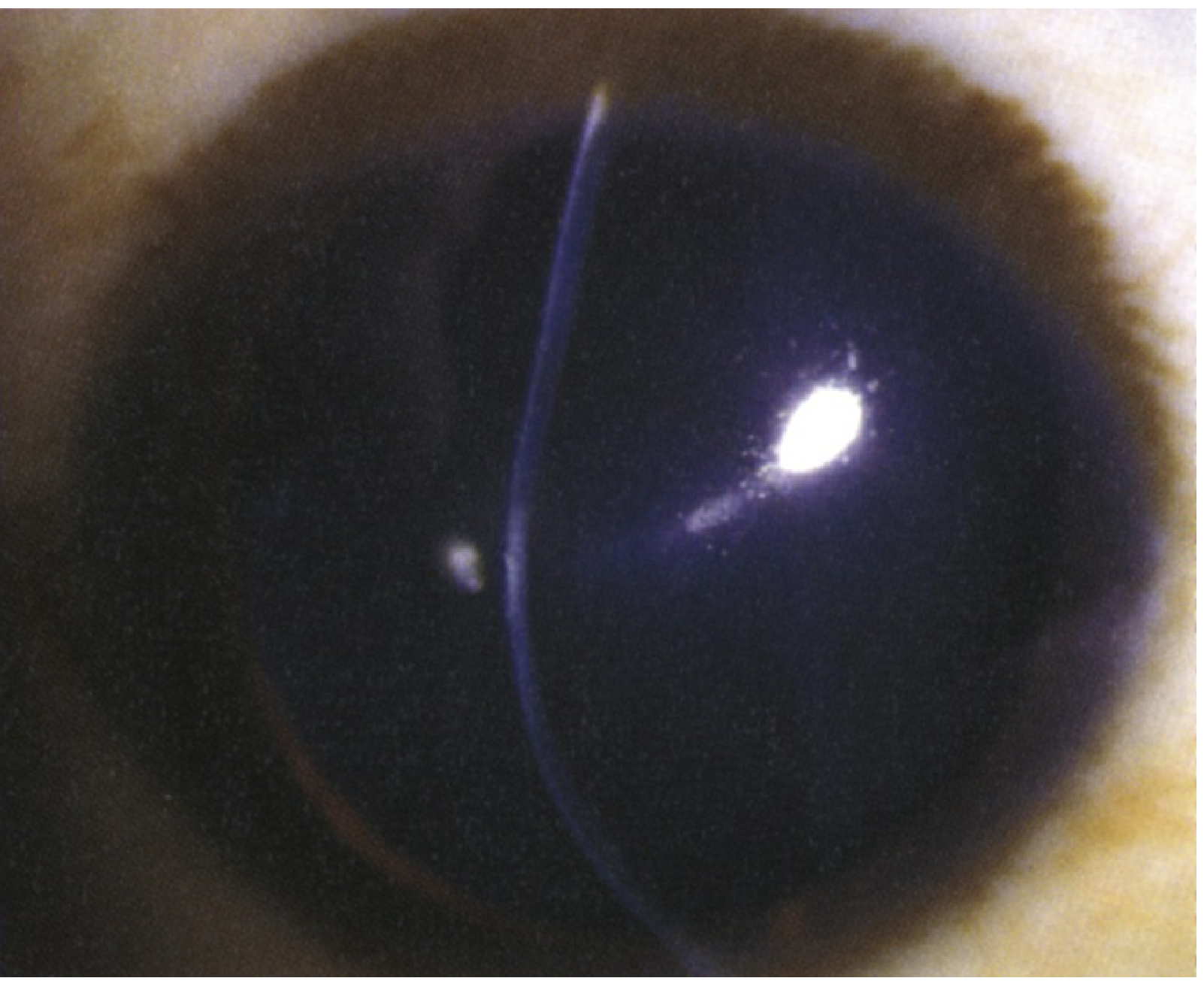
- Keratoconus - hallmark signs:
- Vogt striae - vertical tension lines in the posterior cornea (deep stroma)
- Fleischer ring - iron deposits (haemosiderin) in the epithelium at the base of the cone; best seen with cobalt blue light
- Munson sign - the lower lid bows outward (V-shape) when the patient looks down, due to the conical protrusion
- Apical scarring - superficial stromal opacity at the cone apex
- Acute corneal hydrops - sudden corneal oedema from rupture of Descemet membrane; presents as a white/grey corneal opacity with marked vision loss
Keratometry / Topography Signs:
- Egg-shaped or irregular mires (keratoscope rings) in keratoconus
- Scissor (waterdrop) reflex on retinoscopy in irregular astigmatism
- Oil droplet reflex on direct ophthalmoscopy at 0.5 m distance in keratoconus
4. Investigations
Refractive Assessment
- Subjective refraction - determines the full spectacle correction (sphere + cylinder + axis); the cylinder power and axis define the nature and severity of the astigmatism
- Retinoscopy - objective measurement:
- Regular astigmatism: the reflex moves differently in different meridians; neutralized with a cylindrical lens
- Irregular astigmatism: a "scissor" or "waterdrop" reflex that cannot be neutralized with a simple cylinder
- Cycloplegic refraction - important in children to reveal any latent hypermetropic component accompanying the astigmatism
- Autorefractor - quick objective screen; less reliable for axis determination in irregular astigmatism
Keratometry
- Manual or automated keratometry (ophthalmometry) - measures curvature of the central 3 mm of the cornea in two principal meridians; gives K-readings in dioptres or mm of radius:
- Regular astigmatism: symmetric oval mires
- Keratoconus: egg-shaped or irregular mires; steepened readings (often >47 D)
- Essential before contact lens fitting and refractive surgery
Corneal Topography and Tomography
- Videokeratography (corneal topography) - colour-coded map of the corneal surface:
- Regular astigmatism: symmetric "bow-tie" pattern (equal opposite areas of steepening)
- Keratoconus: inferior steepening, asymmetric bow-tie, progression from symmetric to inferotemporal displacement
- Detects forme fruste (subclinical) keratoconus - a contraindication to LASIK
- Scheimpflug tomography (Pentacam) - maps both anterior and posterior corneal surfaces plus pachymetry:
- Shows posterior corneal elevation and thinning - more sensitive than topography for early keratoconus
- Measures thinnest point location and corneal volume
- Corneal OCT - produces epithelial map; useful for detecting subclinical keratoconus
Visual Acuity
- Snellen / LogMAR VA - reduced BCVA despite optimal spectacle correction suggests irregular astigmatism
- Hard (RGP) contact lens over-refraction - if VA improves with an RGP lens over the spectacle correction, this confirms irregular astigmatism as the cause of reduced BCVA
In Children
- Prescribing threshold: A cylinder of 1.50 D or more should be prescribed in children, especially in anisometropia after age 18 months (to prevent meridional amblyopia)
- Visual evoked potentials (VEPs) - used to assess amblyopia severity when behavioural testing is unreliable
5. Management
A. Optical Correction (Regular Astigmatism)
Spectacles - Cylindrical (toric) lenses
- The standard first-line treatment
- A cylindrical lens corrects by adding power only in the affected meridian
- Prescription written as: Sphere / Cylinder × Axis (e.g., +1.00 / -1.50 × 90)
- Children: prescribe cylinders of 1.50 D or more after age 18 months (especially with anisometropia) to prevent meridional amblyopia
- Full correction is generally well-tolerated
Soft Toric Contact Lenses
- Incorporate a cylindrical correction with orientation stabilization (ballasting, prism ballast, or truncation)
- Suitable for mild-moderate regular astigmatism
- Cannot correct irregular astigmatism
Rigid Gas-Permeable (RGP) Contact Lenses
- The RGP lens vaults over the irregular corneal surface; the tear film between the lens and cornea fills in the irregularities, creating a smooth spherical refracting surface
- The treatment of choice for irregular astigmatism (keratoconus, post-graft astigmatism, corneal scarring)
- Also used when spectacles cause aniseikonia in high anisometropic astigmatism
Scleral Contact Lenses / Hybrid Lenses
- Scleral lenses vault the entire cornea; useful when standard RGPs cannot be tolerated
- Particularly valuable in advanced keratoconus and post-surgical irregular astigmatism
B. Refractive Surgery (Regular Astigmatism)
| Procedure | Range | Notes |
|---|---|---|
| PRK / LASEK (surface ablation) | Up to ~3 D of astigmatism | No corneal flap; longer recovery |
| LASIK | Up to 5 D of astigmatism | Faster recovery; flap complications possible; contraindicated in keratoconus |
| SMILE | Myopic astigmatism | Small-incision lenticule extraction |
| Limbal relaxing incisions (LRI) / Arcuate keratotomy | Small degrees, especially peri-cataract surgery | Paired arcuate incisions in the steep meridian flatten it; can be combined with compression sutures in the perpendicular meridian for large post-keratoplasty astigmatism |
| Toric IOL | Any degree; used at cataract surgery | Incorporates astigmatic correction in the lens implant; postoperative rotation of the implant off-axis is a recognized complication |
| Conductive keratoplasty (CK) | Hypermetropic astigmatism | Radiofrequency applied to corneal periphery |
Preoperative screening (mandatory before any corneal refractive surgery):
- Corneal topography to exclude irregular astigmatism and keratoconus
- Corneal pachymetry to ensure adequate residual stromal bed
- Corneal OCT for epithelial mapping
C. Management of Irregular Astigmatism (Keratoconus Pathway)
- Advise cessation of eye rubbing - chronic eye rubbing is a major risk factor for progression
- Glasses - for mild cases with low regular component
- Soft contact lenses - mild keratoconus only
- RGP or scleral contact lenses - the mainstay for moderate disease; corrects irregular astigmatism optically
- Intracorneal ring segments (ICRS) - implanted within the corneal stroma; reshape the cornea and may allow return to contact lens wear in mild-moderate keratoconus
- Corneal collagen cross-linking (CXL) - the only treatment that halts disease progression:
- Creates new covalent bonds between collagen fibrils using riboflavin drops + UV-A light (365 nm)
- FDA-approved protocol: 9 mm epithelial debridement; riboflavin for 30 minutes; UV-A for 30 minutes
- Approved for age ≥14 years; can be used off-label for younger children
- Should be offered for actively progressive keratoconus; does not improve BCVA
- Penetrating keratoplasty (PK) or Deep Anterior Lamellar Keratoplasty (DALK) - for advanced disease with contact lens intolerance or unsatisfactory vision; DALK preserves the host endothelium; outcomes may be compromised by residual astigmatism requiring contact lenses post-graft
- Post-keratoplasty astigmatism: RGP lenses; arcuate keratotomy ± compression sutures
6. Complications
| Complication | Mechanism | Notes |
|---|---|---|
| Meridional amblyopia | Image blur in one meridian during the critical period of visual development from uncorrected astigmatism (usually >1 D) | Unilateral or bilateral; a form of refractive amblyopia. Treated with optical correction + patching if VA differs by ≥2 Snellen lines |
| Anisometropic amblyopia | Large difference in astigmatic error between the two eyes causes the more astigmatic eye to always have a blurred image | Treatment: full optical correction + occlusion of the better eye |
| Asthenopia / Accommodative fatigue | Chronic ciliary muscle effort trying to partially compensate for astigmatic blur | Headaches, eyestrain; relieved by correct spectacle prescription |
| Keratoconus progression | Untreated or undetected progressive corneal ectasia | May lead to acute corneal hydrops; risk of corneal perforation with minor trauma; end-stage requires corneal transplantation |
| Acute corneal hydrops (keratoconus) | Rupture of Descemet membrane; aqueous enters stroma → sudden oedema | Presents as a white corneal opacity with acute vision loss; usually self-resolves in ~3 months; treated with cycloplegics and hypertonic saline |
| Strabismus | Astigmatism (particularly hypermetropic) can cause accommodative esotropia | Less common as the primary cause than pure hypermetropia |
| Post-LASIK ectasia | Failure to detect subclinical keratoconus preoperatively; residual stroma too thin post-ablation | Progressive irregular astigmatism post-surgery; treated with CXL, RGP lenses, or keratoplasty |
| Toric IOL malrotation | Rotation of toric IOL away from the intended axis | Residual astigmatism; may require surgical repositioning |
Sources: Goldman-Cecil Medicine, 27th ed., Ch. 391; Kanski's Clinical Ophthalmology: A Systematic Approach, 10th ed., Ch. 8 & 7; The Wills Eye Manual, 8th ed., Section 4.24; Kanski's Ch. 18 (amblyopia & strabismus)
Causes and clinical features of conjunctivitis.
Conjunctivitis is inflammation of the conjunctiva, classified broadly as infectious (bacterial, viral, chlamydial) or non-infectious (allergic, toxic, autoimmune). Common features include red eye, discharge, and lid swelling. Viral causes (especially adenovirus) produce watery discharge and follicles; bacterial causes produce purulent discharge; allergic conjunctivitis produces itching and papillae.
Searching sources...
Finding Sources
Finding Sources
Excellent results. Now let me read the key sections in parallel - Kanski's comprehensive conjunctivitis section, Tintinalli's Emergency Medicine classification, the Wills Eye Manual, and the Goldman-Cecil section:
Reading File
Reading File
Reading File
Reading File
Reading File
Reading File
Reading File
Loading Image
Now let me get the viral, chlamydial, and allergic conjunctivitis sections from Kanski's:
Reading File
Reading File
Reading File
Reading File
Now let me get the viral conjunctivitis and allergic conjunctivitis sections from Kanski's:
Finding Sources
Reading File
Reading File
I now have comprehensive content from multiple authoritative textbooks. Let me compile the full structured answer.
Conjunctivitis
Definition
Conjunctivitis is inflammation of the conjunctiva - the transparent mucous membrane covering the anterior sclera (bulbar conjunctiva) and lining the inner surface of the eyelids (palpebral conjunctiva). It is the most common cause of the "red eye" and can result from infectious, allergic, toxic, or mechanical causes. It usually involves the entire conjunctiva and is associated with discharge. Vision is typically normal unless there is corneal involvement (keratoconjunctivitis).
Diagnostic Algorithm: Follicles vs. Papillae
The single most useful bedside distinction is whether the conjunctiva shows follicles or papillae on slit lamp / eversion examination:
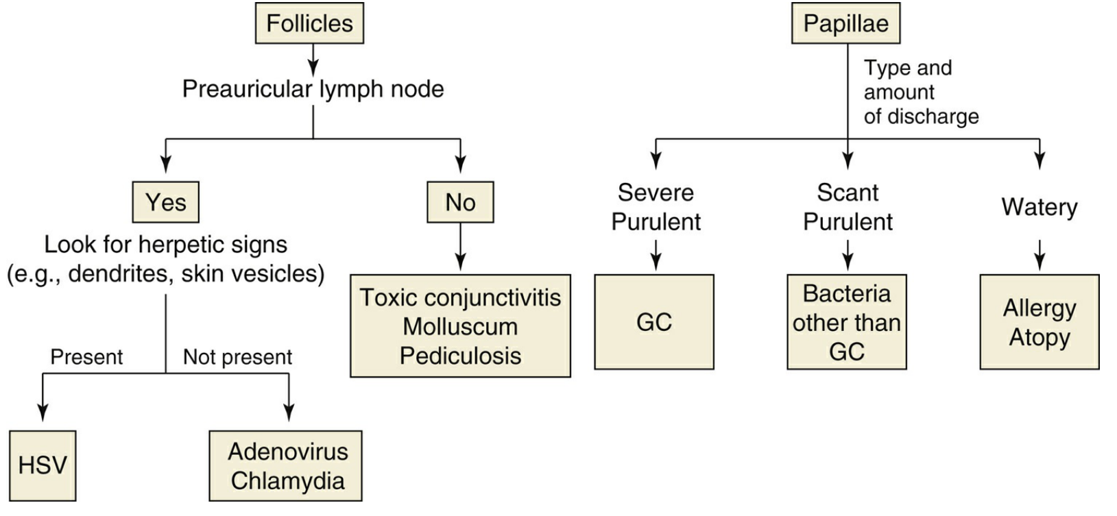
| Feature | Follicles | Papillae |
|---|---|---|
| Structure | Discrete, translucent, rice-grain elevations; blood vessels run around them | Mounds of tissue with a central vascular core; blood vessels run through them |
| Histology | Subepithelial lymphoid germinal centres | Folds of hyperplastic epithelium with fibrovascular cores and inflammatory cell infiltration |
| Location | Most prominent in the fornices; can only form where conjunctiva is loosely attached | Form in palpebral conjunctiva and limbal bulbar conjunctiva (where it is firmly attached) |
| Causes | Viral, chlamydial, toxic (topical drug hypersensitivity), Parinaud oculoglandular syndrome | Bacterial, allergic, contact lens wear, blepharitis, superior limbic keratoconjunctivitis |
Causes and Clinical Features
1. BACTERIAL CONJUNCTIVITIS
A. Acute Bacterial Conjunctivitis (Non-Gonococcal)
Causes (most common isolates):
- Staphylococcus aureus - most common in adults
- Streptococcus pneumoniae - common in children
- Haemophilus influenzae - common in children; risk of otitis and systemic spread
- Moraxella catarrhalis
Clinical Features:
Symptoms:
- Usually bilateral (one eye 1-2 days before the other)
- Painless mucopurulent discharge
- Eyelids stuck together on waking - the classic complaint
- Gritty / foreign-body sensation
- Mild photophobia
- Vision usually normal
Signs:
- Diffuse conjunctival injection (hyperaemia) involving tarsal and forniceal conjunctiva
- Discharge initially watery → rapidly becomes mucopurulent
- Eyelid oedema and erythema (more marked in severe cases)
- Papillary reaction on the tarsal conjunctiva
- Superficial corneal punctate epithelial erosions may occur
- Preauricular lymphadenopathy usually absent (except in gonococcal/meningococcal)
- About 60% are self-limiting within 1-2 weeks
B. Hyperacute (Gonococcal) Conjunctivitis
Cause: Neisseria gonorrhoeae (sexually transmitted; can invade intact corneal epithelium - uniquely dangerous among bacteria)
Clinical Features:
- Hyperacute onset (within 12-24 hours)
- Profuse purulent discharge (copious, cream-coloured pus)
- Marked conjunctival injection and severe chemosis
- Marked eyelid swelling and erythema
- Preauricular lymphadenopathy (present, unlike other bacterial)
- Corneal ulceration that can rapidly progress to perforation
- Systemic disseminated gonococcal infection may occur
Key investigation: Gram stain shows Gram-negative kidney-shaped intracellular diplococci. Culture on enriched media (chocolate agar, Thayer-Martin).
C. Meningococcal Conjunctivitis
- Rare; usually affects children
- Hyperacute picture similar to gonococcal
- Life-threatening risk: up to 30% of cases develop systemic meningococcal disease without treatment
- Requires urgent systemic prophylaxis (IM benzylpenicillin or ceftriaxone)
2. VIRAL CONJUNCTIVITIS
The most common type overall; caused by adenovirus in 90% of cases. Highly contagious - can survive on dry surfaces for weeks and is spread via contact with ocular/respiratory secretions and fomites (towels, slit lamp).
A. Non-Specific Acute Follicular Conjunctivitis (most common form)
Cause: Adenovirus (multiple serotypes)
Clinical Features:
- Often preceded by upper respiratory tract infection (sore throat, coryzal symptoms)
- Unilateral onset then contralateral eye involved 1-2 days later (usually less severely)
- Watery (serous) discharge
- Redness, irritation, itching, mild photophobia
- Follicles on inferior palpebral conjunctiva - the hallmark
- Occasional small subconjunctival haemorrhages
- Preauricular lymphadenopathy - tender; the most useful distinguishing feature from bacterial
- Vision normal unless keratitis present
B. Pharyngoconjunctival Fever (PCF)
Cause: Adenovirus serovars 3, 4, 7; spread by respiratory droplets
- Same ocular features as above plus prominent sore throat and fever
- Keratitis in ~30%
- Common in children; family clusters
C. Epidemic Keratoconjunctivitis (EKC)
Cause: Adenovirus serovars 8, 19, 37 - the most severe adenoviral eye disease
- Severe follicular conjunctivitis
- Keratitis in ~80% - punctate corneal epithelial erosions staining with fluorescein; later subepithelial infiltrates (nummular keratitis) that cause prolonged visual blurring
- Marked photophobia
- Pseudomembranes on the tarsal conjunctiva in severe cases
- Large preauricular lymph node
- Can cause symblepharon and subconjunctival scarring in severe cases
D. Acute Haemorrhagic Conjunctivitis
Cause: Enterovirus 70, Coxsackievirus A24 (tropical/subtropical epidemics)
- Rapid onset and resolution (1-2 weeks)
- Subconjunctival haemorrhages are the hallmark - often extensive
- Follicular conjunctivitis, watery discharge, eyelid swelling
E. Herpes Simplex Virus (HSV) Conjunctivitis
- Usually unilateral - important distinguishing feature
- Associated skin vesicles on lids or periorbital skin
- Follicular conjunctivitis
- Dendrites on cornea (pathognomonic) - detected with fluorescein staining
- Tender preauricular lymphadenopathy
F. Molluscum Contagiosum
- Caused by poxvirus; peak age 2-4 years
- Chronic follicular conjunctivitis from viral particle shedding by lid/lash margin lesions
- Examine the eyelash line carefully for the characteristic umbilicated, pearly-white lid nodule
- Conjunctivitis resolves after treatment of the skin lesion
G. Systemic Viral Infections
- Varicella, measles, mumps - associated follicular conjunctivitis
- Varicella-zoster (ophthalmic shingles) - conjunctivitis plus skin vesicles in V1 distribution
- COVID-19 (SARS-CoV-2) - conjunctivitis in a minority of cases; viral RNA can be isolated in tears
3. CHLAMYDIAL CONJUNCTIVITIS
A. Adult Inclusion Conjunctivitis
Cause: Chlamydia trachomatis serovars D-K; an oculo-genital infection affecting 5-20% of sexually active young adults; transmitted by autoinoculation from genital secretions (or eye-to-eye spread in ~10%); incubation ~1 week
Clinical Features:
Symptoms:
- Subacute onset of unilateral or bilateral redness, watering, and discharge
- Untreated, becomes chronic and may persist for several months
- Always ask about sexual exposure
Signs:
- Watery or mucopurulent discharge
- Tender preauricular lymphadenopathy
- Large follicles most prominent in the inferior fornix and upper tarsal conjunctiva
- Superficial punctate keratitis (common)
- Peripheral corneal infiltrates after 2-3 weeks
- Chronic cases develop papillae (less follicles) with mild conjunctival scarring and superior corneal pannus
Systemic associations:
- Males: non-gonococcal urethritis (often asymptomatic), epididymitis, Reiter syndrome trigger
- Females: urethritis, dysuria, discharge; risk of PID and infertility; Fitz-Hugh-Curtis perihepatitis
B. Trachoma
Cause: C. trachomatis serovars A, B, Ba, C; the world's leading cause of preventable irreversible blindness; associated with poverty, overcrowding, poor hygiene, and recurrent infection cycles
Active Stage (children):
- Mixed follicular and papillary conjunctivitis
- Mucopurulent discharge
- Follicles on the superior tarsal conjunctiva (WHO TF: ≥5 follicles on everted upper lid)
- Papillary hypertrophy (WHO TI: intense inflammation obscuring >half of deep tarsal vessels)
- Superior corneal pannus and punctate keratitis
Cicatricial Stage (adults - from repeated infection):
- Conjunctival scarring - Herbert's pits (scarred limbal follicles), Arlt's line (linear tarsal scar)
- Trichiasis - eyelashes turn inward and scratch the cornea
- Entropion - eyelid margin turns inward
- Corneal scarring and blindness (from repeated abrasion + secondary bacterial infection)
4. ALLERGIC CONJUNCTIVITIS
Mechanism: Type I (immediate) hypersensitivity - IgE-mediated mast cell degranulation; some forms also involve Type IV (delayed) hypersensitivity.
Key distinguishing feature: ITCHING - the dominant symptom that differentiates allergic from infectious conjunctivitis.
A. Acute Allergic Conjunctivitis
- Cause: Acute exposure to environmental allergen (e.g., pollen)
- Typical in young children after playing outside in spring/summer
- Acute intense itching and watering
- Dramatic chemosis (conjunctival oedema) - alarming to parents
- Self-limiting within hours; cool compresses ± single drop of adrenaline 0.1% for severe chemosis
B. Seasonal Allergic Conjunctivitis ("Hay Fever Eyes")
- Allergens: Tree and grass pollens; worse in spring/summer
- Redness, watering, itching, sneezing, nasal discharge
- Mild papillary reaction, variable chemosis, lid oedema
- Normal vision
C. Perennial Allergic Conjunctivitis
- Allergens: House dust mites, animal dander, fungal spores; symptoms year-round, worse in autumn
- Milder and more chronic than seasonal form
D. Vernal Keratoconjunctivitis (VKC)
- Affects young males in hot/dry climates; seasonal (spring/summer) exacerbations
- Giant papillae on upper tarsal conjunctiva ("cobblestone" appearance)
- Limbal Horner-Trantas dots (white chalky deposits of eosinophil debris at the limbus)
- Intense itching, thick ropy mucous discharge, photophobia
- Shield ulcer on superior cornea - a serious complication
E. Atopic Keratoconjunctivitis (AKC)
- Occurs in older patients with atopic dermatitis
- Year-round, severe; may lead to conjunctival scarring, keratoconus, cataract, and corneal vascularization
5. NEONATAL CONJUNCTIVITIS (Ophthalmia Neonatorum)
Defined as conjunctival inflammation within the first 4 weeks of life; the most common neonatal infection (up to 10% of neonates).
| Timing of onset | Cause |
|---|---|
| First few days | Chemical (prophylactic drops - silver nitrate) |
| First week | N. gonorrhoeae - most dangerous; rapid corneal ulceration |
| End of first week | Staphylococci and other bacteria |
| 1-2 weeks | Herpes simplex virus (HSV-2) |
| 1-3 weeks | Chlamydia trachomatis - most common severe cause |
Gonococcal neonatal conjunctivitis:
- Hyperacute purulent discharge; risk of corneal perforation and blindness
- Treat systemically (ceftriaxone) + topical + co-treat for Chlamydia
Chlamydial neonatal conjunctivitis:
- Also causes pneumonitis, rhinitis, otitis (systemic Chlamydia)
- Treat with oral erythromycin for 2 weeks
HSV neonatal conjunctivitis:
- May be associated with encephalitis (life-threatening)
- Treat with high-dose IV acyclovir
6. OTHER CAUSES
| Cause | Features |
|---|---|
| Toxic / Chemical | From topical medications (aminoglycosides, preservatives), chlorine in pools; follicular reaction; history of drop use; resolve on discontinuation |
| Contact lens-related | Overwear, solution hypersensitivity, tight lens syndrome; papillary reaction; manage by discontinuing lens |
| Superior Limbic Keratoconjunctivitis (SLK) | Inflammation of superior bulbar and tarsal conjunctiva; associated with thyroid disease and dry eye; papillary reaction superiorly |
| Parinaud Oculoglandular Syndrome | Unilateral granulomatous conjunctivitis + large ipsilateral preauricular/cervical lymphadenopathy; causes: cat-scratch disease (Bartonella), tularaemia, TB, sarcoidosis |
| Stevens-Johnson Syndrome | Severe immune-mediated mucous membrane and skin disease; membranous/pseudomembranous conjunctivitis; risk of symblepharon, dry eye, blindness |
Summary: Key Distinguishing Features at a Glance
| Feature | Bacterial | Viral | Chlamydial | Allergic |
|---|---|---|---|---|
| Discharge | Mucopurulent | Watery/serous | Watery or mucopurulent | Watery / ropy mucus |
| Laterality | Bilateral | Unilateral → bilateral | Uni or bilateral | Bilateral |
| Itching | Absent/mild | Absent/mild | Absent | Dominant |
| Follicles | Absent | Present | Present (large, inferior) | Absent |
| Papillae | Present | Absent | Late/chronic | Present |
| Preauricular node | Usually absent | Present | Present (tender) | Absent |
| Chemosis | Moderate | Mild | Mild | Marked |
| Corneal involvement | Punctate erosions | Punctate keratitis (EKC) | Punctate keratitis + infiltrates | Shield ulcer (VKC) |
| URTI / systemic | No | Yes (PCF, EKC) | Genital STI | Hayfever/atopy |
Sources: Kanski's Clinical Ophthalmology: A Systematic Approach, 10th ed., Ch. 6; Tintinalli's Emergency Medicine, 9th ed., Ch. 241; The Wills Eye Manual, 8th ed.; Goldman-Cecil Medicine, 27th ed., Ch. 391
Discuss the etiology and clinical features of keratitis, corneal ulcers and corneal abrasions.
Keratitis is inflammation of the cornea; when it progresses to epithelial/stromal breakdown it becomes a corneal ulcer. Causes include bacterial (Pseudomonas, Staphylococcus), viral (HSV - the commonest cause of infectious blindness in developed countries), fungal (Aspergillus, Fusarium), parasitic (Acanthamoeba), and non-infectious (exposure, neurotrophic). Corneal abrasion is a superficial epithelial defect from mechanical trauma. All present with pain, redness, photophobia, and reduced vision.
Searching sources...
Finding Sources
Finding Sources
Reading File
Reading File
Reading File
Reading File
Reading File
This is a shared conversation. Sign in to Orris to start your own chat.