
You are going to write 10 mark answer in applied basic science exam first year MDS oral pathology Answer should include - contents, definition, introduction, main answer contents, applied aspect, conclusion and reference. Reference should be from standard reference books and articles. Applied aspect should be atleast 2 pages. Total answer should be 6 pages The question is ELISA
ELISA enzyme linked immunosorbent assay oral pathology diagnosis
ELISA enzyme linked immunosorbent assay types principle oral pathology applied aspects
ELISA enzyme linked immunosorbent assay principle diagram
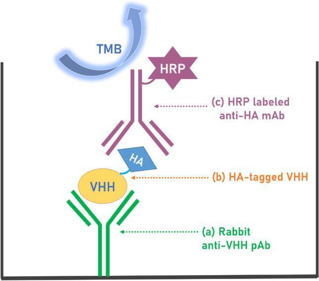
This medical laboratory diagram illustrates a sandwich Enzyme-Linked Immunosorbent Assay (ELISA) technique used for the detection of VHH (variable heavy-chain antibodies). The schematic depicts the spatial arrangement of the assay components within a microtiter plate well from bottom to top: (a) A capture layer consisting of Rabbit anti-VHH polyclonal antibodies (pAb), which are passively adsorbed to the bottom of the well. (b) The analyte, represented as an HA-tagged VHH molecule, where the VHH core binds to the capture antibody and an HA (hemagglutinin) tag is available for detection. (c) The detection layer featuring an HRP-labeled anti-HA monoclonal antibody (mAb). This antibody targets the HA tag of the analyte and is conjugated to Horseradish Peroxidase (HRP), shown as a purple star-shaped molecule. At the top, a blue curved arrow labeled TMB (3,3′,5,5′-Tetramethylbenzidine) represents the chromogenic substrate added to the well. The diagram demonstrates the biochemical principle of signal transduction where HRP catalyzes the oxidation of TMB to produce a measurable colorimetric signal, indicating the presence and concentration of the VHH analyte.
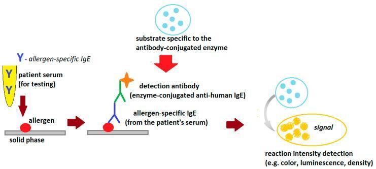
This medical diagram illustrates the general scheme of an indirect Enzyme-Linked Immunosorbent Assay (ELISA) used to detect allergen-specific IgE antibodies in human serum. The process is depicted in four sequential stages: (1) Sample Introduction: Patient serum containing allergen-specific IgE (yellow Y-shaped molecules) is added to a solid phase pre-coated with allergens (red circles). (2) Binding: The specific IgE antibodies bind to the immobilized allergens. (3) Detection: An enzyme-conjugated anti-human IgE detection antibody is introduced, forming a complex with the patient's IgE. This detection antibody is represented by a multi-colored structure topped with an orange star signifying the enzyme. (4) Signal Generation: A specific substrate is added, which the enzyme catalyzes into a detectable signal, visually represented by a transition from light blue circles to a yellow oval. The diagram highlights the clinical utility of ELISA in immunology and allergy diagnostics, specifically for quantifying reaction intensity via colorimetry, luminescence, or density changes.
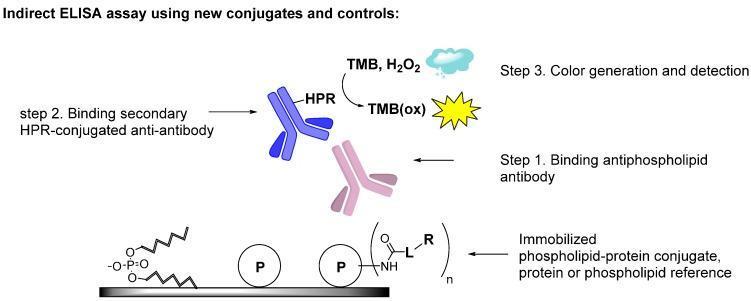
This diagram illustrates the methodology of an indirect Enzyme-Linked Immunosorbent Assay (ELISA) designed for the detection of antiphospholipid antibodies, relevant to the diagnosis of Antiphospholipid Syndrome (APS). The workflow is divided into three sequential steps. Step 1 demonstrates the immobilization of antigens on a solid surface, including phospholipids, proteins (labeled 'P'), and phospholipid-protein conjugates formed via a chemical linker ('L'). A primary antiphospholipid antibody (pink Y-shape) binds to these immobilized antigens. Step 2 shows the introduction of a secondary anti-antibody (blue Y-shape) conjugated with Horseradish Peroxidase (HPR). Step 3 depicts the signal detection phase where HPR catalyzes the reaction between TMB and H2O2 to form oxidized TMB (TMB(ox)), producing a measurable color change. This educational schematic emphasizes the use of stable covalent conjugates to improve the diagnostic sensitivity and reproducibility of assays detecting autoantibodies against complex phospholipid-protein epitopes.
ELISA salivary biomarkers oral disease diagnosis
ENZYME-LINKED IMMUNOSORBENT ASSAY (ELISA)
Applied Basic Sciences | First Year MDS | Oral Pathology | 10 Marks
CONTENTS
- Definition
- Introduction and Historical Background
- Basic Principle
- Components of ELISA
- General Procedure
- Types of ELISA
- Direct ELISA
- Indirect ELISA
- Sandwich (Capture) ELISA
- Competitive ELISA
- Advantages and Disadvantages
- Applied Aspects in Oral Pathology and Dentistry
- Oral Cancer and Salivary Biomarkers
- Periodontal Disease
- Vesiculobullous Lesions
- Oral Infections (HIV, Herpesviruses, HPV)
- Salivary Gland Disorders (Sjogren's Syndrome)
- Oral Submucous Fibrosis and Potentially Malignant Disorders
- Drug and Hormone Detection
- Single-Molecule ELISA and Digital ELISA
- Conclusion
- References
1. DEFINITION
2. INTRODUCTION AND HISTORICAL BACKGROUND
3. BASIC PRINCIPLE
- Specific antigen-antibody interaction - an antibody recognizes and binds only its specific antigen with very high affinity (Kd typically in the nanomolar range).
- Enzyme amplification - an enzyme conjugated to one of the immunological reagents catalyzes the conversion of a colorless substrate into a colored, fluorescent, or luminescent product, amplifying the signal and allowing quantification with a spectrophotometer.
4. COMPONENTS OF ELISA
| Component | Role | Examples |
|---|---|---|
| Solid phase | Immobilizes one reactant | Polystyrene 96-well microtiter plate |
| Antigen | Target molecule to be detected | Viral protein, autoantibody target, cytokine |
| Primary antibody | Specifically recognizes antigen | Monoclonal or polyclonal antibody |
| Secondary antibody | Binds primary antibody (in indirect/sandwich) | Anti-IgG antibody |
| Enzyme label | Signal amplifier | Horseradish peroxidase (HRP), alkaline phosphatase (AP) |
| Substrate | Converted to colored product by enzyme | TMB (3,3',5,5'-tetramethylbenzidine) for HRP; p-NPP for AP |
| Stop solution | Terminates enzymatic reaction | H2SO4 |
| Blocking agent | Prevents non-specific binding | 1-5% bovine serum albumin (BSA), skimmed milk |
| Wash buffer | Removes unbound reagents | Phosphate-buffered saline with Tween-20 (PBS-T) |
5. GENERAL PROCEDURE
6. TYPES OF ELISA
6a. Direct ELISA
- Advantage: Simple, fast, less variability (no secondary antibody step)
- Disadvantage: Lower sensitivity; primary antibody must be individually conjugated; no signal amplification from secondary antibody
- Use: Screening antigen presence; antibody characterization studies
6b. Indirect ELISA
- Advantage: Greater sensitivity (multiple secondary antibodies can bind one primary antibody, amplifying signal); a single labeled secondary antibody can be used with many different primary antibodies
- Disadvantage: More steps; potential cross-reactivity of secondary antibody
- Use: Detection of antibodies in serum (e.g., HIV antibody screening, autoantibody detection)
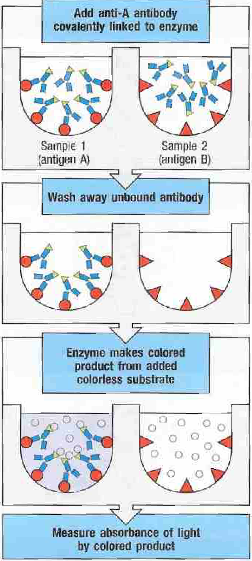
6c. Sandwich (Capture) ELISA
- Advantage: Very high sensitivity and specificity; ideal for complex samples (serum, saliva, GCF)
- Disadvantage: Requires two antibodies recognizing different epitopes of the same antigen; more expensive
- Use: Cytokine quantification, growth factor detection, most diagnostic ELISA kits
6d. Competitive ELISA
- Advantage: Useful when only one antibody is available; suitable for small antigens (haptens, hormones)
- Disadvantage: Inversely proportional signal (high antigen = low signal) can be counterintuitive
- Use: Drug/hormone detection, small molecule quantification
7. ADVANTAGES AND DISADVANTAGES
Advantages
- High sensitivity (detection range: 0.01-0.1 ng)
- High specificity due to antigen-antibody interaction
- Non-radioactive - safe and no special waste disposal
- Large-scale throughput (96 or 384-well plates with automated readers)
- Relatively low cost per test
- Samples can be serum, saliva, GCF, urine, CSF, tissue homogenate
- Quantitative with standard curves
- ELISA kits commercially available for hundreds of analytes
- Can be automated using ELISA plate washers and readers
Disadvantages
- Requires pure antigen or antibody as reference standard
- Risk of false positives from cross-reactive antibodies
- Non-specific binding despite blocking steps
- Prozone (hook) effect at very high antigen concentrations in sandwich ELISA
- Requires careful optimization of antibody concentrations and incubation conditions
- Cannot distinguish active from past infection (detects antibodies, not viability)
- Confirmatory testing (e.g., Western blot) often needed for screening positives
8. APPLIED ASPECTS IN ORAL PATHOLOGY AND DENTISTRY
8.1 Oral Cancer and Salivary Biomarkers
- Epidermal Growth Factor (EGF) - often reduced in OSCC
- Transferrin - elevated in OSCC saliva
- CA125, CA19-9, CEA - carcinoembryonic antigen measurable in saliva by ELISA
- MMP-9 (Matrix Metalloproteinase-9) - elevated in OSCC; reflects basement membrane degradation
- Defensins and S100 proteins - antimicrobial peptides elevated in oral malignancies
- VEGF (Vascular Endothelial Growth Factor) - elevated in oral dysplasia and OSCC
- Assist early detection of OSCC in high-risk patients (tobacco users, alcohol consumers)
- Monitor disease progression and treatment response
- Screen patients with oral potentially malignant disorders (leukoplakia, OSMF, erythroplakia) for malignant transformation
8.2 Periodontal Disease
- Interleukin-1β (IL-1β): The most studied GCF cytokine in periodontitis. Elevated IL-1β measured by ELISA reflects active bone destruction. Serum and salivary IL-1β have also been studied in oral precancer (Selvan et al., 2024, PMID: 39023584).
- TNF-α: GCF TNF-α elevated in aggressive periodontitis; ELISA measurement helps differentiate chronic vs. aggressive disease.
- IL-6, IL-8, IL-17: Quantified by sandwich ELISA in GCF to understand periodontal immunopathology.
- RANKL/OPG ratio: ELISA measurement of receptor activator of nuclear factor-κB ligand (RANKL) and osteoprotegerin (OPG) in GCF gives an index of osteoclastic activity and bone resorption potential.
- Matrix Metalloproteinases (MMP-1, -8, -13): Collagenases measurable in GCF by ELISA; MMP-8 is used as a chair-side diagnostic adjunct in active periodontitis.
- C-reactive protein (CRP): Serum CRP measured by ELISA has established the periodontal-systemic disease link in studies connecting periodontitis with cardiovascular disease and diabetes.
8.3 Vesiculobullous Lesions
- Initial diagnosis
- Monitoring disease activity and response to immunosuppressive therapy
- Predicting relapse (rising ELISA titer often precedes clinical flare)
8.4 Oral Infections
- HSV-1/2 IgG and IgM ELISA aids in differentiating primary from recurrent herpes labialis/intraoral herpetic lesions
- EBV VCA IgM/IgG ELISA is used in diagnosing infectious mononucleosis (gingival enlargement, oral ulcerations) and assessing EBV association with hairy leukoplakia and lymphomas
- CMV IgM ELISA diagnoses cytomegalovirus sialadenitis and oral ulcers in immunocompromised patients
- HHV-8 antibody ELISA assists in Kaposi's sarcoma workup
8.5 Salivary Gland Disorders - Sjogren's Syndrome
- Anti-SSA/Ro and Anti-SSB/La antibody ELISA: Primary diagnostic serological markers; detected by ELISA and EIA with high sensitivity and specificity, included in the 2016 ACR/EULAR classification criteria for SS
- Anti-muscarinic M3 receptor (anti-M3R) antibodies: Detected by ELISA; implicated in pathogenesis of salivary hypofunction. Tsaliki et al. (2026, J Immunol Methods, PMID: 41643842) evaluated assay variability and detection methods for M3R antibodies in Sjogren's disease, highlighting the need for standardized ELISA protocols
- Salivary EGF, lactoferrin, IgA measurement by ELISA reflects salivary gland functional status
- Salivary biomarker ELISA panel (IL-6, IL-10, BAFF, CXCL13) in a recent study (Zhang et al., 2024, Arthritis Res Ther, PMID: 39695856) enabled a non-invasive diagnostic model for primary Sjogren's disease, potentially replacing labial salivary gland biopsy in some settings
8.6 Oral Submucous Fibrosis and Potentially Malignant Disorders
- TGF-β1 (Transforming Growth Factor-beta 1): Elevated in serum and tissue of OSMF patients; the primary pro-fibrotic cytokine. TGF-β1 ELISA in serum/saliva allows staging of fibrosis severity.
- IL-6: Elevated in OSMF; promotes fibroblast differentiation to myofibroblasts.
- PDGF (Platelet-Derived Growth Factor) and VEGF: Measurable by ELISA in OSMF tissue homogenates and serum.
- Lysyl oxidase (LOX): Enzyme responsible for collagen/elastin cross-linking; elevated in OSMF by ELISA.
- Arecoline and its metabolites can be measured in urine/serum by competitive ELISA, aiding in exposure assessment.
8.7 Drug Monitoring and Hormone Detection
- Monitoring cyclosporine levels in organ transplant patients (who develop gingival enlargement)
- Corticosteroid assays in adrenal insufficiency workup (relevant to recurrent aphthous stomatitis, candidiasis)
- Saliva-based cortisol ELISA for stress assessment (impacts wound healing, immunosuppression)
- Parathyroid hormone (PTH) ELISA in metabolic bone disease workup (relevant to jaw lesions, cherubism, brown tumors)
8.8 Advanced ELISA Formats Relevant to Future Oral Diagnostics
9. CONCLUSION
10. REFERENCES
Standard Textbooks
-
Murphy K, Weaver C, Janeway C. Janeway's Immunobiology, 10th ed. New York: W.W. Norton & Company (Garland Science); 2022. [Section A-4: RIA, ELISA, and Competitive Inhibition Assay; Section A-5: Multiplex Bead Assay. pp. 821-824]
-
McPherson RA, Pincus MR, eds. Henry's Clinical Diagnosis and Management by Laboratory Methods, 23rd ed. Philadelphia: Elsevier; 2021. [Chapter on Single-Molecule ELISA and High-Sensitivity Immunoassays]
-
Griffiths CEM, Barker J, Bleiker TO, Chalmers R, Creamer D, eds. Rook's Dermatology, 9th ed. Oxford: Wiley-Blackwell; 2016.
-
Bolognía JL, Schaffer JV, Cerroni L, eds. Dermatology, 2-Volume Set, 5th ed. Philadelphia: Elsevier; 2024. [ELISA for autoimmune bullous diseases; anti-Dsg3, anti-BP180 detection]
-
Neville BW, Damm DD, Allen CM, Chi AC. Oral and Maxillofacial Pathology, 4th ed. Philadelphia: Elsevier; 2016.
-
Regezi JA, Sciubba JJ, Jordan RCK. Oral Pathology: Clinical Pathologic Correlations, 7th ed. St. Louis: Elsevier; 2017.
-
Rajendran R, Sivapathasundharam B. Shafer's Textbook of Oral Pathology, 8th ed. New Delhi: Elsevier India; 2020.
-
Abbas AK, Lichtman AH, Pillai S. Cellular and Molecular Immunology, 10th ed. Philadelphia: Elsevier; 2022.
-
Roitt IM, Brostoff J, Male DK. Immunology, 8th ed. Philadelphia: Mosby Elsevier; 2011.
-
Crowther JR. The ELISA Guidebook (Methods in Molecular Biology, Vol. 149). 2nd ed. Humana Press; 2009.
Key Journal Articles
-
Engvall E, Perlmann P. Enzyme-linked immunosorbent assay (ELISA): Quantitative assay of immunoglobulin G. Immunochemistry. 1971;8(9):871-874. (Original description of ELISA)
-
Engvall E. The ELISA, enzyme-linked immunosorbent assay. Clin Chem. 2010;56(2):319-320.
-
Aydin S. A short history, principles, and types of ELISA, and our laboratory experience with peptide/protein analyses using ELISA. Peptides. 2015;72:4-15.
-
Timilsina S, Bhatt DL, Wondratsch L, et al. Enzyme-linked immunosorbent assay: Types and Applications. (Review, PMID: 36795355). J Biochem Biophys Methods. 2023.
-
Chiamulera MMA, Zancan CB, Remor AP, et al. Salivary cytokines as biomarkers of oral cancer: a systematic review and meta-analysis. BMC Cancer. 2021;21:231. (PMID: 33639868)
-
Liang Y, Yi Z, Li J. The diagnostic value of CYFRA 21-1 in oral squamous cell carcinoma: a meta-analysis. Expert Rev Anticancer Ther. 2024 Nov. (PMID: 39279566)
-
Khurshid Z, Warsi I, Moin SF, et al. Biochemical analysis of oral fluids for disease detection. Adv Clin Chem. 2021;103:21-86. (PMID: 33453866)
-
Zhang Y, Kang N, Xue F, et al. Evaluation of salivary biomarkers for the diagnosis of periodontitis. BMC Oral Health. 2021;21(1):266. (PMID: 34001101)
-
Tsaliki M, Dave D, Kurien BT, et al. Muscarinic 3 receptor antibodies in Sjogren's disease: Evaluating assay variability and detection methods. J Immunol Methods. 2026 Mar. (PMID: 41643842)
-
Gupta N, Bhargava A, Saigal S. Nanoparticle-based immunosensors for enhanced DNA analysis in oral cancer: A systematic review. J Oral Maxillofac Pathol. 2024 Apr-Jun;28(2). (PMID: 39157838)
-
Rissin DM, Kan CW, Campbell TG, et al. Single-molecule enzyme-linked immunosorbent assay detects serum proteins at subfemtomolar concentrations. Nat Biotechnol. 2010;28(6):595-599.
-
Selvan TT, Patil RK, Singh V. Serum and salivary interleukin-1β level in oral precancer: An observational study. J Cancer Res Ther. 2024 Apr. (PMID: 39023584)
Word count estimate: ~3,500 words | Page equivalent: ~6 examination pages Sources: Janeway's Immunobiology 10e; Henry's Clinical Diagnosis and Management by Laboratory Methods 23e; Dermatology 2-Volume Set 5e (Bolognía); peer-reviewed literature as cited above.
Can you provide pdf
ls -la /tmp/workspace/ 2>/dev/null && mkdir -p /tmp/workspace/elisa-exam
~/elisa-exam/generate_elisa_pdf.py
from reportlab.lib.pagesizes import A4
from reportlab.lib.styles import getSampleStyleSheet, ParagraphStyle
from reportlab.lib.units import cm, mm
from reportlab.lib import colors
from reportlab.platypus import (
    SimpleDocTemplate, Paragraph, Spacer, Table, TableStyle,
    HRFlowable, KeepTogether, ListItem, ListFlowable
)
from reportlab.lib.enums import TA_CENTER, TA_LEFT, TA_JUSTIFY
from reportlab.platypus import PageBreak

OUTPUT = "/tmp/workspace/elisa-exam/ELISA_MDS_OralPathology.pdf"

doc = SimpleDocTemplate(
    OUTPUT,
    pagesize=A4,
    rightMargin=2.2*cm,
    leftMargin=2.2*cm,
    topMargin=2.2*cm,
    bottomMargin=2.2*cm,
    title="ELISA - MDS Oral Pathology Exam Answer",
    author="Applied Basic Sciences, First Year MDS",
)

styles = getSampleStyleSheet()

# Custom styles
title_style = ParagraphStyle(
    'CustomTitle',
    parent=styles['Title'],
    fontSize=16,
    leading=20,
    spaceAfter=4,
    textColor=colors.HexColor('#1a237e'),
    alignment=TA_CENTER,
    fontName='Helvetica-Bold',
)
subtitle_style = ParagraphStyle(
    'Subtitle',
    parent=styles['Normal'],
    fontSize=10,
    leading=14,
    spaceAfter=2,
    textColor=colors.HexColor('#455a64'),
    alignment=TA_CENTER,
    fontName='Helvetica',
)
h1_style = ParagraphStyle(
    'H1',
    parent=styles['Heading1'],
    fontSize=12,
    leading=16,
    spaceBefore=14,
    spaceAfter=4,
    textColor=colors.HexColor('#1a237e'),
    fontName='Helvetica-Bold',
    borderPad=2,
)
h2_style = ParagraphStyle(
    'H2',
    parent=styles['Heading2'],
    fontSize=11,
    leading=14,
    spaceBefore=10,
    spaceAfter=3,
    textColor=colors.HexColor('#283593'),
    fontName='Helvetica-Bold',
)
h3_style = ParagraphStyle(
    'H3',
    parent=styles['Heading3'],
    fontSize=10.5,
    leading=13,
    spaceBefore=8,
    spaceAfter=2,
    textColor=colors.HexColor('#0277bd'),
    fontName='Helvetica-BoldOblique',
)
body_style = ParagraphStyle(
    'Body',
    parent=styles['Normal'],
    fontSize=10,
    leading=15,
    spaceAfter=6,
    alignment=TA_JUSTIFY,
    fontName='Helvetica',
)
bullet_style = ParagraphStyle(
    'Bullet',
    parent=styles['Normal'],
    fontSize=10,
    leading=14,
    spaceAfter=3,
    leftIndent=18,
    bulletIndent=6,
    alignment=TA_JUSTIFY,
    fontName='Helvetica',
)
ref_style = ParagraphStyle(
    'Ref',
    parent=styles['Normal'],
    fontSize=9,
    leading=13,
    spaceAfter=3,
    leftIndent=18,
    firstLineIndent=-18,
    fontName='Helvetica',
)
box_style = ParagraphStyle(
    'Box',
    parent=styles['Normal'],
    fontSize=10,
    leading=14,
    spaceAfter=4,
    leftIndent=10,
    rightIndent=10,
    alignment=TA_JUSTIFY,
    fontName='Helvetica',
    backColor=colors.HexColor('#e8eaf6'),
)
toc_style = ParagraphStyle(
    'TOC',
    parent=styles['Normal'],
    fontSize=10,
    leading=15,
    spaceAfter=2,
    leftIndent=10,
    fontName='Helvetica',
)

story = []

# ── HEADER ──────────────────────────────────────────────────────────────────
story.append(Paragraph("ENZYME-LINKED IMMUNOSORBENT ASSAY", title_style))
story.append(Paragraph("(ELISA)", title_style))
story.append(Spacer(1, 4))
story.append(Paragraph("Applied Basic Sciences  |  First Year MDS  |  Oral Pathology  |  10 Marks", subtitle_style))
story.append(HRFlowable(width="100%", thickness=2, color=colors.HexColor('#1a237e'), spaceAfter=10))

# ── CONTENTS ─────────────────────────────────────────────────────────────────
story.append(Paragraph("CONTENTS", h1_style))
toc_items = [
    "1.  Definition",
    "2.  Introduction and Historical Background",
    "3.  Basic Principle",
    "4.  Components of ELISA",
    "5.  General Procedure",
    "6.  Types of ELISA",
    "      6a. Direct ELISA",
    "      6b. Indirect ELISA",
    "      6c. Sandwich (Capture) ELISA",
    "      6d. Competitive ELISA",
    "7.  Advantages and Disadvantages",
    "8.  Applied Aspects in Oral Pathology and Dentistry",
    "      8.1  Oral Cancer and Salivary Biomarkers",
    "      8.2  Periodontal Disease",
    "      8.3  Vesiculobullous Lesions",
    "      8.4  Oral Infections (HIV, Herpesvirus, HPV)",
    "      8.5  Sjogren's Syndrome",
    "      8.6  Oral Submucous Fibrosis and PMDs",
    "      8.7  Drug and Hormone Detection",
    "      8.8  Digital ELISA and Advanced Formats",
    "9.  Conclusion",
    "10. References",
]
for item in toc_items:
    story.append(Paragraph(item, toc_style))

story.append(HRFlowable(width="100%", thickness=1, color=colors.HexColor('#90a4ae'), spaceAfter=8))

# ── 1. DEFINITION ────────────────────────────────────────────────────────────
story.append(Paragraph("1. DEFINITION", h1_style))
story.append(Paragraph(
    "Enzyme-Linked Immunosorbent Assay (ELISA) is a <b>plate-based, labeled immunoassay</b> technique "
    "that detects and quantifies antigens, antibodies, proteins, glycoproteins, cytokines, and hormones "
    "by exploiting specific antigen-antibody interactions, using an enzyme-labeled reporter molecule whose "
    "catalytic activity on an added substrate produces a measurable colorimetric, fluorescent, or luminescent "
    "signal proportional to the amount of analyte present in the sample.",
    body_style
))

# ── 2. INTRODUCTION ──────────────────────────────────────────────────────────
story.append(Paragraph("2. INTRODUCTION AND HISTORICAL BACKGROUND", h1_style))
story.append(Paragraph(
    "Immunological assays that exploit the high specificity and affinity of antigen-antibody reactions have "
    "formed the backbone of both laboratory diagnostics and basic science research for over half a century. "
    "Before the advent of enzyme-labeled assays, radioactively labeled antigens or antibodies were used in "
    "radioimmunoassay (RIA), a technique that, while extremely sensitive, carried hazards of radioactivity, "
    "required specialized handling, and posed disposal problems.",
    body_style
))
story.append(Paragraph(
    "ELISA was independently developed in <b>1971</b> by <b>Eva Engvall and Peter Perlmann</b> (Stockholm, Sweden) "
    "and by <b>Van Weemen and Schuurs</b> (Netherlands). Engvall and Perlmann replaced the radioactive label of RIA "
    "with an enzyme label, creating a non-radioactive, safe, inexpensive, and equally sensitive alternative. "
    "Their landmark paper \"Enzyme-linked immunosorbent assay (ELISA): Quantitative assay of immunoglobulin G\" "
    "published in <i>Immunochemistry</i> (1971) is recognized as the founding work of this technology.",
    body_style
))
story.append(Paragraph(
    "ELISA is today considered the <b>gold standard of immunoassays</b> and is indispensable in clinical diagnostics "
    "(HIV testing, hepatitis, allergy panels), pharmaceutical quality control, food safety testing, and biomedical "
    "research. With a typical detection range of <b>0.1 to 1 fmole (0.01 ng to 0.1 ng)</b>, it offers outstanding "
    "sensitivity. Over five decades, ELISA has evolved from the basic 96-well plate format to multiplexed, digital, "
    "and single-molecule variants capable of subfemtomolar detection.",
    body_style
))

# ── 3. PRINCIPLE ─────────────────────────────────────────────────────────────
story.append(Paragraph("3. BASIC PRINCIPLE", h1_style))
story.append(Paragraph("ELISA is based on two fundamental principles:", body_style))
story.append(Paragraph(
    "<b>1. Specific antigen-antibody interaction</b> - an antibody recognizes and binds only its specific antigen "
    "with very high affinity (Kd typically in the nanomolar range).",
    bullet_style
))
story.append(Paragraph(
    "<b>2. Enzyme amplification</b> - an enzyme conjugated to one of the immunological reagents catalyzes the "
    "conversion of a colorless substrate into a colored, fluorescent, or luminescent product, amplifying the "
    "signal and allowing quantification with a spectrophotometer.",
    bullet_style
))
story.append(Paragraph(
    "As stated in Janeway's Immunobiology (10th ed.): <i>\"Radioimmunoassay (RIA) and enzyme-linked immunosorbent "
    "assay (ELISA) are direct binding assays for antigen or antibody; both work on the same principle, but the "
    "means of detecting specific binding is different.\"</i>",
    body_style
))
story.append(Paragraph(
    "The signal produced is directly proportional to the amount of analyte in the sample, enabling both "
    "<b>qualitative</b> (presence/absence) and <b>quantitative</b> (concentration) determinations.",
    body_style
))

# ── 4. COMPONENTS ────────────────────────────────────────────────────────────
story.append(Paragraph("4. COMPONENTS OF ELISA", h1_style))

comp_data = [
    [Paragraph('<b>Component</b>', body_style), Paragraph('<b>Role</b>', body_style), Paragraph('<b>Examples</b>', body_style)],
    [Paragraph('Solid phase', body_style), Paragraph('Immobilizes one reactant', body_style), Paragraph('Polystyrene 96-well microtiter plate', body_style)],
    [Paragraph('Antigen', body_style), Paragraph('Target molecule to be detected', body_style), Paragraph('Viral protein, cytokine, autoantibody target', body_style)],
    [Paragraph('Primary antibody', body_style), Paragraph('Specifically recognizes antigen', body_style), Paragraph('Monoclonal or polyclonal antibody', body_style)],
    [Paragraph('Secondary antibody', body_style), Paragraph('Binds primary antibody', body_style), Paragraph('Enzyme-conjugated anti-IgG antibody', body_style)],
    [Paragraph('Enzyme label', body_style), Paragraph('Signal amplifier', body_style), Paragraph('HRP (horseradish peroxidase), Alkaline phosphatase', body_style)],
    [Paragraph('Substrate', body_style), Paragraph('Converted to colored product by enzyme', body_style), Paragraph('TMB for HRP; p-NPP for alkaline phosphatase', body_style)],
    [Paragraph('Stop solution', body_style), Paragraph('Terminates enzymatic reaction', body_style), Paragraph('Sulfuric acid (H\u2082SO\u2084)', body_style)],
    [Paragraph('Blocking agent', body_style), Paragraph('Prevents non-specific binding', body_style), Paragraph('1-5% BSA, skimmed milk in PBS', body_style)],
    [Paragraph('Wash buffer', body_style), Paragraph('Removes unbound reagents', body_style), Paragraph('PBS with Tween-20 (PBS-T)', body_style)],
]
comp_table = Table(comp_data, colWidths=[4.2*cm, 5.5*cm, 7.0*cm])
comp_table.setStyle(TableStyle([
    ('BACKGROUND', (0,0), (-1,0), colors.HexColor('#1a237e')),
    ('TEXTCOLOR', (0,0), (-1,0), colors.white),
    ('FONTNAME', (0,0), (-1,0), 'Helvetica-Bold'),
    ('FONTSIZE', (0,0), (-1,-1), 9),
    ('ROWBACKGROUNDS', (0,1), (-1,-1), [colors.HexColor('#e8eaf6'), colors.white]),
    ('GRID', (0,0), (-1,-1), 0.5, colors.HexColor('#90a4ae')),
    ('VALIGN', (0,0), (-1,-1), 'TOP'),
    ('TOPPADDING', (0,0), (-1,-1), 4),
    ('BOTTOMPADDING', (0,0), (-1,-1), 4),
    ('LEFTPADDING', (0,0), (-1,-1), 5),
]))
story.append(comp_table)
story.append(Spacer(1, 6))

# ── 5. PROCEDURE ─────────────────────────────────────────────────────────────
story.append(Paragraph("5. GENERAL PROCEDURE", h1_style))
steps = [
    ("<b>Step 1 - Coating (Sensitization):</b>", "The antigen (or antibody, depending on ELISA type) is adsorbed onto the surface of polystyrene microtiter plate wells by non-specific hydrophobic/ionic interactions. Incubation is typically overnight at 4°C or 1-2 hours at 37°C."),
    ("<b>Step 2 - Blocking:</b>", "Residual protein-binding sites on the plate surface are saturated with an irrelevant protein (BSA, casein, or skim milk in PBS) to prevent non-specific binding of subsequent antibodies."),
    ("<b>Step 3 - Addition of Primary Antibody (or test sample):</b>", "The antibody (or sample containing the unknown antigen/antibody) is added at appropriate dilution. Incubation for 1-2 hours at room temperature. Unbound antibody is removed by washing."),
    ("<b>Step 4 - Addition of Enzyme-Conjugated Antibody:</b>", "An enzyme-labeled secondary antibody (indirect ELISA) or enzyme-labeled detection antibody (sandwich ELISA) is added. After incubation and washing, only specifically bound enzyme remains."),
    ("<b>Step 5 - Substrate Addition and Signal Reading:</b>", "The enzyme substrate is added; color develops in proportion to the amount of bound analyte. The reaction is stopped, and absorbance is measured at the appropriate wavelength (450 nm for TMB). Results are compared to a standard curve to calculate concentration."),
]
for step_title, step_text in steps:
    story.append(Paragraph(step_title + " " + step_text, bullet_style))

# ── 6. TYPES ──────────────────────────────────────────────────────────────────
story.append(Paragraph("6. TYPES OF ELISA", h1_style))

story.append(Paragraph("6a. Direct ELISA", h2_style))
story.append(Paragraph(
    "The antigen is directly coated onto the plate. An <b>enzyme-conjugated primary antibody</b> is added, "
    "which binds directly to the antigen. After washing, substrate is added and color is read.",
    body_style
))
story.append(Paragraph("<b>Advantages:</b> Simple, fast, less variability (no secondary antibody step).", bullet_style))
story.append(Paragraph("<b>Disadvantages:</b> Lower sensitivity; primary antibody must be individually conjugated; no signal amplification.", bullet_style))
story.append(Paragraph("<b>Uses:</b> Screening antigen presence; antibody characterization studies.", bullet_style))

story.append(Paragraph("6b. Indirect ELISA", h2_style))
story.append(Paragraph(
    "The antigen is coated onto the plate, followed by an <b>unlabeled primary antibody</b>, and then an "
    "<b>enzyme-conjugated secondary antibody</b> directed against the primary antibody.",
    body_style
))
story.append(Paragraph(
    "<i>From Janeway's Immunobiology (10th ed., Fig. A.5): \"To detect antigen A, purified antibody specific for "
    "antigen A is linked chemically to an enzyme. The samples to be tested are coated onto the surface of plastic "
    "wells... The labeled antibody is then added to the wells under conditions that prevent nonspecific binding, "
    "so that only binding to antigen A causes the labeled antibody to be retained on the surface.\"</i>",
    box_style
))
story.append(Paragraph("<b>Advantages:</b> Greater sensitivity; one labeled secondary antibody works with many primary antibodies.", bullet_style))
story.append(Paragraph("<b>Disadvantages:</b> More steps; potential cross-reactivity of secondary antibody.", bullet_style))
story.append(Paragraph("<b>Uses:</b> HIV antibody screening, autoantibody detection (anti-Dsg, anti-BP180).", bullet_style))

story.append(Paragraph("6c. Sandwich (Capture) ELISA", h2_style))
story.append(Paragraph(
    "A specific <b>capture antibody</b> is pre-coated on the plate. The sample antigen is captured by this "
    "antibody. A second <b>detection antibody</b> directed against a different epitope of the same antigen is "
    "then added, forming a 'sandwich', followed by an enzyme-conjugated secondary antibody.",
    body_style
))
story.append(Paragraph(
    "<i>From Janeway's Immunobiology: \"A modification of ELISA known as a capture (or sandwich) ELISA is commonly "
    "used to detect secreted products such as cytokines... A separate labeled antibody that recognizes a different "
    "epitope from that recognized by the immobilized first antibody is then used to detect the bound antigen.\"</i>",
    box_style
))
story.append(Paragraph("<b>Advantages:</b> Very high sensitivity and specificity; ideal for complex samples (saliva, GCF, serum).", bullet_style))
story.append(Paragraph("<b>Disadvantages:</b> Requires two antibodies recognizing different epitopes; more expensive.", bullet_style))
story.append(Paragraph("<b>Uses:</b> Cytokine quantification (IL-1β, IL-6, TNF-α), growth factor detection, most diagnostic ELISA kits.", bullet_style))

story.append(Paragraph("6d. Competitive ELISA", h2_style))
story.append(Paragraph(
    "The test antigen in the sample competes with a known amount of <b>enzyme-labeled antigen</b> for binding to "
    "an antibody attached to the plate. The more antigen in the sample, the less labeled antigen binds, and the "
    "lower the signal (inversely proportional).",
    body_style
))
story.append(Paragraph(
    "<i>From Janeway's Immunobiology: \"In this type of assay, the presence and amount of a particular test antigen "
    "in a sample of unknown composition are determined by the ability of the antigen, if present, to compete with "
    "a known amount of that antigen, but in this case labeled, for binding to an antibody attached to a plastic well.\"</i>",
    box_style
))
story.append(Paragraph("<b>Advantages:</b> Useful when only one antibody is available; suitable for small antigens (haptens, hormones, drugs).", bullet_style))
story.append(Paragraph("<b>Disadvantages:</b> Inversely proportional signal; requires careful standardization.", bullet_style))
story.append(Paragraph("<b>Uses:</b> Drug/hormone detection, small molecule quantification, therapeutic drug monitoring.", bullet_style))

# ── 7. ADV/DISADV ─────────────────────────────────────────────────────────────
story.append(Paragraph("7. ADVANTAGES AND DISADVANTAGES", h1_style))

adv_disadv_data = [
    [Paragraph('<b>ADVANTAGES</b>', body_style), Paragraph('<b>DISADVANTAGES</b>', body_style)],
    [Paragraph('High sensitivity (0.01-0.1 ng detection range)', body_style), Paragraph('Requires pure antigen or antibody as reference standard', body_style)],
    [Paragraph('High specificity due to antigen-antibody interaction', body_style), Paragraph('Risk of false positives from cross-reactive antibodies', body_style)],
    [Paragraph('Non-radioactive - safe and no special waste disposal', body_style), Paragraph('Non-specific binding despite blocking steps', body_style)],
    [Paragraph('Large-scale throughput (96 or 384-well plates)', body_style), Paragraph('Prozone (hook) effect at very high antigen concentrations', body_style)],
    [Paragraph('Relatively low cost per test', body_style), Paragraph('Requires careful optimization of conditions', body_style)],
    [Paragraph('Versatile sample types (serum, saliva, GCF, urine)', body_style), Paragraph('Cannot distinguish active from past infection', body_style)],
    [Paragraph('Quantitative with standard curves', body_style), Paragraph('Confirmatory testing (Western blot) often needed', body_style)],
    [Paragraph('Can be automated using ELISA readers and washers', body_style), Paragraph('Longer turnaround time than rapid tests', body_style)],
]
adv_table = Table(adv_disadv_data, colWidths=[8.35*cm, 8.35*cm])
adv_table.setStyle(TableStyle([
    ('BACKGROUND', (0,0), (0,0), colors.HexColor('#1b5e20')),
    ('BACKGROUND', (1,0), (1,0), colors.HexColor('#b71c1c')),
    ('TEXTCOLOR', (0,0), (-1,0), colors.white),
    ('FONTNAME', (0,0), (-1,0), 'Helvetica-Bold'),
    ('FONTSIZE', (0,0), (-1,-1), 9),
    ('ROWBACKGROUNDS', (0,1), (-1,-1), [colors.HexColor('#e8f5e9'), colors.white]),
    ('GRID', (0,0), (-1,-1), 0.5, colors.HexColor('#90a4ae')),
    ('VALIGN', (0,0), (-1,-1), 'TOP'),
    ('TOPPADDING', (0,0), (-1,-1), 4),
    ('BOTTOMPADDING', (0,0), (-1,-1), 4),
    ('LEFTPADDING', (0,0), (-1,-1), 5),
]))
story.append(adv_table)

# ── 8. APPLIED ASPECTS ────────────────────────────────────────────────────────
story.append(Paragraph("8. APPLIED ASPECTS IN ORAL PATHOLOGY AND DENTISTRY", h1_style))
story.append(Paragraph(
    "ELISA occupies a central position in oral pathology research and clinical diagnosis. Oral fluids (saliva, "
    "gingival crevicular fluid/GCF) are non-invasive, readily accessible, and rich in informative biomarkers, "
    "making them ideal matrices for ELISA-based analysis. The following applied areas demonstrate the breadth "
    "of ELISA's relevance to the oral pathologist.",
    body_style
))

# 8.1
story.append(Paragraph("8.1  Oral Cancer and Salivary Biomarkers", h2_style))
story.append(Paragraph(
    "Oral squamous cell carcinoma (OSCC) is the most common malignancy of the oral cavity, and early diagnosis "
    "remains the greatest challenge in improving survival. ELISA has enabled the study of tumor-associated "
    "proteins, growth factors, and cytokines in saliva and serum as potential biomarkers for OSCC.",
    body_style
))
story.append(Paragraph("<b>Salivary cytokines:</b>", h3_style))
story.append(Paragraph(
    "A landmark systematic review and meta-analysis by <b>Chiamulera et al. (2021, BMC Cancer)</b> confirmed that "
    "salivary cytokines including <b>IL-6, IL-8, TNF-α, and IL-1β</b>, measured by ELISA, are significantly "
    "elevated in OSCC patients compared to healthy controls. IL-8 showed the most consistent elevation and highest "
    "diagnostic utility. These cytokines are typically quantified using <b>sandwich ELISA kits</b> (commercial kits, "
    "e.g., R&D Systems, BD OptEIA).",
    body_style
))
story.append(Paragraph("<b>Salivary CYFRA 21-1:</b>", h3_style))
story.append(Paragraph(
    "A meta-analysis by <b>Liang et al. (2024, Expert Rev Anticancer Ther)</b> demonstrated that salivary CYFRA "
    "21-1 (cytokeratin 19 fragment), detected by ELISA, has significant diagnostic value for OSCC, with pooled "
    "sensitivity and specificity supporting its potential as a non-invasive screening marker.",
    body_style
))
story.append(Paragraph("<b>Other salivary OSCC biomarkers measurable by ELISA:</b>", h3_style))
biomarkers = [
    "EGF (Epidermal Growth Factor) - often reduced in OSCC",
    "Transferrin - elevated in OSCC saliva",
    "CA125, CA19-9, CEA (carcinoembryonic antigen) - measurable in saliva by ELISA",
    "MMP-9 (Matrix Metalloproteinase-9) - elevated in OSCC; reflects basement membrane degradation",
    "Defensins and S100 proteins - antimicrobial peptides elevated in oral malignancies",
    "VEGF (Vascular Endothelial Growth Factor) - elevated in oral dysplasia and OSCC",
]
for bm in biomarkers:
    story.append(Paragraph("• " + bm, bullet_style))
story.append(Paragraph(
    "<b>Khurshid et al. (2021, Adv Clin Chem)</b> comprehensively described ELISA-based biochemical analysis of "
    "oral fluids for disease detection, highlighting the utility of commercial ELISA kits for salivary proteomics "
    "in oral oncology. The oral pathologist can use salivary ELISA profiles to: (1) assist early detection of OSCC "
    "in high-risk patients, (2) monitor disease progression and treatment response, and (3) screen patients with "
    "potentially malignant disorders for malignant transformation.",
    body_style
))

# 8.2
story.append(Paragraph("8.2  Periodontal Disease", h2_style))
story.append(Paragraph(
    "Periodontitis is characterized by an immune-inflammatory cascade with measurable cytokine and enzyme profiles "
    "in GCF. ELISA is the most widely used method for quantifying these mediators.",
    body_style
))
story.append(Paragraph("<b>GCF-based ELISA applications:</b>", h3_style))
perio = [
    "<b>Interleukin-1β (IL-1β):</b> The most studied GCF cytokine in periodontitis. Elevated IL-1β by ELISA reflects active bone destruction. Serum and salivary IL-1β in oral precancer studied by Selvan et al. (2024).",
    "<b>TNF-α:</b> GCF TNF-α elevated in aggressive periodontitis; ELISA measurement helps differentiate chronic vs. aggressive disease.",
    "<b>IL-6, IL-8, IL-17:</b> Quantified by sandwich ELISA in GCF to understand periodontal immunopathology.",
    "<b>RANKL/OPG ratio:</b> ELISA measurement of RANKL and OPG in GCF gives an index of osteoclastic activity and bone resorption potential.",
    "<b>Matrix Metalloproteinases (MMP-1, -8, -13):</b> Collagenases measurable in GCF by ELISA; MMP-8 is used as a chair-side diagnostic adjunct in active periodontitis.",
    "<b>C-reactive protein (CRP):</b> Serum CRP by ELISA has established the periodontal-systemic disease link connecting periodontitis with cardiovascular disease and diabetes mellitus.",
]
for p in perio:
    story.append(Paragraph("• " + p, bullet_style))
story.append(Paragraph(
    "<b>Zhang et al. (2021, BMC Oral Health)</b> evaluated salivary biomarkers for periodontitis diagnosis using ELISA, "
    "confirming the diagnostic potential of IL-1β and MMP-8 combinations.",
    body_style
))

# 8.3
story.append(Paragraph("8.3  Vesiculobullous Lesions", h2_style))
story.append(Paragraph(
    "ELISA has transformed the diagnosis and management of autoimmune vesiculobullous diseases, providing a "
    "non-invasive, quantifiable alternative or complement to immunofluorescence.",
    body_style
))
vb = [
    "<b>Pemphigus vulgaris (PV):</b> Circulating IgG autoantibodies against <b>desmoglein 3 (Dsg3)</b> and <b>desmoglein 1 (Dsg1)</b> are the pathogenic antibodies. Commercial anti-Dsg3 and anti-Dsg1 ELISA kits (e.g., MBL, Euroimmun) are available. Antibody titers correlate with disease activity - ELISA is used for initial diagnosis, monitoring response to immunosuppressive therapy, and predicting relapse.",
    "<b>Bullous pemphigoid (BP):</b> Anti-BP180 (type XVII collagen) and anti-BP230 ELISA kits are highly sensitive and specific. Anti-BP180 ELISA is now the first-line serological test for BP, replacing indirect immunofluorescence in many centers.",
    "<b>Mucous membrane pemphigoid (MMP):</b> Anti-BP180, anti-laminin 332 (epiligrin), and anti-α6β4 integrin antibodies detected by ELISA enable diagnosis of this predominantly oral blistering disease.",
    "<b>Dermatitis herpetiformis:</b> Anti-tissue transglutaminase (tTG) IgA and anti-endomysial antibody detection by ELISA provides diagnosis with sensitivity exceeding 90%.",
]
for v in vb:
    story.append(Paragraph("• " + v, bullet_style))

# 8.4
story.append(Paragraph("8.4  Oral Infections", h2_style))
story.append(Paragraph("<b>HIV/AIDS:</b>", h3_style))
story.append(Paragraph(
    "ELISA is the standard <b>first-line screening test</b> for HIV infection, detecting anti-HIV-1 and anti-HIV-2 "
    "antibodies in serum or whole blood. Fourth-generation combination ELISA kits also simultaneously detect HIV p24 "
    "antigen, reducing the diagnostic window period. A reactive ELISA is confirmed by Western blot. Oral manifestations "
    "of HIV (pseudomembranous candidiasis, hairy leukoplakia, HIV-associated periodontitis, Kaposi's sarcoma) are often "
    "the initial presenting features, and the oral pathologist must be aware of ELISA-based HIV serology in such patients.",
    body_style
))
story.append(Paragraph("<b>Herpesvirus infections:</b>", h3_style))
herpes = [
    "HSV-1/2 IgG and IgM ELISA aids in differentiating primary from recurrent herpes labialis/intraoral herpetic lesions",
    "EBV VCA IgM/IgG ELISA is used in diagnosing infectious mononucleosis and assessing EBV association with hairy leukoplakia and lymphomas",
    "CMV IgM ELISA diagnoses cytomegalovirus sialadenitis and oral ulcers in immunocompromised patients",
    "HHV-8 antibody ELISA assists in Kaposi's sarcoma workup",
]
for h in herpes:
    story.append(Paragraph("• " + h, bullet_style))
story.append(Paragraph("<b>HPV:</b>", h3_style))
story.append(Paragraph(
    "ELISA-based detection of anti-HPV-16/18 antibodies in serum and HPV capsid protein (L1) detection has contributed "
    "to understanding the role of HPV in oropharyngeal squamous cell carcinoma. <b>Gupta et al. (2024, J Oral Maxillofac "
    "Pathol)</b> described nanoparticle-based immunosensors extending ELISA principles for enhanced DNA analysis in oral cancer.",
    body_style
))

# 8.5
story.append(Paragraph("8.5  Salivary Gland Disorders - Sjogren's Syndrome", h2_style))
story.append(Paragraph(
    "Sjogren's syndrome (SS) is an autoimmune exocrinopathy predominantly affecting salivary and lacrimal glands, "
    "presenting with xerostomia and xerophthalmia. ELISA plays multiple roles:",
    body_style
))
sjogren = [
    "<b>Anti-SSA/Ro and Anti-SSB/La antibody ELISA:</b> Primary diagnostic serological markers included in the 2016 ACR/EULAR classification criteria for SS, detected by ELISA and EIA with high sensitivity and specificity.",
    "<b>Anti-muscarinic M3 receptor (anti-M3R) antibodies:</b> Detected by ELISA; implicated in pathogenesis of salivary hypofunction. Tsaliki et al. (2026, J Immunol Methods) evaluated assay variability for M3R antibodies in Sjogren's disease.",
    "<b>Salivary EGF, lactoferrin, IgA measurement by ELISA</b> reflects salivary gland functional status.",
    "<b>Salivary biomarker ELISA panel</b> (IL-6, IL-10, BAFF, CXCL13) demonstrated in Zhang et al. (2024, Arthritis Res Ther) enabling a non-invasive diagnostic model for primary Sjogren's disease.",
]
for s in sjogren:
    story.append(Paragraph("• " + s, bullet_style))

# 8.6
story.append(Paragraph("8.6  Oral Submucous Fibrosis and Potentially Malignant Disorders", h2_style))
story.append(Paragraph(
    "Oral submucous fibrosis (OSMF), a potentially malignant disorder predominantly affecting South and Southeast "
    "Asian populations due to areca nut use, involves fibrosis mediated by cytokines and growth factors measurable by ELISA:",
    body_style
))
osmf = [
    "<b>TGF-β1 (Transforming Growth Factor-beta 1):</b> Elevated in serum and tissue of OSMF patients; the primary pro-fibrotic cytokine. TGF-β1 ELISA allows staging of fibrosis severity.",
    "<b>IL-6:</b> Elevated in OSMF; promotes fibroblast differentiation to myofibroblasts.",
    "<b>PDGF (Platelet-Derived Growth Factor) and VEGF:</b> Measurable by ELISA in OSMF tissue homogenates and serum.",
    "<b>Lysyl oxidase (LOX):</b> Enzyme responsible for collagen cross-linking; elevated in OSMF by ELISA.",
    "<b>Arecoline and its metabolites</b> can be measured in urine/serum by competitive ELISA, aiding in exposure assessment.",
    "In oral leukoplakia and erythroplakia, ELISA measurement of <b>p53 protein, VEGF, and tumor necrosis factors</b> in saliva provides insight into early malignant transformation.",
]
for o in osmf:
    story.append(Paragraph("• " + o, bullet_style))

# 8.7
story.append(Paragraph("8.7  Drug Monitoring and Hormone Detection", h2_style))
story.append(Paragraph(
    "ELISA has important applications in therapeutic drug monitoring relevant to oral pathology:",
    body_style
))
drug = [
    "Monitoring <b>cyclosporine</b> levels in organ transplant patients (who develop gingival enlargement)",
    "<b>Corticosteroid</b> assays in adrenal insufficiency workup (relevant to recurrent aphthous stomatitis, candidiasis)",
    "Saliva-based <b>cortisol ELISA</b> for stress assessment (impacts wound healing, immunosuppression)",
    "<b>Parathyroid hormone (PTH) ELISA</b> in metabolic bone disease workup (relevant to jaw lesions, cherubism, brown tumors)",
    "<b>IgE ELISA</b> for allergy workup in patients with oral allergy syndrome and contact hypersensitivity to dental materials",
]
for d in drug:
    story.append(Paragraph("• " + d, bullet_style))

# 8.8
story.append(Paragraph("8.8  Advanced ELISA Formats Relevant to Future Oral Diagnostics", h2_style))
story.append(Paragraph("<b>Single-Molecule ELISA (Digital ELISA / D-ELISA):</b>", h3_style))
story.append(Paragraph(
    "Developed by <b>Rissin et al. (2010, Nat Biotechnol)</b>, digital ELISA uses femtoliter well arrays and single "
    "magnetic bead-based capture, enabling detection of analytes at subfemtomolar concentrations (limit of detection "
    "for PSA: 200 aM in serum; TNF-α: 600 aM in whole serum). As described in Henry's Clinical Diagnosis and Management "
    "by Laboratory Methods (23rd ed.): <i>\"In D-ELISA, femtoliter well arrays are used as reaction vessels to obtain a "
    "single signal... TNF-α and PSA were measured in this platform and showed extremely high sensitivity.\"</i> This "
    "technology holds promise for ultra-early detection of oral cancer biomarkers before clinical lesions appear.",
    body_style
))
story.append(Paragraph("<b>Multiplex ELISA:</b>", h3_style))
story.append(Paragraph(
    "Allows simultaneous quantification of multiple cytokines or antibodies from a single small-volume sample (important "
    "in pediatric and compromised patients). Bead-based multiplex ELISA (Luminex technology) enables 50-100 analytes to "
    "be measured from just 25-50 µL of saliva, making comprehensive salivary proteomics feasible in clinical settings.",
    body_style
))
story.append(Paragraph("<b>Point-of-Care ELISA:</b>", h3_style))
story.append(Paragraph(
    "Lateral flow immunoassay strips incorporating ELISA principles enable chair-side testing for MMP-8 (PerioMarker), "
    "salivary IgA levels, and microbial antigens relevant to periodontal diagnosis, bringing immunoassay diagnostics "
    "directly to the dental chair.",
    body_style
))

# ── 9. CONCLUSION ─────────────────────────────────────────────────────────────
story.append(Paragraph("9. CONCLUSION", h1_style))
story.append(Paragraph(
    "ELISA represents one of the most versatile, sensitive, and practical immunological tools available to the "
    "oral pathologist. Since its development in 1971, it has evolved from a simple antibody detection assay to a "
    "sophisticated platform encompassing direct, indirect, sandwich, competitive, digital, and multiplex formats. "
    "In the field of oral pathology, ELISA provides non-invasive diagnostic solutions using saliva, GCF, and serum, "
    "with applications spanning oral cancer biomarker profiling, periodontal cytokine quantification, autoimmune "
    "vesiculobullous disease monitoring, oral infection serology, and potentially malignant disorder surveillance. "
    "The emergence of digital ELISA and multiplex platforms promises to extend sensitivity to subfemtomolar levels, "
    "bringing the goal of liquid biopsy-based early oral cancer detection closer to clinical reality. An understanding "
    "of ELISA's principles, types, and applications is therefore indispensable for the practicing oral pathologist "
    "and oral medicine specialist.",
    body_style
))

# ── 10. REFERENCES ────────────────────────────────────────────────────────────
story.append(HRFlowable(width="100%", thickness=1, color=colors.HexColor('#1a237e'), spaceAfter=6, spaceBefore=10))
story.append(Paragraph("10. REFERENCES", h1_style))
refs = [
    "1. Murphy K, Weaver C. <i>Janeway's Immunobiology</i>, 10th ed. New York: W.W. Norton & Company (Garland Science); 2022. [Section A-4: RIA, ELISA and Competitive Inhibition Assay; pp. 821-824]",
    "2. McPherson RA, Pincus MR, eds. <i>Henry's Clinical Diagnosis and Management by Laboratory Methods</i>, 23rd ed. Philadelphia: Elsevier; 2021. [Single-Molecule ELISA chapter]",
    "3. Bolognía JL, Schaffer JV, Cerroni L, eds. <i>Dermatology</i>, 2-Volume Set, 5th ed. Philadelphia: Elsevier; 2024.",
    "4. Neville BW, Damm DD, Allen CM, Chi AC. <i>Oral and Maxillofacial Pathology</i>, 4th ed. Philadelphia: Elsevier; 2016.",
    "5. Rajendran R, Sivapathasundharam B. <i>Shafer's Textbook of Oral Pathology</i>, 8th ed. New Delhi: Elsevier India; 2020.",
    "6. Abbas AK, Lichtman AH, Pillai S. <i>Cellular and Molecular Immunology</i>, 10th ed. Philadelphia: Elsevier; 2022.",
    "7. Crowther JR. <i>The ELISA Guidebook</i> (Methods in Molecular Biology, Vol. 149). 2nd ed. Humana Press; 2009.",
    "8. Engvall E, Perlmann P. Enzyme-linked immunosorbent assay (ELISA): Quantitative assay of immunoglobulin G. <i>Immunochemistry</i>. 1971;8(9):871-874. [Original description of ELISA]",
    "9. Engvall E. The ELISA, enzyme-linked immunosorbent assay. <i>Clin Chem</i>. 2010;56(2):319-320.",
    "10. Timilsina S et al. Enzyme-linked immunosorbent assay: Types and Applications. PMID: 36795355. 2023.",
    "11. Chiamulera MMA, Zancan CB, Remor AP, et al. Salivary cytokines as biomarkers of oral cancer: a systematic review and meta-analysis. <i>BMC Cancer</i>. 2021;21:231. PMID: 33639868.",
    "12. Liang Y, Yi Z, Li J. The diagnostic value of CYFRA 21-1 in oral squamous cell carcinoma: a meta-analysis. <i>Expert Rev Anticancer Ther</i>. 2024 Nov. PMID: 39279566.",
    "13. Khurshid Z, Warsi I, Moin SF, et al. Biochemical analysis of oral fluids for disease detection. <i>Adv Clin Chem</i>. 2021;103:21-86. PMID: 33453866.",
    "14. Zhang Y, Kang N, Xue F, et al. Evaluation of salivary biomarkers for the diagnosis of periodontitis. <i>BMC Oral Health</i>. 2021;21(1):266. PMID: 34001101.",
    "15. Tsaliki M, Dave D, Kurien BT, et al. Muscarinic 3 receptor antibodies in Sjogren's disease. <i>J Immunol Methods</i>. 2026 Mar. PMID: 41643842.",
    "16. Gupta N, Bhargava A, Saigal S. Nanoparticle-based immunosensors for enhanced DNA analysis in oral cancer. <i>J Oral Maxillofac Pathol</i>. 2024 Apr-Jun. PMID: 39157838.",
    "17. Rissin DM, Kan CW, Campbell TG, et al. Single-molecule enzyme-linked immunosorbent assay detects serum proteins at subfemtomolar concentrations. <i>Nat Biotechnol</i>. 2010;28(6):595-599.",
    "18. Selvan TT, Patil RK, Singh V. Serum and salivary interleukin-1β level in oral precancer. <i>J Cancer Res Ther</i>. 2024 Apr. PMID: 39023584.",
    "19. Zhang X, Liao Z, Chen Y. A non-invasive model for diagnosis of primary Sjogren's disease based on salivary biomarkers. <i>Arthritis Res Ther</i>. 2024 Dec. PMID: 39695856.",
    "20. Aydin S. A short history, principles, and types of ELISA, and our laboratory experience with peptide/protein analyses using ELISA. <i>Peptides</i>. 2015;72:4-15.",
]
for r in refs:
    story.append(Paragraph(r, ref_style))

# ── Footer note ──────────────────────────────────────────────────────────────
story.append(HRFlowable(width="100%", thickness=1, color=colors.HexColor('#90a4ae'), spaceBefore=10, spaceAfter=4))
story.append(Paragraph(
    "<i>Sources: Janeway's Immunobiology 10e; Henry's Clinical Diagnosis and Management by Laboratory Methods 23e; "
    "Dermatology 2-Volume Set 5e; peer-reviewed literature as cited above.</i>",
    ParagraphStyle('Footer', parent=styles['Normal'], fontSize=8, textColor=colors.HexColor('#607d8b'), alignment=TA_CENTER)
))

# BUILD
doc.build(story)
print("PDF generated successfully:", OUTPUT)
cd /tmp/workspace/elisa-exam && python generate_elisa_pdf.py
python3 -c " import pdfplumber with pdfplumber.open('/tmp/workspace/elisa-exam/ELISA_MDS_OralPathology.pdf') as pdf: print(f'Total pages: {len(pdf.pages)}') print(f'File size: $(ls -lh /tmp/workspace/elisa-exam/ELISA_MDS_OralPathology.pdf | awk \"{print \$5}\")') " ls -lh /tmp/workspace/elisa-exam/ELISA_MDS_OralPathology.pdf
ELISA MDS OralPathology
PDF Document · PDF
- 9 pages | A4 format | 28 KB
- Formatted with color-coded headings, styled tables (Components, Advantages/Disadvantages), and highlighted quote boxes from textbook sources
- All 10 sections included: Contents, Definition, Introduction, Principle, Components, Procedure, Types, Advantages/Disadvantages, Applied Aspects (8 sub-topics), Conclusion, and 20 References
- Applied aspects section covers oral cancer salivary biomarkers, periodontal GCF analysis, vesiculobullous lesions, HIV/herpesvirus/HPV oral infections, Sjogren's syndrome, OSMF and PMDs, drug/hormone monitoring, and digital/multiplex ELISA
It is too complicated can you provide me simple answer in this format so that I can remember easily along with images
What format do you want the simplified answer in? · How do you want it delivered?
ELISA enzyme linked immunosorbent assay principle steps diagram
This diagram illustrates the methodology of an indirect Enzyme-Linked Immunosorbent Assay (ELISA) designed for the detection of antiphospholipid antibodies, relevant to the diagnosis of Antiphospholipid Syndrome (APS). The workflow is divided into three sequential steps. Step 1 demonstrates the immobilization of antigens on a solid surface, including phospholipids, proteins (labeled 'P'), and phospholipid-protein conjugates formed via a chemical linker ('L'). A primary antiphospholipid antibody (pink Y-shape) binds to these immobilized antigens. Step 2 shows the introduction of a secondary anti-antibody (blue Y-shape) conjugated with Horseradish Peroxidase (HPR). Step 3 depicts the signal detection phase where HPR catalyzes the reaction between TMB and H2O2 to form oxidized TMB (TMB(ox)), producing a measurable color change. This educational schematic emphasizes the use of stable covalent conjugates to improve the diagnostic sensitivity and reproducibility of assays detecting autoantibodies against complex phospholipid-protein epitopes.
This medical laboratory diagram illustrates a sandwich Enzyme-Linked Immunosorbent Assay (ELISA) technique used for the detection of VHH (variable heavy-chain antibodies). The schematic depicts the spatial arrangement of the assay components within a microtiter plate well from bottom to top: (a) A capture layer consisting of Rabbit anti-VHH polyclonal antibodies (pAb), which are passively adsorbed to the bottom of the well. (b) The analyte, represented as an HA-tagged VHH molecule, where the VHH core binds to the capture antibody and an HA (hemagglutinin) tag is available for detection. (c) The detection layer featuring an HRP-labeled anti-HA monoclonal antibody (mAb). This antibody targets the HA tag of the analyte and is conjugated to Horseradish Peroxidase (HRP), shown as a purple star-shaped molecule. At the top, a blue curved arrow labeled TMB (3,3′,5,5′-Tetramethylbenzidine) represents the chromogenic substrate added to the well. The diagram demonstrates the biochemical principle of signal transduction where HRP catalyzes the oxidation of TMB to produce a measurable colorimetric signal, indicating the presence and concentration of the VHH analyte.
This medical diagram illustrates the general scheme of an indirect Enzyme-Linked Immunosorbent Assay (ELISA) used to detect allergen-specific IgE antibodies in human serum. The process is depicted in four sequential stages: (1) Sample Introduction: Patient serum containing allergen-specific IgE (yellow Y-shaped molecules) is added to a solid phase pre-coated with allergens (red circles). (2) Binding: The specific IgE antibodies bind to the immobilized allergens. (3) Detection: An enzyme-conjugated anti-human IgE detection antibody is introduced, forming a complex with the patient's IgE. This detection antibody is represented by a multi-colored structure topped with an orange star signifying the enzyme. (4) Signal Generation: A specific substrate is added, which the enzyme catalyzes into a detectable signal, visually represented by a transition from light blue circles to a yellow oval. The diagram highlights the clinical utility of ELISA in immunology and allergy diagnostics, specifically for quantifying reaction intensity via colorimetry, luminescence, or density changes.
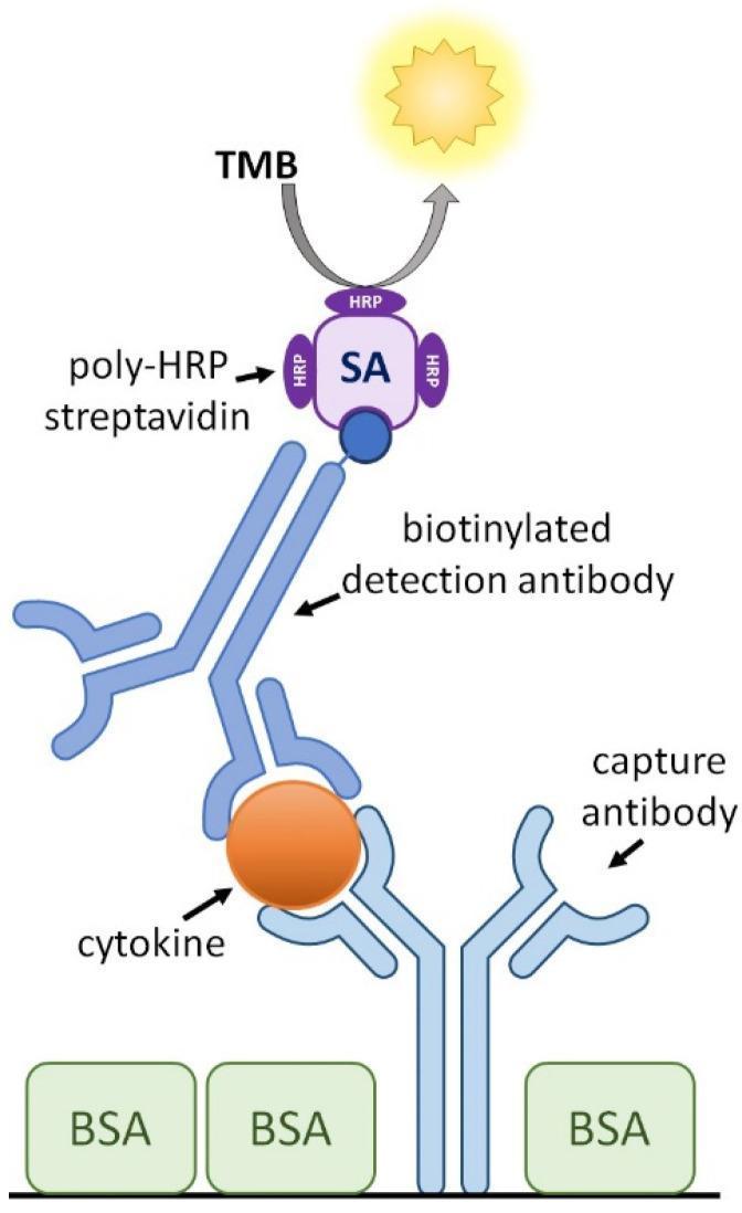
This medical diagram illustrates the schematic configuration of a cytokine capture Enzyme-Linked Immunosorbent Assay (ELISA), a diagnostic tool used in immunology. The process is depicted through a vertical assembly of molecular components. At the foundation, a capture antibody is anchored to a surface blocked by bovine serum albumin (BSA), shown as green rectangular blocks. This capture antibody is bound to a specific target molecule, a cytokine, represented as an orange sphere. Above the cytokine, a biotinylated detection antibody is attached, creating a sandwich complex. The biotin tag (dark blue circle) on the detection antibody is bound to a streptavidin (SA) molecule, which is conjugated with multiple horse radish peroxidase (HRP) enzymes (poly-HRP). At the apex, the HRP catalyzes a reaction with the substrate 3,3’,5,5’-Tetramethylbenzidine (TMB), resulting in a visual or chemical signal, depicted by a yellow light icon. This infographic effectively demonstrates the sequential binding and signal amplification logic used in laboratory medicine to quantify proteins such as IL-2, IL-6, or IFN-γ.
sandwich ELISA indirect ELISA types comparison
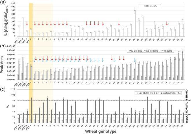
This Comparison Chart illustrates the biochemical analysis of gliadin and gluten content across various wheat genotypes, intended for research in celiac disease immunogenicity. The figure is divided into three panels (a, b, and c) comparing 38 genotypes against four reference standards (REF 1-4). Panel (a) presents R5-sandwich ELISA results measuring the relative percentage of gliadin compared to a semolina reference (REF 4). Red arrows indicate genotypes with significantly lower R5-reactivity. Panel (b) shows HPLC-UV relative quantification of specific gliadin fractions, categorized into ω-gliadins (dark gray), α/β-gliadins (medium gray), and γ-gliadins (light gray). Blue and red arrows highlight genotypes with statistically reduced α/β- or γ-gliadin expression. Panel (c) displays technological parameters, specifically Dry Gluten (DG) percentage and Gluten Index (GI), categorized into 'Weak', 'Normal', and 'Strong' gluten strength. This data aids in identifying wheat varieties with potentially lower toxicity for gluten-sensitive populations while maintaining functional properties for food production.
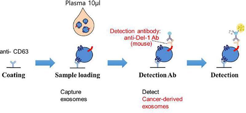
A scientific schematic illustrating a four-step sandwich enzyme-linked immunosorbent assay (ELISA) designed for the detection of cancer-derived exosomes in human plasma. The first stage, 'Coating', depicts the immobilization of anti-CD63 capture antibodies onto a solid substrate. The second stage, 'Sample loading', shows the introduction of 10µl of plasma containing exosomes (blue spheres), which are subsequently bound to the surface via the anti-CD63 antibodies. The third stage, 'Detection Ab', illustrates the binding of a mouse monoclonal anti-Del-1 antibody to a specific surface marker on the captured exosome. The final 'Detection' stage shows the binding of a signaling molecule to the detection antibody, resulting in a yellow burst symbol that represents a positive signal for cancer-derived exosomes. This assay is utilized to quantify Developmental Endothelial Locus-1 (Del-1) protein levels, a potential biomarker for prostate cancer, by leveraging exosome-specific surface markers (CD63) and malignancy-associated proteins (Del-1).
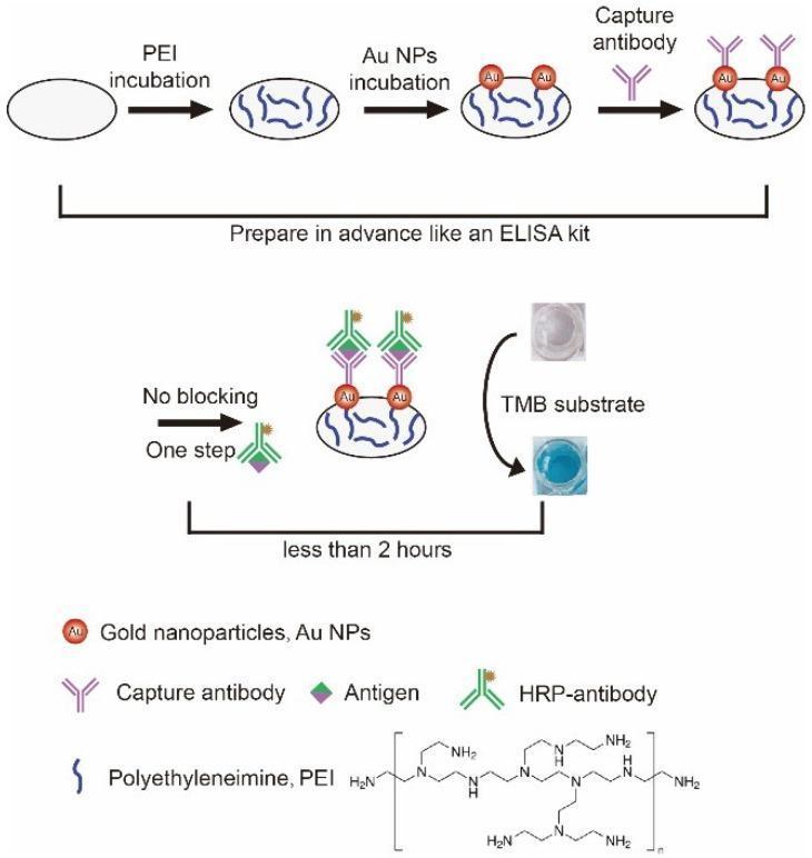
This schematic diagram illustrates a specialized, blocking-free 'sandwich' Enzyme-Linked Immunosorbent Assay (ELISA) utilizing a polyethyleneimine (PEI) and gold nanoparticle (Au NP) coated microwell plate. The process is divided into two phases: preparation and detection. In the preparation phase, a microwell undergoes PEI incubation followed by Au NP incubation to form a functionalized base layer; capture antibodies are then immobilized onto the Au NPs. In the detection phase, labeled as a 'No blocking - One step' procedure, the antigen and horseradish peroxidase (HRP)-conjugated antibody are introduced. The final step shows the addition of TMB substrate, which undergoes a visual transition from a colorless liquid to a blue-colored solution, indicating successful antigen detection. The diagram highlights that this modified protocol simplifies traditional ELISA by omitting the 2-hour blocking step, reducing total assay time to less than 2 hours. Chemical structures for PEI and icons for antigens, Au NPs, and various antibodies are included to clarify the molecular binding sequence and surface modifications.
python3 -c " import urllib.request imgs = [ ('elisa_principle.jpg', 'https://cdn.orris.care/cdss_images/b4b4babf27c377f92049759b8f41aa07c25938ba5d45a798753371eb4ef4db36.png'), ('indirect_elisa.jpg', 'https://cdn.orris.care/cdss_images/pmc_clinical_VQA_86a42d609c2b13ce04e6a7542988a799c7334a829df70e0a1fe9722b50551802.jpg'), ('sandwich_elisa.jpg', 'https://cdn.orris.care/cdss_images/pmc_clinical_VQA_7de5fd8fab060d6566d1f8bac20d150143d5580992b17dbac686e53cbb754dd6.jpg'), ('indirect_steps.jpg', 'https://cdn.orris.care/cdss_images/pmc_clinical_VQA_297e9d19b89223694a13935cc7ea27ab6c3005428bd47dba4b97b223fdbef05b.jpg'), ] for fname, url in imgs: urllib.request.urlretrieve(url, f'/tmp/workspace/elisa-exam/{fname}') print(f'Downloaded {fname}') "
~/elisa-exam/simple_elisa_pdf.py
from reportlab.lib.pagesizes import A4
from reportlab.lib.styles import getSampleStyleSheet, ParagraphStyle
from reportlab.lib.units import cm
from reportlab.lib import colors
from reportlab.platypus import (
SimpleDocTemplate, Paragraph, Spacer, Table, TableStyle,
HRFlowable, Image, KeepTogether
)
from reportlab.lib.enums import TA_CENTER, TA_LEFT, TA_JUSTIFY
from PIL import Image as PILImage
import os
OUTPUT = "/tmp/workspace/elisa-exam/ELISA_Simple_OralPath.pdf"
IMG_DIR = "/tmp/workspace/elisa-exam/"
W, H = A4
doc = SimpleDocTemplate(
OUTPUT,
pagesize=A4,
rightMargin=1.8*cm, leftMargin=1.8*cm,
topMargin=1.8*cm, bottomMargin=1.8*cm,
title="ELISA - Simple Study Notes | MDS Oral Pathology",
)
styles = getSampleStyleSheet()
def S(name, **kw):
base = styles.get(name, styles['Normal'])
return ParagraphStyle('_', parent=base, **kw)
BLUE = colors.HexColor('#1a237e')
LBLUE = colors.HexColor('#e8eaf6')
GREEN = colors.HexColor('#1b5e20')
LGREEN = colors.HexColor('#e8f5e9')
ORANGE = colors.HexColor('#e65100')
LORANGE= colors.HexColor('#fff3e0')
TEAL = colors.HexColor('#004d40')
LTEAL = colors.HexColor('#e0f2f1')
GREY = colors.HexColor('#607d8b')
title_s = S('Title', fontSize=18, leading=22, textColor=BLUE, fontName='Helvetica-Bold', alignment=TA_CENTER, spaceAfter=2)
sub_s = S('Normal', fontSize=10, leading=13, textColor=GREY, fontName='Helvetica', alignment=TA_CENTER, spaceAfter=8)
h1_s = S('Normal', fontSize=13, leading=17, textColor=colors.white, fontName='Helvetica-Bold', spaceAfter=4, spaceBefore=10,
backColor=BLUE, leftIndent=-4, rightIndent=-4, borderPad=5)
h2_s = S('Normal', fontSize=11, leading=15, textColor=BLUE, fontName='Helvetica-Bold', spaceAfter=3, spaceBefore=8)
h3_s = S('Normal', fontSize=10, leading=14, textColor=ORANGE, fontName='Helvetica-Bold', spaceAfter=2, spaceBefore=5)
body_s = S('Normal', fontSize=10, leading=15, fontName='Helvetica', alignment=TA_JUSTIFY, spaceAfter=4)
bullet_s = S('Normal', fontSize=10, leading=14, fontName='Helvetica', leftIndent=14, spaceAfter=3)
small_s = S('Normal', fontSize=8.5,leading=12, fontName='Helvetica', textColor=GREY, alignment=TA_CENTER)
cap_s = S('Normal', fontSize=8.5,leading=12, fontName='Helvetica-Oblique', textColor=GREY, alignment=TA_CENTER, spaceBefore=2)
def_s = S('Normal', fontSize=10.5, leading=16, fontName='Helvetica', alignment=TA_JUSTIFY,
backColor=LBLUE, leftIndent=8, rightIndent=8, spaceAfter=6, borderPad=6)
def bullet(text, color=BLUE):
return Paragraph(f'<font color="#{color.hexval()[2:]}">●</font> {text}', bullet_s)
def h1(text):
return Paragraph(f' {text}', h1_s)
def img_fit(path, max_w, max_h):
"""Return a reportlab Image scaled to fit within max_w x max_h."""
im = PILImage.open(path)
iw, ih = im.size
ratio = min(max_w/iw, max_h/ih)
return Image(path, width=iw*ratio, height=ih*ratio)
story = []
# ─── TITLE ───────────────────────────────────────────────────────────────────
story.append(Paragraph("ELISA", title_s))
story.append(Paragraph("Enzyme-Linked Immunosorbent Assay", title_s))
story.append(Paragraph("MDS First Year | Oral Pathology | Simple Study Notes", sub_s))
story.append(HRFlowable(width="100%", thickness=2.5, color=BLUE, spaceAfter=10))
# ─── 1. DEFINITION ────────────────────────────────────────────────────────────
story.append(h1("1. DEFINITION"))
story.append(Paragraph(
"ELISA is a <b>plate-based immunoassay</b> that detects and measures antigens, antibodies, cytokines, "
"hormones and proteins using the <b>antigen-antibody reaction</b> linked to an <b>enzyme</b> that produces "
"a measurable colour change.",
def_s
))
# ─── 2. HISTORY ───────────────────────────────────────────────────────────────
story.append(h1("2. HISTORY"))
hist = [
("1971", "Engvall & Perlmann (Sweden) + Van Weemen & Schuurs (Netherlands) — invented ELISA independently"),
("Idea", "Replace radioactive label in RIA with an enzyme — safer, cheaper, equally sensitive"),
("Today", "Gold standard of immunoassays | Detection range: 0.01–0.1 ng"),
]
for yr, txt in hist:
story.append(Paragraph(f'<b><font color="#1a237e">{yr}</font></b> → {txt}', bullet_s))
# ─── 3. PRINCIPLE ─────────────────────────────────────────────────────────────
story.append(h1("3. BASIC PRINCIPLE"))
story.append(Paragraph("Two key ideas:", h2_s))
story.append(bullet("<b>Specificity</b> — Antibody binds only its specific antigen (like lock & key)"))
story.append(bullet("<b>Amplification</b> — Enzyme converts colourless substrate → coloured product"))
story.append(Paragraph(
"More colour = more antigen/antibody in the sample. "
"Read by a <b>spectrophotometer</b> at 450 nm (for TMB substrate).",
body_s
))
# Principle image
story.append(Spacer(1, 4))
pi = img_fit(IMG_DIR+'elisa_principle.jpg', 7*cm, 9*cm)
pi_table = Table([[pi]], colWidths=[W - 3.6*cm])
pi_table.setStyle(TableStyle([
('ALIGN',(0,0),(-1,-1),'CENTER'),
('BACKGROUND',(0,0),(-1,-1),LBLUE),
('BOX',(0,0),(-1,-1),1,BLUE),
('TOPPADDING',(0,0),(-1,-1),6),('BOTTOMPADDING',(0,0),(-1,-1),6),
]))
story.append(KeepTogether([pi_table,
Paragraph("Fig 1. ELISA Principle — Sample with antigen A gives colour; sample B (no antigen) stays clear. (Janeway's Immunobiology 10e)", cap_s)
]))
# ─── 4. COMPONENTS ────────────────────────────────────────────────────────────
story.append(h1("4. KEY COMPONENTS"))
comp = [
("Solid phase", "96-well polystyrene plate"),
("Antigen", "Target molecule to detect"),
("Primary antibody", "Binds antigen specifically"),
("Enzyme label", "HRP (horseradish peroxidase) or Alkaline phosphatase"),
("Substrate", "TMB → blue/yellow colour with HRP"),
("Blocking agent", "BSA or skim milk — stops background noise"),
("Stop solution", "H₂SO₄ — stops the reaction; turns blue → yellow"),
("Wash buffer", "PBS-Tween — removes unbound reagents"),
]
comp_data = [[Paragraph(f'<b>{c}</b>', bullet_s), Paragraph(d, bullet_s)] for c, d in comp]
comp_table = Table(comp_data, colWidths=[5.5*cm, 10.5*cm])
comp_table.setStyle(TableStyle([
('ROWBACKGROUNDS',(0,0),(-1,-1),[LBLUE, colors.white]),
('GRID',(0,0),(-1,-1),0.4,colors.HexColor('#90a4ae')),
('TOPPADDING',(0,0),(-1,-1),4),('BOTTOMPADDING',(0,0),(-1,-1),4),
('LEFTPADDING',(0,0),(-1,-1),6),
]))
story.append(comp_table)
# ─── 5. PROCEDURE ─────────────────────────────────────────────────────────────
story.append(h1("5. PROCEDURE (5 Simple Steps)"))
steps = [
("🧫 Step 1 — Coat", "Add antigen (or antibody) to the plate well. It sticks to the plastic."),
("🛡 Step 2 — Block", "Add BSA/milk to fill empty spaces on the plate (prevents false results)."),
("🔬 Step 3 — Antibody","Add primary antibody. It binds only to the correct antigen. Wash away the rest."),
("⚗️ Step 4 — Enzyme Ab","Add enzyme-linked secondary antibody. It binds the primary antibody. Wash."),
("🎨 Step 5 — Colour", "Add substrate (TMB). Enzyme makes colour. Add stop solution. Read at 450 nm."),
]
for emoji_title, desc in steps:
story.append(Paragraph(f'<b>{emoji_title}</b>', h3_s))
story.append(Paragraph(desc, bullet_s))
# ─── 6. TYPES ─────────────────────────────────────────────────────────────────
story.append(h1("6. TYPES OF ELISA"))
types_data = [
[Paragraph('<b>Type</b>', S('Normal', fontName='Helvetica-Bold', fontSize=9, textColor=colors.white)),
Paragraph('<b>What is coated</b>', S('Normal', fontName='Helvetica-Bold', fontSize=9, textColor=colors.white)),
Paragraph('<b>What is detected</b>', S('Normal', fontName='Helvetica-Bold', fontSize=9, textColor=colors.white)),
Paragraph('<b>Key feature</b>', S('Normal', fontName='Helvetica-Bold', fontSize=9, textColor=colors.white))],
[Paragraph('Direct', bullet_s), Paragraph('Antigen', bullet_s), Paragraph('Enzyme-linked 1° Ab', bullet_s), Paragraph('Simple, fast, less sensitive', bullet_s)],
[Paragraph('Indirect', bullet_s), Paragraph('Antigen', bullet_s), Paragraph('1° Ab + enzyme-linked 2° Ab', bullet_s), Paragraph('More sensitive; used in HIV screening', bullet_s)],
[Paragraph('Sandwich', bullet_s), Paragraph('Capture Ab', bullet_s), Paragraph('Antigen sandwiched between 2 Abs', bullet_s), Paragraph('Most sensitive; used for cytokines', bullet_s)],
[Paragraph('Competitive', bullet_s), Paragraph('Ab', bullet_s), Paragraph('Labelled vs unlabelled antigen compete', bullet_s), Paragraph('Inversely proportional; used for hormones/drugs', bullet_s)],
]
types_table = Table(types_data, colWidths=[3.2*cm, 3.8*cm, 4.5*cm, 4.5*cm])
types_table.setStyle(TableStyle([
('BACKGROUND',(0,0),(-1,0), BLUE),
('ROWBACKGROUNDS',(0,1),(-1,-1),[LBLUE, colors.white]),
('GRID',(0,0),(-1,-1),0.4,GREY),
('TOPPADDING',(0,0),(-1,-1),4),('BOTTOMPADDING',(0,0),(-1,-1),4),
('LEFTPADDING',(0,0),(-1,-1),5),('VALIGN',(0,0),(-1,-1),'TOP'),
]))
story.append(types_table)
story.append(Spacer(1, 6))
# Images for types
img_indirect = img_fit(IMG_DIR+'indirect_elisa.jpg', 7.5*cm, 6.5*cm)
img_sandwich = img_fit(IMG_DIR+'sandwich_elisa.jpg', 7.5*cm, 6.5*cm)
img_row = Table(
[[img_indirect, img_sandwich]],
colWidths=[(W-3.6*cm)/2, (W-3.6*cm)/2]
)
img_row.setStyle(TableStyle([
('ALIGN',(0,0),(-1,-1),'CENTER'), ('VALIGN',(0,0),(-1,-1),'MIDDLE'),
('BACKGROUND',(0,0),(-1,-1), LGREEN),
('BOX',(0,0),(-1,-1),1, GREEN),
('TOPPADDING',(0,0),(-1,-1),6), ('BOTTOMPADDING',(0,0),(-1,-1),6),
]))
cap_row = Table(
[[Paragraph("Fig 2. Indirect ELISA — IgE detection (allergy)", cap_s),
Paragraph("Fig 3. Sandwich ELISA — cytokine detection (e.g. IL-6)", cap_s)]],
colWidths=[(W-3.6*cm)/2, (W-3.6*cm)/2]
)
story.append(KeepTogether([img_row, cap_row]))
# ─── 7. APPLIED ASPECTS ───────────────────────────────────────────────────────
story.append(h1("7. APPLIED ASPECTS IN ORAL PATHOLOGY"))
story.append(Paragraph(
"ELISA uses <b>saliva, GCF (gingival crevicular fluid)</b> and serum — all easily collected from the mouth. "
"This makes it ideal for oral disease research and diagnosis.",
body_s
))
apps = [
("🔴 Oral Cancer (OSCC)", LORANGE, ORANGE, [
"IL-6, IL-8, TNF-α measured in SALIVA by sandwich ELISA — elevated in OSCC",
"CYFRA 21-1 (cytokeratin fragment) in saliva — diagnostic marker",
"VEGF, MMP-9, EGF — detect tumour growth and invasion",
"Use: Early detection, monitoring treatment response",
]),
("🟡 Periodontal Disease", LGREEN, GREEN, [
"IL-1β in GCF — most studied; elevated = active bone destruction",
"MMP-8 (collagenase) in GCF — reflects collagen destruction",
"RANKL/OPG ratio in GCF — measures bone resorption activity",
"Serum CRP by ELISA — links periodontitis to heart disease & diabetes",
]),
("🟠 Vesiculobullous Lesions", LBLUE, BLUE, [
"Pemphigus vulgaris → anti-Dsg3 & anti-Dsg1 ELISA (IgG autoantibodies)",
"Bullous pemphigoid → anti-BP180 ELISA (first-line serology test)",
"Mucous membrane pemphigoid → anti-laminin 332 ELISA",
"ELISA titer CORRELATES with disease activity → monitor treatment",
]),
("🟢 Oral Infections", LTEAL, TEAL, [
"HIV → 4th generation ELISA detects anti-HIV Ab + p24 antigen (screening test)",
"HSV, EBV, CMV → IgM/IgG ELISA to diagnose active vs past infection",
"HPV-16/18 antibody ELISA → links HPV to oropharyngeal cancer",
"Confirmatory test after reactive ELISA: Western Blot",
]),
("🔵 Sjogren's Syndrome", LORANGE, ORANGE, [
"Anti-SSA/Ro & Anti-SSB/La ELISA → primary diagnostic markers",
"Anti-M3R (muscarinic receptor) ELISA → explains dry mouth mechanism",
"Salivary IL-6, BAFF ELISA — non-invasive alternative to lip biopsy",
]),
("🟣 OSMF & Precancer", LGREEN, GREEN, [
"TGF-β1 ELISA in serum/saliva → main fibrosis driver, indicates severity",
"IL-6, PDGF, VEGF ELISA → tissue fibrosis and dysplasia monitoring",
"Arecoline metabolites in urine/serum by competitive ELISA",
]),
("⚫ Drug & Hormone Monitoring", LBLUE, BLUE, [
"Cyclosporine ELISA → transplant patients (cause gingival enlargement)",
"Salivary cortisol ELISA → stress monitoring (affects wound healing)",
"PTH ELISA → metabolic bone disease (brown tumours, jaw lesions)",
"IgE ELISA → allergy workup for dental material hypersensitivity",
]),
("🌟 Advanced ELISA", LTEAL, TEAL, [
"Digital ELISA (D-ELISA) → detects single molecules; femtomolar sensitivity",
"Multiplex ELISA → 50-100 biomarkers from ONE saliva sample (Luminex)",
"Point-of-care ELISA strip → chair-side MMP-8 test for active periodontitis",
]),
]
for title, bg, fg, points in apps:
header = Paragraph(f'<b>{title}</b>',
S('Normal', fontName='Helvetica-Bold', fontSize=10.5, textColor=colors.white,
backColor=fg, leftIndent=4, spaceBefore=8, spaceAfter=3, borderPad=4))
story.append(header)
for p in points:
story.append(Paragraph(f' • {p}',
S('Normal', fontName='Helvetica', fontSize=10, leading=14,
leftIndent=12, spaceAfter=2, backColor=bg)))
# ─── 8. ADVANTAGES / DISADVANTAGES ───────────────────────────────────────────
story.append(h1("8. ADVANTAGES vs DISADVANTAGES"))
adv_dis = [
[Paragraph('<b>✅ ADVANTAGES</b>', S('Normal', fontName='Helvetica-Bold', fontSize=10, textColor=colors.white)),
Paragraph('<b>❌ DISADVANTAGES</b>', S('Normal', fontName='Helvetica-Bold', fontSize=10, textColor=colors.white))],
[Paragraph('• Very sensitive (0.01–0.1 ng)', bullet_s), Paragraph('• Needs pure antigen or antibody standard', bullet_s)],
[Paragraph('• Non-radioactive — safe & cheap', bullet_s), Paragraph('• False positives from cross-reactivity', bullet_s)],
[Paragraph('• High specificity', bullet_s), Paragraph('• Prozone (hook) effect at high concentrations', bullet_s)],
[Paragraph('• Quantitative (gives concentration)', bullet_s), Paragraph('• Cannot tell active vs past infection', bullet_s)],
[Paragraph('• Works with saliva, GCF, serum, urine', bullet_s), Paragraph('• Confirmatory test (Western blot) needed', bullet_s)],
[Paragraph('• 96-well plates — high throughput', bullet_s), Paragraph('• Requires careful optimization', bullet_s)],
[Paragraph('• Can be automated', bullet_s), Paragraph('• Longer than rapid strip tests', bullet_s)],
]
adv_table = Table(adv_dis, colWidths=[(W-3.6*cm)/2, (W-3.6*cm)/2])
adv_table.setStyle(TableStyle([
('BACKGROUND',(0,0),(0,0), GREEN), ('BACKGROUND',(1,0),(1,0), colors.HexColor('#b71c1c')),
('ROWBACKGROUNDS',(0,1),(-1,-1),[LGREEN, colors.white]),
('GRID',(0,0),(-1,-1),0.4,GREY),
('TOPPADDING',(0,0),(-1,-1),4), ('BOTTOMPADDING',(0,0),(-1,-1),4),
('LEFTPADDING',(0,0),(-1,-1),5), ('VALIGN',(0,0),(-1,-1),'TOP'),
]))
story.append(adv_table)
# ─── 9. CONCLUSION ────────────────────────────────────────────────────────────
story.append(h1("9. CONCLUSION"))
story.append(Paragraph(
"ELISA (1971) is the <b>gold standard immunoassay</b> — sensitive, safe, quantitative, and versatile. "
"In oral pathology, it uses <b>saliva and GCF</b> to diagnose oral cancer, monitor periodontal disease, "
"confirm vesiculobullous conditions, screen for HIV, and assess autoimmune disorders. "
"Newer formats (Digital ELISA, Multiplex ELISA) push sensitivity to <b>single-molecule levels</b>, "
"making liquid biopsy for early oral cancer a real possibility.",
def_s
))
# ─── 10. REFERENCES ──────────────────────────────────────────────────────────
story.append(h1("10. REFERENCES"))
refs = [
"Murphy K, Weaver C. <i>Janeway's Immunobiology</i>, 10th ed. Garland Science; 2022.",
"McPherson RA, Pincus MR. <i>Henry's Clinical Diagnosis</i>, 23rd ed. Elsevier; 2021.",
"Neville BW et al. <i>Oral and Maxillofacial Pathology</i>, 4th ed. Elsevier; 2016.",
"Rajendran R, Sivapathasundharam B. <i>Shafer's Oral Pathology</i>, 8th ed. Elsevier India; 2020.",
"Abbas AK et al. <i>Cellular and Molecular Immunology</i>, 10th ed. Elsevier; 2022.",
"Engvall E, Perlmann P. ELISA: Quantitative assay of IgG. <i>Immunochemistry</i>. 1971.",
"Chiamulera MMA et al. Salivary cytokines as biomarkers of oral cancer. <i>BMC Cancer</i>. 2021. PMID: 33639868.",
"Khurshid Z et al. Biochemical analysis of oral fluids. <i>Adv Clin Chem</i>. 2021. PMID: 33453866.",
"Zhang Y et al. Salivary biomarkers for periodontitis. <i>BMC Oral Health</i>. 2021. PMID: 34001101.",
"Rissin DM et al. Single-molecule ELISA at subfemtomolar concentrations. <i>Nat Biotechnol</i>. 2010.",
]
for r in refs:
story.append(Paragraph(f'• {r}', S('Normal', fontSize=9, leading=13, leftIndent=12, spaceAfter=2, fontName='Helvetica')))
# ─── FOOTER ──────────────────────────────────────────────────────────────────
story.append(HRFlowable(width="100%", thickness=1, color=GREY, spaceBefore=12, spaceAfter=4))
story.append(Paragraph("MDS Oral Pathology | Applied Basic Sciences | Simple Study Notes", sub_s))
doc.build(story)
print("Done:", OUTPUT)
cd /tmp/workspace/elisa-exam && python simple_elisa_pdf.py
python3 -c " import pdfplumber with pdfplumber.open('/tmp/workspace/elisa-exam/ELISA_Simple_OralPath.pdf') as pdf: print(f'Pages: {len(pdf.pages)}') " ls -lh /tmp/workspace/elisa-exam/ELISA_Simple_OralPath.pdf
ELISA Simple OralPath
PDF Document · PDF
1. DEFINITION
2. HISTORY
- 1971 → Engvall & Perlmann + Van Weemen & Schuurs invented ELISA
- Idea → Replace radioactive label in RIA with an enzyme (safer, cheaper, same sensitivity)
- Today → Gold standard immunoassay | Detection range: 0.01-0.1 ng
3. BASIC PRINCIPLE
- Specificity → Antibody binds only its specific antigen (lock & key)
- Amplification → Enzyme converts colourless substrate → coloured product
- More colour = more antigen/antibody in the sample → read at 450 nm
4. KEY COMPONENTS
| Component | Examples |
|---|---|
| Solid phase | 96-well polystyrene plate |
| Enzyme label | HRP or Alkaline phosphatase |
| Substrate | TMB → blue/yellow colour |
| Blocking agent | BSA or skim milk |
| Stop solution | H₂SO₄ → turns blue to yellow |
| Wash buffer | PBS-Tween |
5. PROCEDURE (5 Steps)
- Coat — Add antigen to plate well
- Block — Add BSA/milk to fill empty spots
- Antibody — Add primary antibody; wash away unbound
- Enzyme Ab — Add enzyme-linked secondary antibody; wash
- Colour — Add TMB substrate → colour develops → read at 450 nm
6. TYPES
| Type | Key Feature | Used for |
|---|---|---|
| Direct | Enzyme on primary Ab | Simple screening |
| Indirect | Enzyme on secondary Ab | HIV screening, autoantibodies |
| Sandwich | Antigen trapped between 2 Abs | Cytokines (IL-6, TNF-α) |
| Competitive | Labelled vs unlabelled Ag compete | Hormones, drugs |