
What is abdominal girth ratio look like
The abdominal girth ratio (also called the waist-to-height ratio or abdominal circumference ratio) is a simple measurement used to assess central obesity and metabolic risk. It compares waist circumference to height, and a ratio above 0.5 is generally considered a marker of increased cardiometabolic risk.
Searching sources...
Reading File
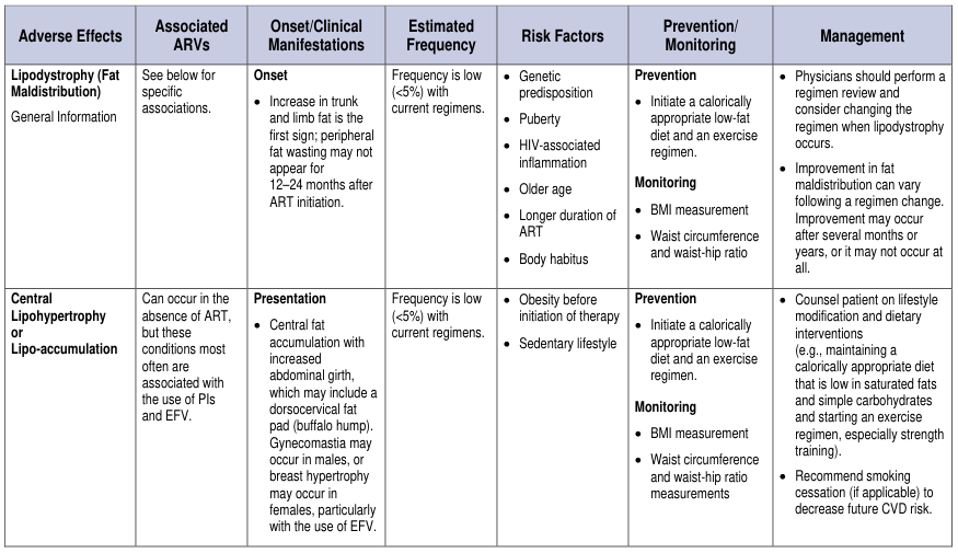
Finding Sources
Searching Images
waist to height ratio obesity measurement diagram
Searching the Web
abdominal girth ratio measurement what does it look like visual
Loading Image
Loading Image
Loading Image
Finding Sources
Reading File
Here is a complete picture of the abdominal girth ratio - what it is, how it looks, and what the numbers mean:
Abdominal Girth (Waist Circumference) - What It Looks Like
The measurement is made by placing a soft tape measure around the bare abdomen at the level of the iliac crest (hip bone) while the person stands upright. The tape should be snug but not compressing the skin. You breathe out naturally - do not suck in - then read the measurement.
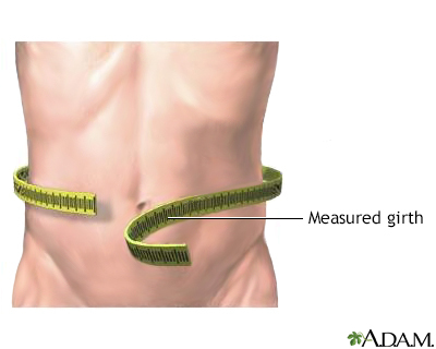
The image above shows exactly how a clinician measures abdominal girth: a tape is wrapped horizontally around the narrowest part of the abdomen, roughly level with the belly button/iliac crest.
The Two Main Ratios
1. Waist-to-Hip Ratio (WHR)
The waist circumference is divided by the hip circumference (measured at the widest point of the buttocks).
Formula: WHR = Waist (cm) / Hip (cm)
| Category | Men | Women |
|---|---|---|
| Average normal | ~0.93 (range 0.75-1.10) | ~0.83 (range 0.70-1.00) |
| Abdominal obesity (WHO) | > 0.90 | > 0.85 |
2. Waist-to-Height Ratio (WHtR)
The waist circumference is divided by total height.
Formula: WHtR = Waist (cm) / Height (cm)
| Result | Interpretation |
|---|---|
| < 0.5 | Healthy range |
| 0.51 - 0.59 | Increased health risk |
| ≥ 0.60 | High health risk |
The NHS rule of thumb: your waist should be less than half your height (ratio < 0.5).
High-Risk Waist Circumference Cutoffs
These thresholds (used in metabolic syndrome criteria) signal abdominal obesity even without a ratio calculation:
- Men: > 102 cm (40 inches)
- Women: > 88 cm (35 inches)
(Source: Basic Medical Biochemistry - A Clinical Approach, 6e; Guyton and Hall Textbook of Medical Physiology)
Why It Matters
Abdominal (central) fat - called visceral fat - sits around internal organs and is metabolically active. Elevated abdominal girth is linked to:
- Metabolic syndrome (with hypertension, high fasting glucose, low HDL, high triglycerides)
- Type 2 diabetes
- Cardiovascular disease
- GERD (increased intra-abdominal pressure reduces lower esophageal sphincter tone)
- Obstructive sleep apnea and asthma in obese individuals
The waist circumference often correlates better with intra-abdominal (visceral) fat and associated risk than BMI alone, which is why it is recommended as a clinical "vital sign" by major guidelines.
Here is an NHS video walkthrough on how to perform the measurement and calculate the ratio:
What about 74-75 -78. As in like above umbilicus-umbilicus-below umbilicus
Those three measurements (74 cm above umbilicus, 75 cm at umbilicus, 78 cm below umbilicus) describe a pattern where the lower abdomen is the widest point - this suggests predominant lower abdominal fat distribution.
Researching further...
Finding Sources
Searching the Web
abdominal girth measurement above at below umbilicus three level clinical significance
Great question - this is a three-level abdominal girth assessment, which gives much more clinical detail than a single measurement. Here is how to interpret your numbers:
Your Three-Level Readings
| Level | Measurement | What it reflects |
|---|---|---|
| Above umbilicus | 74 cm | Upper abdominal / epigastric girth |
| At umbilicus | 75 cm | Mid-abdominal girth (closest to standard waist measurement) |
| Below umbilicus | 78 cm | Lower abdominal / hypogastric girth |
What This Pattern Means
Your below-umbilicus measurement is the largest (78 cm), with a gradual taper going upward. This is called a lower abdominal predominance or infra-umbilical fullness pattern. Clinically, this can indicate:
- Subcutaneous fat accumulation in the lower abdomen - very common, especially in women (gynoid/pear-shaped distribution)
- Pelvic organ fullness - e.g., a full bladder, uterine enlargement, ovarian cysts, or a gravid uterus can push the lower abdominal girth out
- Ascites - free fluid in the abdomen tends to sag to dependent areas (flanks and lower abdomen when standing), making the below-umbilical girth largest. The classic "flanks bulge" sign
- Bowel distension - gas or fecal loading preferentially distends the lower bowel (sigmoid, cecum)
- Post-surgical changes - lower midline scars can alter fat distribution
Compare to Visceral (Central) Fat Pattern
If the umbilical level were the largest, that would be more classic for visceral/central obesity - the "apple" shape strongly linked to metabolic syndrome, where intra-abdominal fat pushes outward most at the mid-abdomen.
Your pattern (74-75-78) shows only a 4 cm difference across all three levels, so the abdomen is fairly uniform - the slight infra-umbilical prominence is mild.
Are These Values Normal?
For context, using standard waist circumference cutoffs (measured at the iliac crest, approximately at or just below the umbilicus):
| Sex | Low risk | High risk |
|---|---|---|
| Women | < 80 cm | ≥ 88 cm |
| Men | < 94 cm | ≥ 102 cm |
At 78 cm, this is in the healthy-to-borderline range for women and well within normal for men - no metabolic risk flag by standard cutoffs.
Clinical Use of Three-Level Measurement
Clinicians measure at three levels (above, at, and below the umbilicus) primarily in the following contexts:
- Monitoring ascites - serial measurements track fluid accumulation or response to diuretics
- Pregnancy monitoring - tracking abdominal growth
- Post-operative bowel distension - tracking ileus resolution
- Abdominal mass surveillance - noting where the girth is increasing can guide where a mass is growing
In routine obesity/metabolic screening, only the single umbilical or iliac crest level is used. The three-level approach is more of a clinical examination or serial monitoring tool.
This is a shared conversation. Sign in to Orris to start your own chat.