
Symptoms of hospital acquire infection
hospital acquired infection symptoms types signs nosocomial
"healthcare-associated infections" AND symptoms
https://www.ncbi.nlm.nih.gov/books/NBK559312
hospital acquired infection nosocomial types diagram
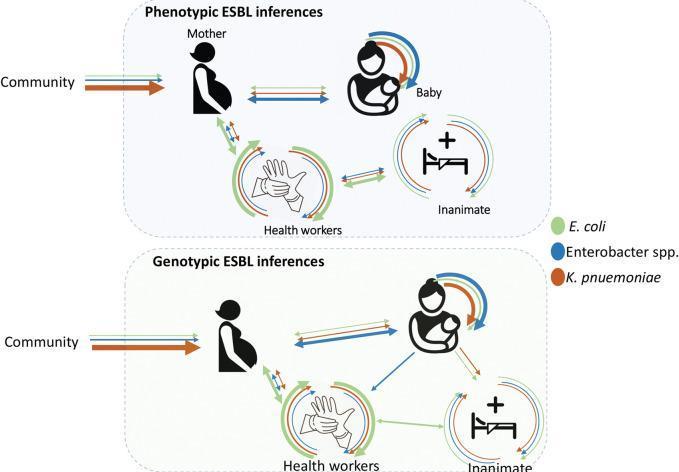
This medical illustration is a pathophysiology diagram depicting the transmission dynamics of Extended-Spectrum Beta-Lactamase (ESBL)-producing bacteria in a maternity ward setting. The diagram is divided into two panels: 'Phenotypic ESBL inferences' (top) and 'Genotypic ESBL inferences' (bottom), used to compare inferred transmission pathways based on different laboratory methods. Four key entities are represented using icons: the mother (representing community entry), the baby, healthcare workers (hands icon), and the hospital environment (inanimate object/bed icon). Colored arrows indicate the potential bidirectional transmission of three specific multidrug-resistant pathogens: E. coli (green), Enterobacter spp. (blue), and K. pneumoniae (orange). The layout illustrates how these bacteria move from the community to the mother and subsequently cycle between patients, medical staff, and inanimate surfaces, highlighting the role of the hospital environment and healthcare providers in the horizontal transmission of nosocomial infections. This visual serves as an educational tool for epidemiology, infection control, and neonatal medicine, focusing on the complex pathways of antimicrobial resistance within specialized clinical departments.
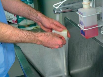
A clinical photograph demonstrating a fundamental infection control procedure: surgical hand antisepsis or clinical handwashing. The image shows a healthcare professional wearing blue scrubs, with arms exposed to the mid-forearm, performing hand hygiene at a stainless steel medical scrub sink. A steady stream of water flows from a chrome faucet. The individual is actively lathering a white rectangular surgical scrub sponge or bar of soap between their hands. To the right of the faucet, a wall-mounted wire rack holds a plastic dispenser bottle containing a reddish-pink antimicrobial solution, likely chlorhexidine gluconate or povidone-iodine. The background shows clinical supply packaging, reinforcing the hospital or surgical setting. This visual illustrates standard precautions, aseptic technique, and the prevention of nosocomial infections in a clinical environment.
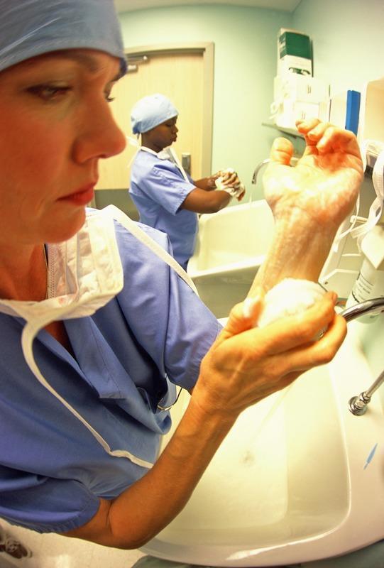
This clinical photograph illustrates the essential medical practice of preoperative surgical hand scrubbing to ensure infection control and hospital hygiene. In the foreground, a healthcare professional is seen meticulously lathering their hands and distal forearms with an antimicrobial soap at a surgical sink. The individual is wearing standard operating room attire, including a blue surgical gown, a matching bouffant cap, and a white surgical mask draped around the neck. A second medical professional in similar surgical scrub attire is visible in the background, also engaged in hand washing at a separate station. The environment features standard medical fixtures, including deep white surgical sinks, chrome gooseneck faucets, and wall-mounted soap or paper towel dispensers. This image serves as an educational resource for demonstrating aseptic techniques, the prevention of nosocomial infections, and the rigorous hygiene protocols required for hospital staff before entering a sterile field.
Hospital-Acquired Infections (HAIs / Nosocomial Infections): Symptoms
Definition
- Was not present or incubating at the time of admission
- Develops 48 hours or more after hospital admission, OR within 30 days after discharge, or within 1 year for infections related to implants/prosthetics
Types and Their Specific Symptoms
1. Surgical Site Infection (SSI)
- Purulent (pus-like) discharge from the wound
- Abscess formation at or near the surgical site
- Spreading redness (cellulitis) around the incision
- Localized pain, warmth, and swelling
- Fever (systemic sign)
2. Catheter-Associated Urinary Tract Infection (CAUTI)
- Burning or painful urination (dysuria)
- Increased frequency or urgency of urination
- Cloudy, foul-smelling, or bloody urine
- Lower abdominal or pelvic pain
- Fever and chills (if infection spreads to kidneys - pyelonephritis)
- Positive urine culture (≥10⁵ bacteria/mL) with or without clinical symptoms
3. Hospital-Acquired Pneumonia (HAP) / Ventilator-Associated Pneumonia (VAP)
- New-onset fever
- Cough with purulent sputum
- Declining oxygen saturation / worsening oxygenation
- Leukocytosis (raised white cell count)
- Coarse breath sounds and rales (crackles) on auscultation
- New or progressive infiltrate on chest X-ray
- In sedated/intubated patients: fever, increasing O₂ requirements, and purulent secretions on endotracheal suctioning
4. Central Line-Associated Bloodstream Infection (CLABSI)
- Fever and/or rigors (chills)
- Inflammation, lymphangitis, or purulent discharge at the catheter insertion site
- At least one positive blood culture
- Can rapidly progress to sepsis: hypotension, tachycardia, altered mental status, organ dysfunction
5. Clostridioides difficile (C. diff) Infection
- Watery diarrhea (hallmark symptom)
- Abdominal pain, cramping, and distension
- Fever
- Nausea, anorexia, dehydration
- In immunocompromised or elderly patients (who may lack typical inflammatory responses): altered mental status, lethargy, fatigue, tachycardia, or hypotension
General / Systemic Symptoms (All HAI Types)
| Symptom | Significance |
|---|---|
| Fever (>38°C) | Most common, often the earliest sign |
| Rigors/Chills | Suggests bacteremia or septicemia |
| Tachycardia | Sign of systemic response |
| Hypotension | Progressing toward septic shock |
| Leukocytosis or Leukopenia | Inflammatory or overwhelmed immune response |
| Altered mental status | Especially in elderly - often first sign |
| Fatigue, malaise, anorexia | Non-specific systemic signs |
| Localized redness, warmth, swelling, pain | At site of infection or device insertion |
| Purulent discharge | At wounds, lines, or tubes |
Sources of Hospital Infection
- Patients - infected wounds, respiratory/urinary infections, skin conditions
- Hospital staff - nasal/skin carriers of S. aureus, throat carriers of Streptococci
- Environment - dust, linen, bed clothes, door handles, sinks, and air
Common Causative Pathogens
| Pathogen | Common HAI Type |
|---|---|
| Staphylococcus aureus (incl. MRSA) | Bloodstream, surgical site |
| Escherichia coli | Urinary tract |
| Enterococci | Blood, UTI, wound |
| Pseudomonas aeruginosa | Respiratory, kidney, UTI |
| Acinetobacter baumannii | Ventilator-associated pneumonia |
| Clostridioides difficile | Gastrointestinal |
| Klebsiella pneumoniae | Bloodstream, UTI |
Key Risk Factors That Increase Susceptibility
- Invasive procedures and indwelling devices (catheters, ventilators, central lines)
- Recent antibiotic use (disrupts normal flora)
- Immunosuppression (chemotherapy, transplant medications, HIV)
- Advanced age or very young age
- Prolonged hospital stay
- Surgery (especially abdominal or cardiac)