
Pneumatic torniquets
pneumatic tourniquet complications orthopedic surgery
pneumatic tourniquet surgical limb orthopedic
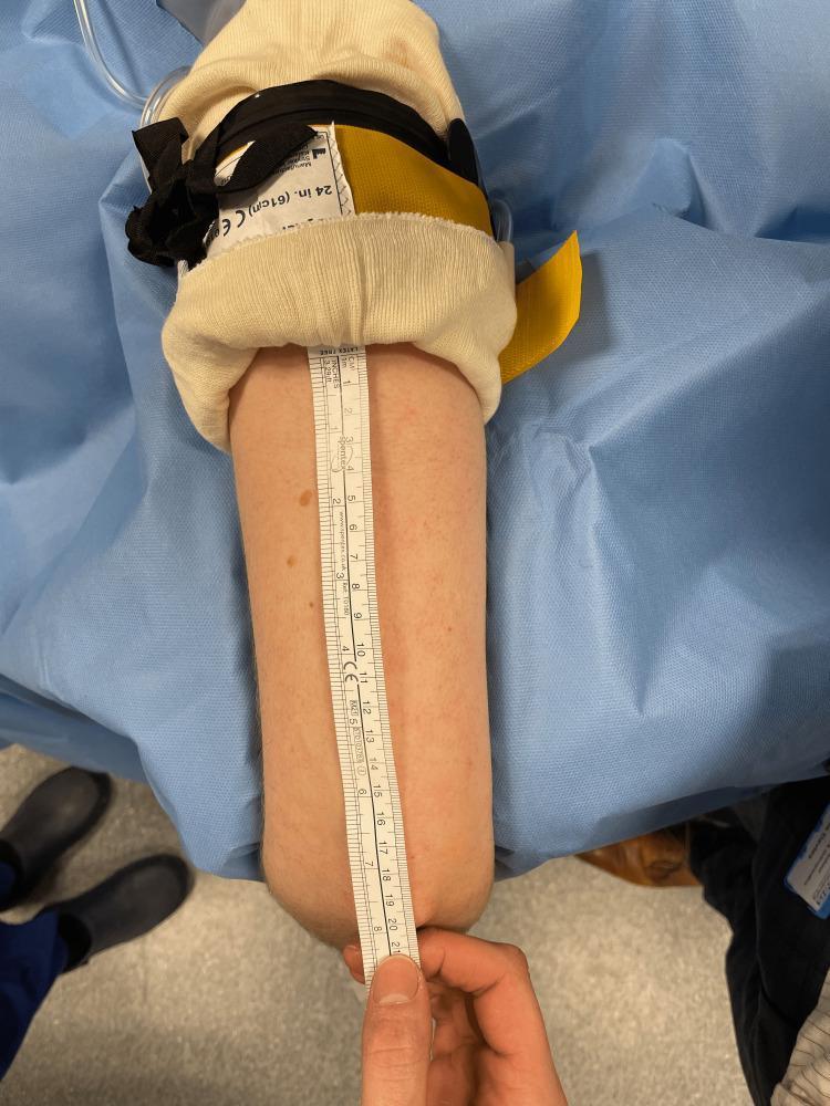
A clinical procedural photograph demonstrating the measurement of a surgical field on a human upper limb. The arm is positioned on a sterile light-blue surgical drape. At the proximal end, a Sterile Standard Tourniquet (SST) is applied over cream-colored protective padding. The pneumatic tourniquet features a black constricting band, black securing straps, a yellow fabric component, and a visible label marked '24 in. (61cm) CE'. A white rigid medical ruler is placed vertically along the posterior aspect of the arm to measure the distance from the distal edge of the sterile tourniquet to the tip of the olecranon (elbow). The ruler shows measurements in both centimeters and inches, indicating a usable surgical field length of approximately 20 cm. This image illustrates perioperative protocols for determining available sterile surgical space when using sterile pneumatic tourniquet systems in orthopedic surgery.
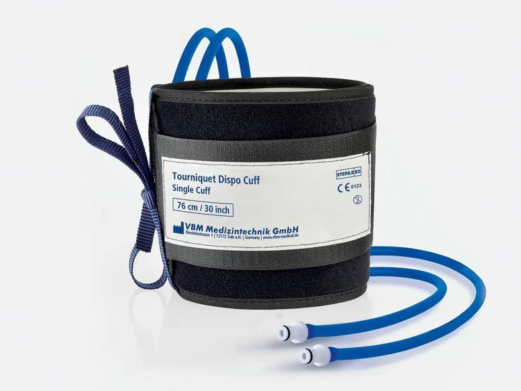
This clinical photograph displays a sterile standard pneumatic (SST) tourniquet cuff designed for surgical use. The device is a black, disposable synthetic fabric cuff featuring a centralized white label. The label identifies the product as a 'Tourniquet Dispo Cuff, Single Cuff' with a specified length of 76 cm (30 inches), and includes manufacturer details for VBM Medizintechnik GmbH. Regulatory and safety symbols are present on the label, including the 'STERILE EO' mark indicating ethylene oxide sterilization, a 'CE 0123' conformity mark, and a 'do not reuse' (single-use) icon. Integral to the pneumatic system, two blue inflation tubes extend from the base of the cuff, terminating in white and gray luer-lock style connectors for attachment to a pressure regulator. A dark blue fabric fastening strap is attached to the lateral side for securing the cuff around a patient's limb. This equipment is primarily used in orthopedic surgery to create a bloodless operative field by occluding distal blood flow.
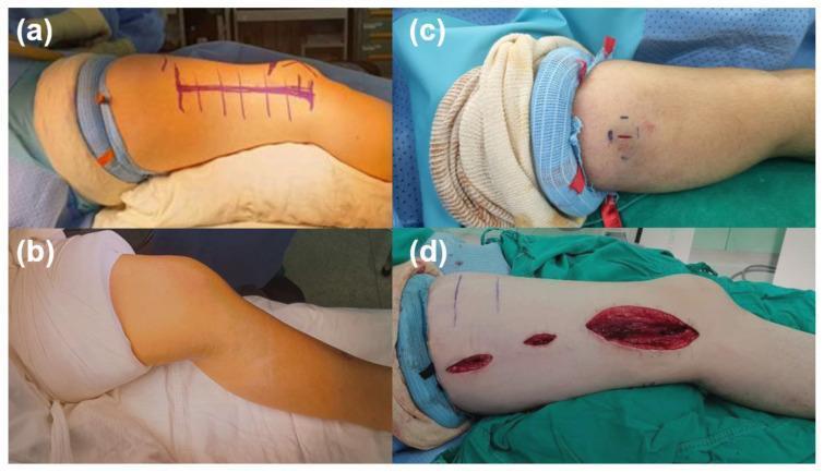
A four-panel clinical photograph comparison demonstrating surgical site exposure provided by different tourniquet types in pediatric orthopedic surgery. Panel (a) illustrates a sterile silicone ring tourniquet applied to the upper thigh, showing its narrow profile (approximately 2-3 cm) and excellent proximal limb exposure for a planned femur incision. Panel (b) shows a conventional pneumatic tourniquet on the proximal thigh, which is significantly wider (8-16 cm) and limits surgical access to the upper thigh. Panel (c) demonstrates a sterile silicone ring tourniquet applied to the upper arm for the excision of a distal humerus region lesion (pilomatricoma), showing minimal interference with the operative field. Panel (d) shows the intraoperative application of the silicone ring tourniquet on the proximal thigh during an open reduction and internal fixation of a femur shaft fracture, highlighting its ability to maintain a bloodless field and provide maximal exposure for extensive longitudinal incisions. The images emphasize the educational objective of choosing appropriate hemostatic devices to optimize the surgical field in pediatric patients with short limb lengths.
Pneumatic Tourniquets
Components and Equipment
- Cuff - an inner inflatable bladder fully enclosed in an outer casing (to prevent "blowout" and pressure loss)
- Pressure source - compressed gas cylinder or wall supply
- Pressure gauge/regulator - aneroid or digital; must be calibrated frequently (discrepancy >20 mm Hg requires repair/replacement; gauges have been found to be up to 300 mm Hg off calibration)
- Alarm system - modern automatic units self-test on startup
Application Technique
- Padding: Apply at least two layers of orthopaedic wool/felt (e.g., Webril) smoothly around the limb. Wrinkles must be avoided - they can cause blisters, skin pinching, and necrosis.
- Application site: Upper arm (near axilla) for the upper limb; mid/upper thigh for the lower limb. A well-padded proximal calf tourniquet is safe for foot and ankle surgery.
- Exsanguination: Elevate the limb for 2-5 minutes, or wrap with a 10 cm elastic bandage from the fingertips proximally just distal to the tourniquet.
- Inflation: Inflate to the appropriate pressure (see below).
- Skin preparation precaution: Apply a circumferential adhesive-backed plastic drape just distal to the tourniquet to prevent prep solutions from running underneath - otherwise chemical burns can result.
- Campbell's Operative Orthopaedics 15th Ed 2026
Cuff Selection
- Wide cuffs are more effective at lower inflation pressures than narrow ones
- Curved (contoured) cuffs fit conical extremities better and require significantly lower arterial occlusion pressures than straight (rectangular) cuffs
- Straight tourniquets on conical thighs should be avoided, especially in muscular or obese individuals (risk of skin slough at the upper margin, particularly in the gluteal fold region)
- Recommended widths: 10 cm for the arm; 15 cm or wider for the leg
Pressure Guidelines
| Extremity | Pressure |
|---|---|
| Upper extremity | 50-100 mm Hg above systolic, or 135-255 mm Hg |
| Lower extremity | Double systolic, or 175-305 mm Hg; leg 250-350 mm Hg |
| Foot/ankle (calf cuff) | 201-250 mm Hg most commonly used |
| Thigh cuff (foot/ankle surgery) | 251-351 mm Hg most commonly used |
Duration and Timing
- General maximum: 2 hours (absolute maximum 3 hours; recovery takes 5-7 days)
- Tourniquet >90 minutes is a risk factor for wound healing complications in elective foot and ankle surgery
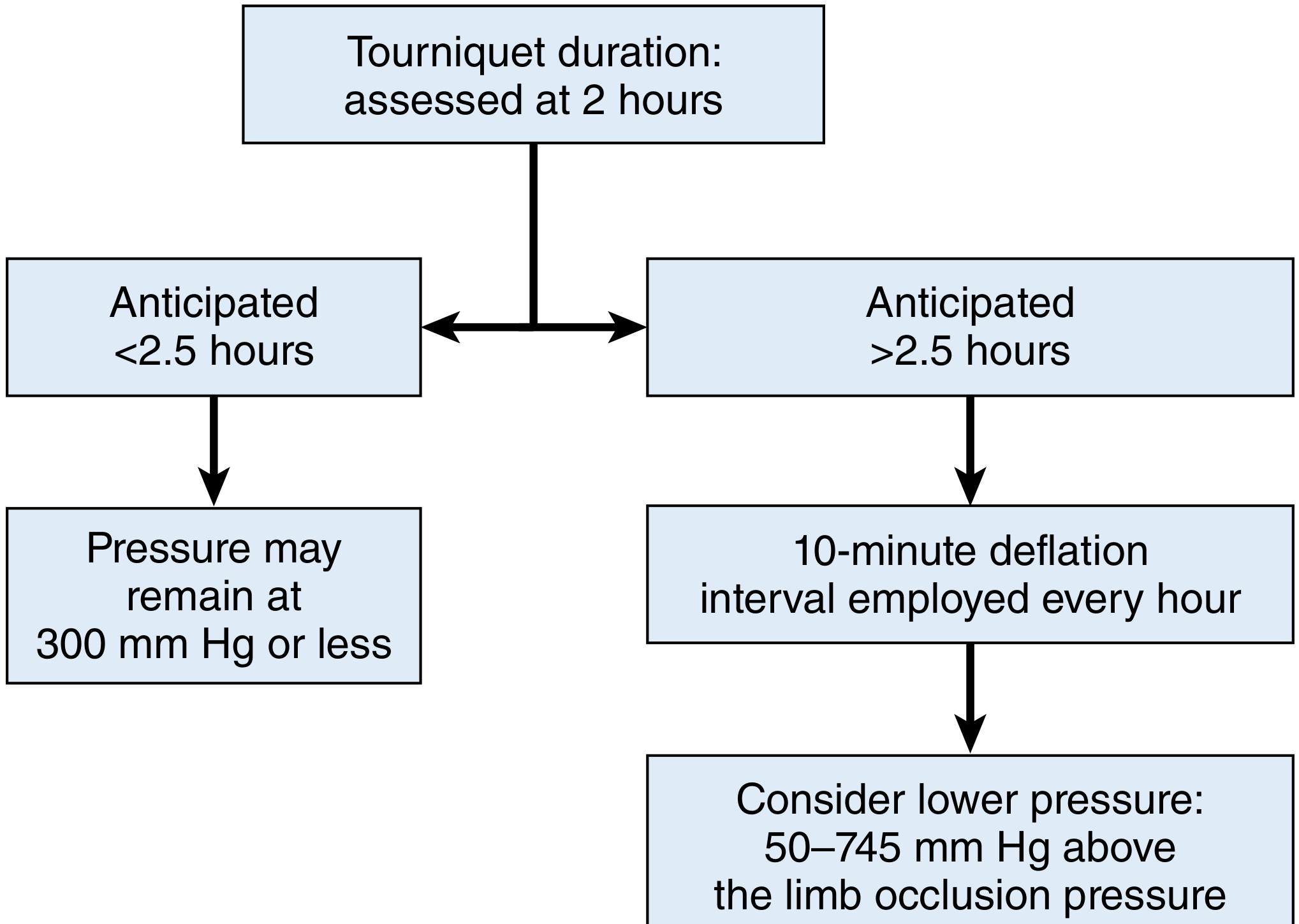
- For longer procedures (anticipated >2.5 hours): employ a 10-minute deflation interval every hour, then consider reducing pressure to 50-75 mm Hg above LOP
Contraindications and Cautions
- Compromised limb vascularity (peripheral vascular disease, questionable blood supply to the foot)
- Severe infections (may spread organisms proximally)
- Sickle cell disease (sickling risk under ischemia)
- DVT (risk of embolization)
- Severe hypertension (difficult to achieve adequate occlusion)
- Use with caution in diabetic neuropathy, severe atherosclerosis, and in patients on anticoagulants
Complications
| Complication | Notes |
|---|---|
| Tourniquet paralysis (nerve injury) | Commonest serious complication; due to direct compression + ischemia; usually neurapraxia, recovers in days-weeks |
| Muscle injury | Ischemia beneath and distal to the cuff; worse with higher pressures and longer duration |
| Post-tourniquet syndrome | Edema, stiffness, pallor, weakness, and heaviness after deflation |
| Tourniquet pain | Aching pain even under regional anesthesia; requires supplemental sedation or GA for long cases |
| Chemical burns | From skin prep running under the cuff |
| Skin blistering/necrosis | From wrinkles in padding or straight cuff on conical limb |
| DVT/PE | Venous stasis during ischemia; hypercoagulable state post-deflation |
| Cardiac effects | Release of tourniquet causes sudden drop in SVR and bolus of acidic, hypoxic, hyperkalemic blood - can cause hypotension |
| Compartment syndrome | Rare; from reperfusion injury |
- Campbell's Operative Orthopaedics 15th Ed 2026
Forearm Tourniquet (Alternative to Upper Arm)
- Allows the local anesthetic dose to be reduced to almost half that required with an upper arm tourniquet
- Lower frequency and severity of tourniquet pain (especially for procedures <25 minutes, or distal to the wrist with regional block)
- Associated with longer sensory block and prolonged postoperative analgesia
Ten Rules of Pneumatic Tourniquet Use (Braithwaite & Klenerman's Modification of Bruner's Rules)
| Rule | Recommendation |
|---|---|
| Application | Only to a healthy limb; with caution to an unhealthy limb |
| Cuff size | Arm: 10 cm; Leg: 15 cm (or wider for large legs) |
| Site | Upper arm; mid/upper thigh ideally |
| Padding | At least two layers of orthopaedic felt |
| Pressure | 50-100 mm Hg above systolic (arm); double systolic (thigh); or arm 200-250 mmHg, leg 250-350 mmHg |
| Duration | Generally ≤2 hours; absolute max 3 hours |
| Temperature | Avoid heating (e.g., surgical lights); cool and keep moist when feasible |
| Documentation | Record duration and pressure; calibrate weekly; equipment maintenance every 3 months |
- Campbell's Operative Orthopaedics 15th Ed 2026, Table 2.6
Advantage Over Esmarch Tourniquet
Key principle: The greatest danger in tourniquet use is an improperly calibrated gauge - gauges have been found to be 300 mm Hg off calibration in documented tourniquet injuries. Routine calibration is non-negotiable.
From millers for Anaesthesia
Pneumatic Tourniquets - Miller's Anesthesia 10e (2-Volume Set)
Purpose and Basic Settings
- Reduce intraoperative blood loss
- Provide a bloodless field (e.g., for cement fixation in TKA)
- Miller's Anesthesia 10e, Hip and Knee Arthroplasties
Haemodynamic Effects
On Inflation
- Blood from the exsanguinated limb is "autotransfused" centrally - raises preload transiently
- MAP and SVR may rise
On Deflation (clinically important)
-
Mean arterial blood pressure drops significantly after tourniquet release
-
Two mechanisms:
- Release of metabolites from the ischemic limb into systemic circulation
- Decrease in peripheral vascular resistance as the limb reperfuses
-
In prolonged cases, the ischemic limb accumulates lactate, CO2, K⁺, and acid - release causes a sudden metabolic acidaemia
-
In replantation surgery, lactic acidosis from prolonged tourniquet time is particularly problematic in patients with underlying lung disease; controlled ventilation is advisable to compensate for metabolic acidosis
-
Miller's Anesthesia 10e, Hip and Knee Arthroplasties; Extremity Replantation
Tourniquet Pain
-
Tourniquet pain typically begins after 60 minutes of inflation
-
It occurs even in the presence of a regional anaesthetic that is adequate for the surgery
-
Mechanism: caused by unblocking of unmyelinated C-fibres during recession of a neuraxial block (C-fibres are more resistant to local anaesthetic block than A-delta fibres and persist longer)
-
Management:
- Addition of opioids to spinal or epidural anaesthesia may ameliorate tourniquet pain
- Blocking T2 dermatome helps decrease tourniquet pain in shoulder/elbow surgery (where the tourniquet sits on the upper arm)
- Dual-cuff technique (for IVRA/Bier block): a second tourniquet is placed distal to the first; inflated 15 minutes after the proximal cuff, then the proximal cuff is deflated - the distal cuff now sits over anaesthetised skin, reducing tourniquet pain
-
Miller's Anesthesia 10e, Hip and Knee Arthroplasties; Hand Surgery; Shoulder and Elbow Surgery
Nerve Injury from Tourniquet
- When prolonged tourniquet inflations are required, deflating the tourniquet for 30 minutes of reperfusion may reduce neural ischemia before re-inflation
Post-Deflation Bleeding
- Bleeding begins immediately on deflation and can continue for the next 24 hours
- This must be factored into fluid and transfusion management
Intravenous Regional Anaesthesia (Bier Block) - Tourniquet Protocol
- Exsanguinate the arm with an elastic bandage (Esmarch)
- Inflate the upper arm tourniquet to 50-100 mm Hg above systolic (often to 250 mm Hg)
- Inject preservative-free local anaesthetic (lidocaine; prilocaine preferred in UK/Europe for its high therapeutic index) into a hand vein
- Onset: approximately 6-8 minutes
- Apply second (distal) cuff 15 minutes later, inflate it, then deflate the proximal cuff - reduces tourniquet pain by positioning the active cuff over anaesthetised skin
- Do not deflate before 30 minutes - risk of LAST (systemic local anaesthetic toxicity) from bulk drug release into circulation
- Adjuvants (ketorolac, clonidine, dexamethasone, dexmedetomidine) can reduce tourniquet pain and improve block quality, though routine use of ketorolac is now controversial
Key safety point: Complications occur when the tourniquet fails during initial injection or if deflated too early (<30 minutes), both of which risk LAST.
- Miller's Anesthesia 10e, Hand Surgery; Intravenous Regional Anaesthesia
Contraindication - Specific Patient Populations
| Condition | Tourniquet Consideration |
|---|---|
| Ehlers-Danlos Syndrome (EDS) | Pneumatic tourniquet avoided unless absolutely necessary (friable skin, vascular fragility, bleeding risk) |
| Sickle cell / haemoglobinopathies | Tourniquet placement worsens hypoxemia risk; regional anaesthesia (especially neuraxial) preferred to reduce sickling |
| Preexisting sciatic neuropraxia, neuropathic pain, or vascular disease in the operative leg | Operation can be performed without a tourniquet |
| Replantation (prolonged tourniquet >90-120 min) | Reperfusion intervals should be considered; controlled ventilation to buffer metabolic acidosis |
- Miller's Anesthesia 10e
Foot and Ankle Surgery
- Thigh or leg tourniquets commonly used to limit blood loss
- Regional anaesthesia combining sciatic + femoral nerve blocks is sufficient for all procedures below the knee that do not require a thigh tourniquet
- If a thigh tourniquet is needed, the femoral nerve territory must also be blocked
Summary - Key Anaesthetic Points
| Issue | Miller's Recommendation |
|---|---|
| Inflation pressure | Systolic BP + 50-100 mm Hg |
| Max duration | 3 hours |
| Tourniquet pain | Begins ~60 min; C-fibres responsible; add opioids to neuraxial block |
| On deflation | Drop in MAP; release of ischemic metabolites |
| Nerve injury | Ischemia + mechanical compression; reperfuse 30 min if prolonged |
| IVRA safety | Never deflate <30 min; dual-cuff technique reduces pain |
| Replantation | Controlled ventilation if prolonged tourniquet (lactic acidosis risk) |
- Miller's Anesthesia, 2-Volume Set, 10th Edition (2023)
Possible questions about blood for md Anaesthesia osce exam
Which blood topics should I focus on? · What format do you want?
Blood - MD Anaesthesia OSCE: Structured Notes with Likely Questions
TOPIC 1: Blood Products - Components and Indications
❓ Likely OSCE Questions
- "What are the components of a unit of PRBC and how do they differ from whole blood?"
- "What solutions can and cannot be co-administered with PRBCs? Why?"
- "What are the indications for FFP, cryoprecipitate, and platelets?"
Key Facts
- Haematocrit: ~57-60% (plasma removed)
- pH: 6.79 | pCO2: 79 mmHg | K⁺: 20.5 mmol/L | Lactate: 9.4 mmol/L
- Same Hb content as whole blood, with less plasma
- Suitable for most indications except severe haemorrhage (where whole blood preferred)
- 0.9% saline, 5% dextrose/0.9% saline, 5% dextrose/0.45% saline, Normosol-R (pH 7.4)
- Lactated Ringer's CONTRAINDICATED - contains Ca²⁺ which reverses citrate anticoagulation and may cause clotting
- Hypotonic solutions cause RBC swelling and haemolysis - avoid
| Product | Content | Shelf life | Temp | Trigger/Indication |
|---|---|---|---|---|
| PRBCs | RBCs, minimal plasma | 42 days | 1-6°C | Hb <7-8 g/dL (or <10 in ischaemic heart disease) |
| FFP | All clotting factors, fibrinogen, albumin | 1 year (frozen) | Frozen | INR >1.5-2, coagulopathy, reversal of warfarin, TTP |
| Cryoprecipitate | Fibrinogen, Factor VIII, vWF, Factor XIII, fibronectin | 1 year (frozen) | Frozen | Fibrinogen <1.5 g/L, haemophilia A, vWD, DIC |
| Platelets | Platelets in plasma | 5 days | 20-24°C with agitation | Count <50 × 10⁹/L for surgery; <10 × 10⁹/L prophylactic |
| Fresh Whole Blood | All components intact | <24 h (optimal) | Not cooled | Massive haemorrhage; military setting |
TOPIC 2: Transfusion Triggers and Decision-Making
❓ Likely OSCE Questions
- "When do you transfuse? What is your haemoglobin trigger?"
- "How do you assess need for further transfusion intraoperatively?"
- "How do you measure intraoperative blood loss?"
Key Facts
- General: Hb 7 g/dL (restrictive threshold - well supported by evidence)
- Ischaemic heart disease / elderly / TBI: Hb 8-10 g/dL
- Asymptomatic, haemodynamically stable: can tolerate lower
- Trend in vital signs
- Measured and anticipated blood loss
- Volume of IV fluids given
- Current Hb/Haematocrit
- Surgical concerns (ongoing bleeding, procedure type)
- Visual + gravimetric (weight of dry vs. blood-soaked gauze)
- Anesthesiologists consistently overestimate blood loss by up to 40%
- No gold standard method exists
- Continuous non-invasive Hb monitoring available (Masimo SpHb - pulse co-oximetry)
TOPIC 3: Massive Transfusion Protocol (MTP)
❓ Likely OSCE Questions
- "What is massive transfusion? Define it."
- "Describe your MTP. What ratios do you use?"
- "What is the lethal triad in trauma? How do you prevent it?"
- "What is damage control resuscitation (DCR)?"
- "What is the role of tranexamic acid in trauma?"
Key Facts
-
10 units PRBCs in 24 hours, OR
-
4 units in 1 hour, OR replacement of entire blood volume
- Hypothermia + Acidosis + Coagulopathy
- Each perpetuates the others; must be broken simultaneously
- Some hospitals: 4 units O-negative uncrossmatched PRBCs + 4 units thawed AB plasma + 1 unit platelets available in ~5 minutes
| Ratio | Evidence |
|---|---|
| 1:1:1 (plasma:platelets:RBCs) | PROPPR trial - faster haemostasis, fewer deaths from exsanguination at 24 h |
| 1:1:2 | Similar 24-h and 30-day mortality in PROPPR, but slower haemostasis |
| Current practice | 1:1:1 most commonly adopted |
- DCR model - empiric ratios (approximating whole blood); used in Phase 1 (uncontrolled haemorrhage)
- Goal-directed haemostatic resuscitation - point-of-care viscoelastic monitoring (TEG/ROTEM) + targeted haemostatic concentrates; used in Phase 2 (controlled haemorrhage)
- Hypocalcaemia is common and associated with death in massive haemorrhage
- Citrate in stored blood chelates calcium; give calcium chloride or gluconate empirically
- May precede blood product administration - supplement early
- Tranexamic acid (TXA): antifibrinolytic; shown to reduce mortality in trauma if given within 3 hours of injury (CRASH-2 trial). Also used in elective surgery (joint arthroplasty) to reduce blood loss.
- Fibrinogen concentrate / Cryoprecipitate: when fibrinogen <1.5 g/L; most US centres use cryo due to cost
- Prothrombin Complex Concentrate (PCC): contains factors II, VII, IX, X; for oral anticoagulant reversal and traumatic intracranial haemorrhage
- rFVIIa: licensed for haemophilia with inhibitors; use in trauma not routinely recommended (thromboembolism risk, no mortality benefit in large RCT)
TOPIC 4: Complications of Blood Transfusion
❓ Likely OSCE Questions
- "A patient develops fever and rigors during transfusion - what do you do?"
- "What is TRALI? How does it differ from TACO?"
- "What is an acute haemolytic transfusion reaction under GA? How do you recognise and manage it?"
- "What are the infective risks of blood transfusion?"
- "What is TRIM?"
Key Facts
- Most common cause: human error (wrong blood to wrong patient) - >half the errors occur AFTER blood leaves the blood bank
- Incidence: 1:1,200 - 1:190,000
| Feature | Conscious patient | Under GA |
|---|---|---|
| Symptoms | Fever, chills, chest/flank pain, nausea | Masked by anaesthesia |
| First sign | Fever/rigors | Haemoglobinuria (presenting sign) |
| Other signs | - | Hypotension, bleeding diathesis |
- Free Hb appears in plasma when capacity of haptoglobin exceeded (haptoglobin binds ~100 mg Hb/100 mL plasma)
- Haemoglobinuria occurs when plasma Hb >150 mg/dL
- Stop the transfusion immediately
- Maintain IV access - give IV fluids
- Check identity labels (patient vs. blood bag)
- Send: urine for haemoglobin, blood for direct antiglobulin test (DAT/Coombs), serum haptoglobin, plasma Hb, bilirubin
- Maintain urine output (fluids ± frusemide/mannitol)
- Treat hypotension, coagulopathy/DIC
- Notify blood bank
- TRALI
- TACO
- Bacterial contamination (from platelets)
| Feature | TRALI | TACO |
|---|---|---|
| Mechanism | Immune-mediated (donor antibodies vs. recipient WBCs) | Volume overload |
| Timing | During or within 6 h of transfusion | During or within 6 h |
| CXR | Bilateral infiltrates | Cardiomegaly, pulmonary oedema |
| JVP/CVP | Normal or low | Elevated |
| BNP | Low/normal | Elevated |
| Treatment | Supportive, O2, ICU, may need intubation | Diuresis, O2 |
| Mortality | ~5-10% | Lower |
- Fever ± chills, cough after transfusion
- Caused by pyrogenic cytokines from donor leukocytes
- Leukoreduction has reduced incidence
- Rule out haemolytic reaction with DAT first
- Treat with paracetamol
- Minor (urticaria) - slow transfusion + antihistamine; do not need to stop
- Anaphylactic - occurs in IgA-deficient patients receiving IgA-containing products (anti-IgA antibodies)
- After only a few mL of blood; treat with epinephrine
- Future transfusions: use washed RBCs (IgA removed)
- Donor lymphocytes engraft and attack recipient tissues
- Risk: immunocompromised patients, directed donations from first/second-degree relatives (shared HLA)
- Features: rash, leucopenia, thrombocytopenia → sepsis → death
- Prevention: irradiation of blood products
- Allogeneic blood has nonspecific immunosuppressive effect on recipient
- May relate to cancer recurrence, postoperative infections, viral activation
- Mechanism unclear; universal leukoreduction may reduce this
| Virus | Current risk per unit |
|---|---|
| HIV | ~1:1.5 million |
| HCV | ~1:1.2 million |
| HBV | ~1:280,000 |
| HTLV | ~1:641,000 |
| West Nile virus | Very low (NAT tested) |
TOPIC 5: Autologous Blood & Cell Salvage
❓ Likely OSCE Questions
- "What are the techniques of autologous blood conservation?"
- "When is cell salvage indicated? What are its contraindications?"
- "What are the complications of intraoperative cell salvage?"
Key Facts
- Preoperative autologous donation (PAD) - patient donates own blood before elective surgery
- Acute normovolaemic haemodilution (ANH) - blood drawn at induction, replaced with crystalloid/colloid, reinfused at end
- Intraoperative cell salvage (ICS) - blood suctioned from field, washed, reinfused
- Postoperative cell salvage - blood from drains collected and reinfused (must reinfuse within 6 hours; upper limit ~1400 mL unprocessed)
- Active infection/sepsis at operative site
- Malignancy (relative - use leukodepletion filter)
- Bowel contamination of field
- Use of topical haemostatics not approved for re-infusion
- Hypervolaemia, bacterial contamination, hypotension
- Non-immune and immune haemolysis
- Febrile non-haemolytic reactions, allergic reactions
- DIC, coagulopathy
- Air embolus
- Reactions from reinfused anticoagulants (heparin) or other contaminants
TOPIC 6: O-Negative Universal Donor Blood
❓ Likely OSCE Questions
- "When and why do you use O-negative blood? What are the risks?"
- "Can you switch back to type-specific blood after emergency O-negative transfusion?"
Key Facts
- Type O blood lacks A and B antigens - cannot be haemolysed by recipient anti-A or anti-B
- Available in emergency in ~5 minutes from blood bank cache
- Prefer O-negative PRBCs over O-negative whole blood - less plasma, almost free of haemolytic anti-A/anti-B antibodies
- If whole blood must be used: blood bank must confirm it is free of haemolytic antibodies
- Risk of detectable haemolysis: only 0.1% in large series (10,916 uncrossmatched units)
TOPIC 7: Fresh Whole Blood
❓ Likely OSCE Questions
- "What is fresh whole blood? What are its advantages in massive haemorrhage?"
Key Facts
- "Fresh" = stored <24-48 hours at 2-6°C (definitions vary)
- Retains coagulation factors, platelets, and RBC function better than stored components
- Whole blood stored >24 hours at 4°C has less haemostatic effect than blood stored <6 hours (decreased platelet aggregability)
- Extensively used by US military in Iraq/Afghanistan - shown effective in treating massive haemorrhage
- Where available, effectively approximates 1:1:1 component therapy as a single product
TOPIC 8: Coagulopathy of Trauma (Acute Traumatic Coagulopathy - ATC)
❓ Likely OSCE Questions
- "What is acute traumatic coagulopathy? How does it differ from dilutional coagulopathy?"
- "What is the role of viscoelastic testing (TEG/ROTEM) in managing coagulopathy?"
Key Facts
- ATC is an early, endogenous coagulopathy that occurs with shock + trauma - separate from the later dilutional coagulopathy from fluids
- Caused by: tissue injury + shock → activated protein C → hyperfibrinolysis + anticoagulation
- Worsened by: hypothermia + acidosis (both impair enzyme activity of clotting cascade)
- Allows targeted replacement rather than empiric ratios
- Identifies: clot initiation (PT/APTT), clot strength (fibrinogen, platelets), fibrinolysis
- Guides: FFP, cryoprecipitate, platelets, TXA, PCC based on specific deficits
HIGH-YIELD OSCE SUMMARY TABLE
| Topic | Key Number/Fact |
|---|---|
| PRBC haematocrit | 57-60% |
| PRBC plasma K⁺ | 20.5 mmol/L |
| Transfusion trigger (general) | Hb 7 g/dL |
| Transfusion trigger (IHD/elderly) | Hb 8-10 g/dL |
| MTP ratio | 1:1:1 (plasma:platelets:RBCs) |
| Max tourniquet duration | 3 hours |
| Emergency O-neg blood availability | ~5 min |
| Platelet storage | 20-24°C, agitation, 5 days |
| Platelet bacterial contamination rate | ~1:2,500 units |
| First sign of haemolytic reaction under GA | Haemoglobinuria |
| Most common cause of ABO incompatibility death | Human error (wrong patient) |
| IgA-deficient anaphylaxis - use | Washed RBCs |
| GVHD prevention | Irradiation of blood products |
| TXA: must give within | 3 hours of injury |
| PROPPR trial: 1:1:1 advantage | Faster haemostasis, fewer 24-h exsanguination deaths |
| Cannot co-administer with PRBCs | Lactated Ringer's (contains Ca²⁺) |
| Postoperative cell salvage time limit | Reinfuse within 6 hours |
Osce questions on electrolytes
Electrolytes - MD Anaesthesia OSCE: Structured Notes with Likely Questions
TOPIC 1: SODIUM
❓ Likely OSCE Questions
- "What is the normal range of serum sodium? What controls it?"
- "Classify hyponatraemia. How do you approach it?"
- "What are the dangers of rapid correction of hyponatraemia?"
- "What is SIADH? How does it differ from other causes of hyponatraemia?"
- "When do you give hypertonic saline and how?"
Normal Physiology
- Normal serum Na⁺: 138-142 mEq/L
- Na⁺ is the dominant ECF cation - the prime determinant of ECF volume
- Total body Na⁺: ~4000 mmol; only 10% intracellular (ICF:ECF ratio 1:15)
- Controls serum Na⁺:
- Hypothalamic osmoreceptors → ADH release → water retention
- Atrial volume sensing → ANP release → natriuresis
- Juxtaglomerular apparatus → RAAS activation → Na⁺ retention
- 99.5% of filtered Na⁺ is reabsorbed (mainly at PCT)
- Daily requirement: adults 1-1.5 mEq/kg/day
Hyponatraemia (Na⁺ < 135 mEq/L)
| Type | Serum osmolality | Causes |
|---|---|---|
| Hypo-osmolar (true) | Low | SIADH, heart failure, cirrhosis, renal failure, hypothyroidism, Addison's |
| Iso-osmolar (pseudohyponatraemia) | Normal | Hyperlipidaemia, hyperproteinaemia (lab artefact) |
| Hyperosmolar | High | Hyperglycaemia, mannitol, contrast media (water moves out of cells) |
| Volume status | ECF | Cause | Urine Na⁺ |
|---|---|---|---|
| Hypovolaemic | ↓ | Diuretics, GI losses, adrenal insufficiency | <20 mEq/L (renal conservation) or >20 (diuretics) |
| Euvolaemic | Normal | SIADH, hypothyroidism, psychogenic polydipsia | >20 mEq/L |
| Hypervolaemic | ↑ | Heart failure, cirrhosis, nephrotic syndrome | <20 mEq/L |
- Plasma osmolality <280 mOsm/kg
- Urine osmolality >100 mOsm/kg (inappropriately concentrated)
- Urine Na⁺ >20 mEq/L (Na⁺ wasting)
- Clinically euvolaemic
- Normal renal, adrenal, and thyroid function
Clinical Features of Hyponatraemia
| Na⁺ (mEq/L) | Symptoms |
|---|---|
| 130-135 | Usually asymptomatic |
| 125-130 | Nausea, malaise |
| <125 | Headache, lethargy, confusion |
| <120 | Seizures, respiratory arrest, cerebral oedema, death |
Correction of Hyponatraemia
⚠️ The most dangerous complication: Osmotic Demyelination Syndrome (ODS) / Central Pontine Myelinolysis (CPM) - caused by TOO RAPID correction.
- Hypovolaemic: 0.9% saline to restore volume (Na⁺ will self-correct as ADH drops)
- SIADH: fluid restriction ± loop diuretics; vasopressin receptor antagonists (vaptans) for refractory cases
- Severe symptomatic (seizures): 3% hypertonic saline - give via central vein (NaCl >7.5% causes endothelial damage)
- Hypo-osmolar hyponatraemia correction
- ↑ Intracranial pressure (reduces cerebral oedema; may be superior to mannitol)
- NOT for TBI without confirmed ↑ICP (not shown beneficial in trials)
Hypernatraemia (Na⁺ > 145 mEq/L)
- Inadequate water intake (elderly, intubated patients, impaired thirst)
- Excess water loss: diabetes insipidus (DI), osmotic diuresis, fever, burns
- Iatrogenic: NaHCO₃ therapy, hypertonic saline, enteral feeds
- Formula: Free water deficit = 0.6 × weight (kg) × [(Na/140) - 1]
- Use 5% dextrose or 0.45% saline
TOPIC 2: POTASSIUM
❓ Likely OSCE Questions
- "What are the ECG changes in hyperkalaemia? How do you treat it urgently?"
- "Why is hypokalaemia dangerous in anaesthesia?"
- "What causes a shift of K⁺ into/out of cells? How does this affect serum K⁺?"
- "What is your threshold for operating on a patient with hypokalaemia/hyperkalaemia?"
Normal Physiology
- Normal serum K⁺: 3.5-5.0 mEq/L
- K⁺ is the dominant ICF cation - 98% intracellular (muscle, liver, RBCs)
- Total body K⁺: ~4000 mmol; only ~70 mEq in ECF
- Vital for resting membrane potential of all excitable tissues
- Na⁺/K⁺-ATPase: exports 3 Na⁺ for 2 K⁺ inward - maintains gradient
- Insulin (stimulates Na⁺/K⁺-ATPase)
- β₂-adrenergic agonists (salbutamol, adrenaline - stimulate Na⁺/K⁺-ATPase) → clinically useful for treatment
- Alkalosis (H⁺ leaves cells, K⁺ enters)
- Glucose (stimulates insulin)
- Acidosis (especially mineral acids - inorganic H⁺ cannot enter cells freely, so H⁺/K⁺ exchange)
- Suxamethonium - depolarises all muscle → K⁺ efflux (~0.5 mEq/L rise normal; 5-10 mEq/L rise in burns, paraplegia, denervation)
- Digoxin (inhibits Na⁺/K⁺-ATPase)
- Cell lysis (haemolysis, rhabdomyolysis, tumour lysis)
- Hyperosmolar states (osmotic drag)
- Beta-blockade
Hypokalaemia (K⁺ < 3.5 mEq/L)
- GI losses: vomiting (alkalosis → kaliuresis), diarrhoea, bowel prep, NG suction, fistulas
- Renal losses: diuretics (thiazides, loop), hyperaldosteronism, Cushing's, RTA, Mg²⁺ deficiency
- Cellular shift: insulin therapy, β₂-agonists, alkalosis, refeeding syndrome
- Inadequate intake (rare in isolation)
- Muscle weakness, cramps, fatigue, ileus, polyuria (nephrogenic DI)
- ECG changes: T-wave flattening/inversion, prominent U waves, ST depression, widened QRS; at severe levels: VT/VF
- Enhanced sensitivity to non-depolarising muscle relaxants
- Risk of arrhythmias (especially in digitalis patients)
- Impaired respiratory muscle function → delayed extubation
- Paradoxical aciduria in metabolic alkalosis
- Operating threshold: Elective surgery generally safe if K⁺ ≥ 3.0 mEq/L; aim ≥ 3.5 mEq/L in patients on digoxin or with cardiac disease
- Oral KCl preferred for mild-moderate
- IV KCl for severe or unable to take orally: max 20-40 mEq/hour peripherally (peripheral vein limit: 40 mEq/L concentration; central line for higher concentrations/rates)
- Always correct Mg²⁺ first - hypokalaemia refractory to K⁺ replacement without correcting hypomagnesaemia
- Rule of thumb: 1 mEq/L fall in serum K⁺ ≈ 200-400 mEq total body deficit
Hyperkalaemia (K⁺ > 5.5 mEq/L)
- Renal failure (most common)
- ACE inhibitors, ARBs, K⁺-sparing diuretics, NSAIDs
- Suxamethonium (burn, denervation, crush injuries, prolonged immobility)
- Acidosis, rhabdomyolysis, haemolysis, massive blood transfusion
- Adrenal insufficiency, hypoaldosteronism
- Pseudohyperkalaemia (haemolysed sample, thrombocytosis)
- Tall peaked ("tented") T waves - first change (K⁺ ~5.5-6.5)
- Prolonged PR interval (K⁺ ~6.5-7)
- Widened QRS (K⁺ ~7-8)
- Sine wave pattern (K⁺ ~8-9)
- VF / asystole (K⁺ > 9-10)
| Step | Drug | Dose | Mechanism | Onset | Duration |
|---|---|---|---|---|---|
| 1. Membrane stabilisation | Calcium gluconate 10% | 10 mL IV over 2-3 min | Antagonises cardiac membrane effect | 1-3 min | 30-60 min |
| 2. Shift K⁺ into cells | Insulin + Dextrose | 10 units actrapid + 50 mL 50% dextrose | Stimulates Na⁺/K⁺-ATPase | 15-30 min | 4-6 h |
| 3. Shift K⁺ into cells | Salbutamol (nebulised or IV) | 10-20 mg nebulised | β₂-agonist → Na⁺/K⁺-ATPase | 15-30 min | 4-6 h |
| 4. Shift K⁺ into cells | NaHCO₃ (if acidotic) | 50-100 mEq IV | Alkalosis drives K⁺ intracellularly | 15-30 min | Variable |
| 5. Remove K⁺ from body | Frusemide (if urine output) | 40-80 mg IV | Renal K⁺ excretion | 30-60 min | - |
| 5. Remove K⁺ from body | Resonium/Patiromer | Oral/PR | Ion exchange resin | Hours | - |
| 6. Definitive | Dialysis/Haemofiltration | - | Removes K⁺ directly | Immediate (on circuit) | - |
⚠️ Suxamethonium contraindicated in: burns >24-48h, spinal cord injury, prolonged immobility, denervation injuries, severe trauma - risk of hyperkalaemic cardiac arrest.
TOPIC 3: CALCIUM
❓ Likely OSCE Questions
- "What forms does calcium exist in plasma? Which is physiologically active?"
- "What are the signs of hypocalcaemia? Chvostek's and Trousseau's signs?"
- "When does hypocalcaemia occur perioperatively?"
- "How does pH affect ionised calcium?"
Normal Physiology
- Normal total serum Ca²⁺: 2.2-2.6 mmol/L (8.5-10.5 mg/dL)
- Normal ionised Ca²⁺: 1.1-1.3 mmol/L - the physiologically active fraction
- Distribution:
- 40-50% bound to albumin (inactive)
- 5-10% complexed with anions (citrate, phosphate, bicarbonate) (inactive)
- 45-50% ionised (active)
- Alkalosis → more Ca²⁺ binds albumin → ↓ ionised Ca²⁺ → symptoms of hypocalcaemia (e.g., hyperventilation tetany)
- Acidosis → less binding → ↑ ionised Ca²⁺
- PTH ↑: ↑ bone resorption, ↑ renal Ca²⁺ reabsorption, ↑ 1,25-OH-Vit D synthesis → ↑ Ca²⁺
- Calcitonin ↓: ↓ bone resorption → ↓ Ca²⁺
- Vitamin D (1,25-OH): ↑ intestinal Ca²⁺ absorption
Hypocalcaemia (ionised Ca²⁺ < 1.1 mmol/L)
- Massive blood transfusion (citrate in stored blood chelates Ca²⁺) - important! Give CaCl₂/Ca gluconate in MTP
- Post-thyroid/parathyroid surgery (hypoparathyroidism)
- Acute pancreatitis
- Alkalosis (hyperventilation)
- Hypomagnesaemia (impairs PTH secretion and action)
- Rhabdomyolysis, tumour lysis, renal failure
- Vitamin D deficiency
| System | Feature |
|---|---|
| Neuromuscular | Paraesthesias (perioral, fingertips), muscle cramps, tetany, laryngospasm |
| Signs | Chvostek's sign (facial muscle twitch on tapping CN VII at parotid), Trousseau's sign (carpal spasm with BP cuff inflated > systolic for 3 min) |
| Cardiac | Prolonged QT interval, bradycardia, heart block, hypotension, impaired contractility |
| CNS | Confusion, seizures |
- Symptomatic/acute: 10% Calcium gluconate 10-20 mL IV (preferred; 10% CaCl₂ if cardiac arrest - more elemental Ca²⁺ per mL, but more irritant to veins)
- Calcium gluconate contains 2.25 mmol elemental Ca²⁺ per 10 mL; CaCl₂ contains 6.8 mmol per 10 mL
- Chronic: oral calcium + Vitamin D supplements
⚠️ Do NOT give calcium and bicarbonate in the same line - will precipitate as CaCO₃
Hypercalcaemia (total Ca²⁺ > 2.6 mmol/L; severe >3.5 mmol/L)
- Primary hyperparathyroidism (outpatient, usually mild)
- Malignancy (PTHrP secretion, bone mets, haematological malignancy)
- Vitamin D toxicity, sarcoidosis, thyrotoxicosis, immobility (Paget's), thiazides, lithium
- Bones: pain, pathological fractures
- Stones: renal calculi, nephrocalcinosis
- Groans: nausea, vomiting, constipation, peptic ulceration, pancreatitis
- Psychic moans: depression, confusion, psychosis, coma
- Reduced neuromuscular blockade response (increased resistance to NDNMBs? - actually hypercalcaemia ENHANCES ACh release and may antagonise NDNMBs)
- Digitalis toxicity potentiated
- Hypovolaemia (polyuria)
- IV saline hydration (2-4 L/24h) - first step
- Frusemide (after rehydration, promotes calciuresis)
- IV bisphosphonates (zoledronate, pamidronate) - 24-48h to effect
- Calcitonin (rapid but short-lived)
- Steroids (sarcoidosis, vitamin D toxicity, haematological malignancy)
- Dialysis (severe/refractory)
TOPIC 4: MAGNESIUM
❓ Likely OSCE Questions
- "What is the role of magnesium in anaesthesia?"
- "What are the signs of magnesium toxicity during infusion?"
- "When do you use magnesium perioperatively?"
- "Why must you correct magnesium before replacing potassium?"
Normal Physiology
- Normal serum Mg²⁺: 0.7-1.0 mmol/L (1.5-2.5 mEq/L)
- 99% intracellular; only 1% extracellular
- 30% of serum Mg²⁺ bound to albumin; 70% ionised or complexed
- Cofactor for >300 enzyme reactions including Na⁺/K⁺-ATPase
- Regulates K⁺ handling: Mg²⁺ deficiency → renal K⁺ wasting → refractory hypokalaemia
Hypomagnesaemia (Mg²⁺ < 0.7 mmol/L)
- Chronic alcoholism (most common in surgical patients)
- Poor intake, malabsorption, diarrhoea
- Loop diuretics, aminoglycosides, cisplatin, PPIs
- DKA treatment (insulin-driven shift)
- Refeeding syndrome
- Neuromuscular: tremor, muscle weakness, tetany, seizures (similar to hypocalcaemia)
- Cardiac: prolonged QT, broad T-waves, torsades de pointes, VF
- Hypokalaemia and hypocalcaemia refractory to replacement (Mg²⁺ needed for PTH secretion and renal K⁺/Ca²⁺ conservation)
- Tocolysis (premature labour) - uterine relaxation
- Pre-eclampsia/eclampsia - seizure prophylaxis and treatment (loading dose 4-6 g IV over 15-20 min, then 1-2 g/h infusion)
- Analgesia - adjunct in multimodal regimens (NMDA receptor antagonist)
- Bronchospasm - refractory bronchospasm/status asthmaticus
- Arrhythmias - Torsades de pointes (1-2 g IV bolus), digoxin toxicity arrhythmias
- Potentiates NDNMBs - reduces the dose required; prolongs block duration
- Anti-hypertensive (prevents autonomic instability, obtunds laryngoscopy response)
Magnesium Toxicity
Critical for OSCE: know the levels and signs
| Mg²⁺ level (mmol/L) | Sign |
|---|---|
| 1.5-2.5 | Normal therapeutic range |
| 2.5-3.5 | Nausea, flushing, sedation, double vision |
| 3.5-5.0 | Loss of deep tendon reflexes (first clinical sign of toxicity - check patellar reflex) |
| 5.0-6.5 | Somnolence, slurred speech |
| 6.5-7.5 | Respiratory muscle paralysis (most dangerous) |
| >10 | Cardiac arrest |
- Patellar (knee-jerk) reflex - check before each dose; loss = STOP infusion
- Urine output >25-30 mL/h (Mg²⁺ renally excreted)
- Respiratory rate >12/min
- Serum Mg²⁺ levels
TOPIC 5: PHOSPHATE
❓ Likely OSCE Questions
- "What are the consequences of hypophosphataemia?"
- "When does refeeding syndrome occur and why?"
Normal Physiology
- Normal serum phosphate: 0.8-1.5 mmol/L
- 85% in bone as hydroxyapatite; intracellular phosphate essential for ATP synthesis
- Inversely related to calcium (PTH ↑ phosphaturia)
Hypophosphataemia (PO₄ < 0.8 mmol/L)
- Refeeding syndrome (most important perioperatively): glucose infusion → insulin → massive cellular uptake of phosphate, K⁺, Mg²⁺ → plasma levels crash
- Malnutrition, alcohol, malabsorption
- Antacids (bind phosphate in gut)
- DKA recovery (insulin drives intracellular shift)
- Hyperparathyroidism
- Impaired diaphragm/respiratory muscle function → failure to wean from ventilator
- Haemolytic anaemia (RBC membrane dysfunction)
- Impaired leukocyte function → infection
- Cardiomyopathy
- Encephalopathy, seizures
HIGH-YIELD OSCE SUMMARY TABLE
| Electrolyte | Low - key sign | Low - key treatment | High - key sign | High - key treatment |
|---|---|---|---|---|
| Na⁺ | Seizures (Na⁺ <120) | 3% NaCl (slow, max 8-10 mEq/24h) | Confusion, hyperreflexia | Free water replacement (slow) |
| K⁺ | U waves on ECG | KCl IV; fix Mg²⁺ first | Peaked T waves → sine wave | CaGluconate → insulin/dextrose → salbutamol → dialysis |
| Ca²⁺ | Prolonged QT, Trousseau's, Chvostek's, laryngospasm | CaGluconate 10 mL IV | Short QT, "bones/stones/groans/moans" | IV saline → frusemide → bisphosphonates |
| Mg²⁺ | Torsades, refractory ↓K⁺/↓Ca²⁺ | MgSO₄ IV | Loss of patellar reflex → resp arrest | CaGluconate 10 mL IV |
| PO₄ | Failure to wean ventilator | Phosphate replacement | Hypocalcaemia (binds Ca²⁺) | Saline, dialysis |
KEY PERIOPERATIVE RELATIONSHIPS (OSCE GOLD)
| Scenario | Electrolyte disturbance | Mechanism |
|---|---|---|
| Massive transfusion | ↓ Ca²⁺, ↓ Mg²⁺, ↓ K⁺ (then ↑K⁺) | Citrate chelates; stored blood K⁺ release |
| Vomiting (NGT, bowel obstruction) | ↓ K⁺, ↓ Cl⁻, ↓ H⁺ (metabolic alkalosis) | Gastric HCl loss → secondary hyperaldosteronism → renal K⁺ wasting |
| Diarrhoea | ↓ K⁺, ↓ HCO₃⁻ (metabolic acidosis) | Large bowel secretions high in K⁺ and HCO₃⁻ |
| Refeeding | ↓ PO₄, ↓ K⁺, ↓ Mg²⁺ | Insulin-driven cellular uptake |
| Suxamethonium (burns/denervation) | ↑ K⁺ | Upregulation of extrajunctional AChRs → massive depolarisation |
| Hyperventilation | ↓ ionised Ca²⁺ (not total) | Alkalosis → Ca²⁺ binds albumin |
| Acid-base disturbances | K⁺ shifts inversely with pH | Mineral acid: K⁺ rises 0.4-0.6 mEq/L per 0.1 pH unit fall |
| Pre-eclampsia treatment | ↑ Mg²⁺ (therapeutic) | MgSO₄ infusion; monitor patellar reflex |
| Bowel prep | ↓ K⁺, dehydration | Replace with crystalloid + K⁺ supplementation |