
Necrosis in 5 to 7 marks with etiology pathogenesis flow charts digrams H&E digrams where needed with a short for m
necrosis H&E histology types coagulative liquefactive caseous
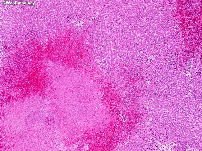
Imaging modality: not relevant; histopathology: Hematoxylin and eosin (H&E) stained liver biopsy. Anatomical location: hepatic parenchyma of the liver, particularly lobular hepatocytes. Visual features: irregular confluent zones of coagulative necrosis with surrounding hemorrhage, producing mottled, pale-to-roseate subcapsular and midzonal areas; the necrotic zones are often well-demarcated from intact parenchyma. Inflammation is disproportionately mild relative to the extent of necrosis, with sparse portal and lobular inflammatory cells. Viral cytopathic effects may be evident as eosinophilic intranuclear inclusions (Cowdry type A) within hepatocytes and occasional multinucleated syncytial cells. The tissue architecture shows disruption of normal hepatic cords, with sinusoidal congestion and hemorrhagic foci. Clinically, these changes reflect fulminant or subfulminant herpes simplex virus hepatitis, a high-mortality entity that can occur in immunocompromised hosts or during disseminated infection. Diagnostic relevance: histology supports HSV in the differential when HSV-specific testing (PCR, immunostaining) confirms viral etiology; the pattern helps distinguish from other viral hepatitides and drug-induced liver injury. Documentation of this pattern informs antiviral management (acyclovir/valacyclovir) and urgent clinical correlation for rapid treatment. Educational use: this image illustrates classic patchy necrosis with hemorrhage and minimal inflammation, aiding histopathology teaching, differential diagnosis, and correlating clinicopathologic findings. Correlation with serology and HSV DNA testing enhances diagnostic confidence significantly.
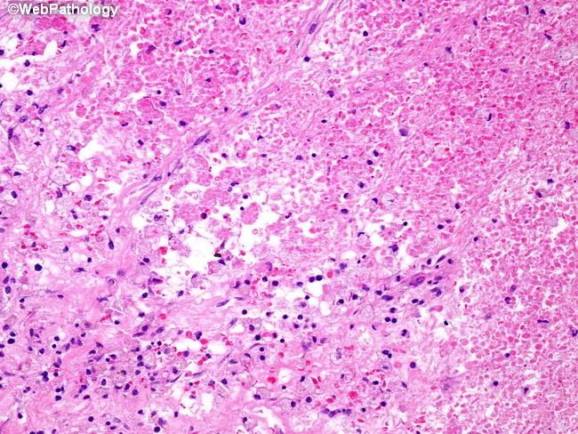
This histopathology image depicts post-chemotherapy therapy-induced changes in a Wilms tumor (nephroblastoma) of the kidney, illustrating how treatment alters tumor architecture in surgical resection specimens. The slide, stained with Hematoxylin and Eosin (H&E), shows extensive coagulative necrosis within tumor tissue, evidenced by homogeneous eosinophilic zones with preservation of basic architectural outlines but loss of viable tumor cells. Interspersed fibrous stroma forms bands and nodules replacing neoplastic blastemal and epithelial elements, consistent with treatment response. Within the fibrotic matrix, numerous hemosiderin-laden macrophages appear as dark intracellular pigment, reflecting prior hemorrhagic components and iron storage. Vascular remodeling and residual inflammatory cells may be present at the periphery of necrotic/fibrotic areas. Overall, the neoplasm is partially or largely regressed with sparse residual viable tumor in some cases, aligning with risk stratification into low, intermediate, or high categories based on residual tumor burden and histologic response. Clinically, this post-therapy histology informs NWTS and SIOP treatment decisions, guiding adjuvant chemotherapy intensity and radiotherapy planning. The image underscores standardized terminology—coagulative necrosis, fibrosis, hemosiderin-laden macrophages, regression—and demonstrates how histologic response correlates with prognosis and postoperative management. This morphologic assessment complements clinical staging and informs multidisciplinary planning for chemotherapy regimens, nephron-sparing considerations, and long-term surveillance and follow-up.
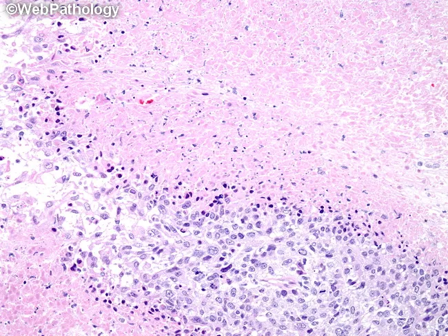
Histopathology: Hematoxylin and eosin (H&E) stained formalin-fixed paraffin-embedded breast tissue section examined by bright-field light microscopy. The specimen shows invasive carcinoma of the breast with conspicuous tumor necrosis. At the inferior/outer aspect, nests of malignant cells display high cellular density, marked atypia, pleomorphism, hyperchromatic nuclei, and conspicuous nucleoli. Mitotic activity is elevated. The tumor front invades into surrounding stromal tissue with irregular, infiltrative borders. Central regions exhibit extensive coagulative necrosis characterized by eosinophilic, acellular debris and paucity of viable tumor cells, while viable tumor nests persist at the advancing edge. A desmoplastic stromal response is variably present. The cellular architecture is consistent with high-grade ductal carcinoma; areas may display basal-like cytologic features. The Ki-67 proliferative index is typically high in such necrotic, rapidly dividing tumors. The necrosis is clinically significant as it correlates with aggressive biology, rapid growth, and potential for early recurrence or progression, particularly in tumors that are not hormone receptor–positive. This histology supports a poor prognostic profile and would prompt comprehensive systemic therapy considerations. Immunohistochemical correlation is recommended to determine ER/PR/HER2 status and to assess basal-like markers for prognostication. Correlative clinical data, including patient age, stage, and receptor profile, strengthens risk stratification and therapeutic planning for personalized management.
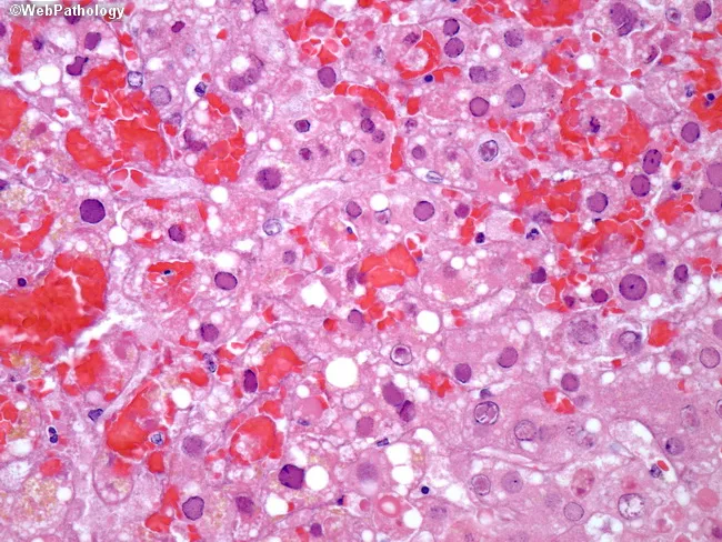
Histology, bright-field light micrographs of liver tissue (liver biopsy) stained with Hematoxylin and Eosin (H&E). The specimen shows hepatic parenchyma with focal to confluent zones of coagulative necrosis within liver lobules, often surrounding viable hepatocytes. Viral cytopathic effects are evident in adjacent cells, including intranuclear inclusions with surrounding clear halos and peripheral chromatin margination producing ground-glass appearing nuclei. Eosinophilic Cowdry type A inclusions or basophilic Cowdry type B inclusions may be present, reflecting HSV-induced nuclear changes. Additional evidence includes scattered enlarged hepatocytes with faint cytoplasmic eosinophilia and occasional cytoplasmic vacuolization; viral particles can be identified in hepatocytes by higher-magnification views (and are detectable with corroborating immunostains). Immunohistochemistry or in situ hybridization can help confirm HSV and exclude other viruses such as CMV, which would display Owl's eye inclusions. The overall pattern is necroinflammation with predominance in periportal to midzonal regions and disruption of normal lobular architecture. Diagnostic significance lies in the combination of characteristic nuclear inclusions, ground-glass nuclei, and necrotizing hepatitis, especially in immunocompromised patients or those with fulminant disease. Clinically, HSV hepatitis requires prompt antiviral therapy and correlation with serology and clinical presentation. Early detection guides antiviral therapy decisions.
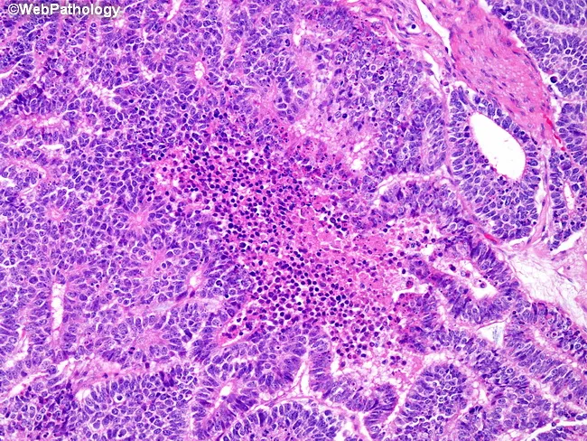
Histopathology, bright-field light microscopy of an H&E-stained renal tumor section (Wilms tumor/nephroblastoma). The image highlights the epithelial component with a focal area of necrosis. The neoplasm displays triphasic morphology: blastemal, stromal, and epithelial elements; here, abortive tubules and glomeruloid structures comprise the epithelial differentiation. The epithelial cells are cuboidal to columnar with hyperchromatic nuclei, scant cytoplasm, and occasional mitotic figures; adjacent blastemal cells are small, hyperchromatic, and densely packed. A discrete center contains coagulative necrosis with eosinophilic, anucleate ghost outlines and karyorrhectic debris, surrounded by a mixed inflammatory infiltrate and viable tumor cells. The surrounding stroma can appear myxoid or fibrous, and residual renal parenchyma is not evident in this field. No overt anaplasia is observed in the region shown. In Wilms tumor, focal necrosis within epithelial components can reflect rapid tumor turnover or sampling of a necrotic focus; it is not unique to this tumor and should be interpreted in the context of overall histology. Clinically, recognition of necrotic epithelial foci aids grading and prognosis, informs differential diagnosis versus other pediatric renal tumors, and supports pathologic staging after nephrectomy or biopsy. This pattern underscores the importance of correlating morphology with clinical data to guide prognosis and therapeutic decisions effectively.
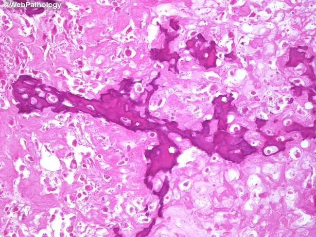
This is a post-treatment osteosarcoma histology slide derived from a limb-sparing surgical resection after neoadjuvant chemotherapy. The imaging modality is bright-field light microscopy of Hematoxylin and Eosin (H&E) stained bone tumor tissue. The specimen demonstrates marked sclerosis with densely packed bony trabeculae and extensive tumor necrosis. In many fields there is cell dropout and a paucity or absence of viable neoplastic cells, reflecting a favorable chemotherapeutic response. Interspersed foci show coagulative necrosis with residual granulation tissue replacing tumor; occasional residual osteoid production may be present. Areas of cartilage formation are evident consistent with chondroblastic osteosarcoma components, which are often relatively chemo-resistant. Overall the lesion displays reduced cellularity, abundant necrosis, and prominent post-treatment remodeling within the metaphyseal bone environment. The diagnostic significance lies in evaluating the degree of tumor response to neoadjuvant chemotherapy, which remains prognostically important but lacks universally accepted, standardized necrosis quantification guidelines. Clinically, these histologic changes inform prognosis, surgical margins assessment, and decisions regarding adjuvant therapy. Potential differential considerations include distinguishing post-therapy necrosis and granulation tissue from residual viable tumor and recognizing heterogeneity of response within tumor foci. Correlation with preoperative imaging and clinical course is essential for accurate interpretation.
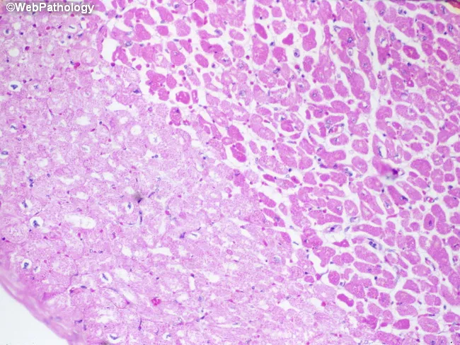
Imaging modality: Light microscopy of hematoxylin-eosin (H&E) stained cardiac tissue. Specimen is paraffin-embedded myocardial section, 5 micron thick, imaged with bright-field optics at high magnification. Anatomic region: left ventricular myocardium with an infarct in the left half of the field. The histology demonstrates pallor of necrotic cardiomyocytes (myocytolysis) produced by sarcolemmal disruption with influx of salt and water and cytoplasmic swelling. In this early post-ischemic stage, fibers lose cross-striations and display increased eosinophilia; nuclei are often absent or pyknotic. The infarct border may show waviness of fibers and mild edema; contraction bands may be present at the margin. Neutrophilic infiltration is typically minimal during the first 24 hours but may begin to appear around this window. The combination of coagulative necrosis, myocytolysis, and pallor indicates an acute myocardial infarction approximately one day old. This histologic pattern correlates with ischemia from coronary occlusion, clinical presentation of acute coronary syndrome, and aligns with diagnostic timelines (0-24 h). Clinically the image supports timing of infarct and helps distinguish acute infarction from myocarditis or chronic scar, serving as an educational reference for pathology, medical education, and research on myocardial injury dynamics. Further confirmed by clinical correlation and education.
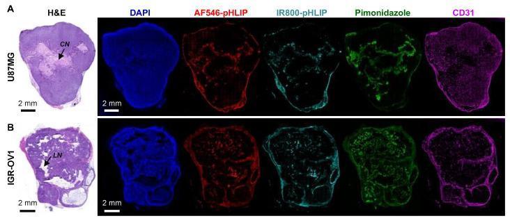
Multi-panel fluorescence microscopy and histological comparison of whole-tumor sections from human cancer xenografts. Row A displays a U87MG (glioblastoma) tumor section, while Row B shows an IGR-OV1 (ovarian cancer) tumor section. The panels compare H&E staining (far left) with multiplexed fluorescence markers including DAPI (blue, nuclei), AF546-pHLIP (red, pH-low insertion peptide), IR800-pHLIP (aqua, pH-low insertion peptide), pimonidazole (green, hypoxia marker), and CD31 (magenta, microvascular marker). In the U87MG tumor, H&E identifies a region of coagulative necrosis (CN) that corresponds spatially with high pHLIP accumulation and pimonidazole-positive hypoxia. In the IGR-OV1 tumor, a region of liquefactive necrosis (LN) shows similar pHLIP and hypoxia localization. Both tumor models demonstrate that pHLIP-based probes preferentially accumulate in hypoxic and necrotic regions, which are characterized by diminished CD31-stained microvascular density. The identical distribution of red (AF546) and aqua (IR800) signals validates the reliability of pHLIP variants for targeting the acidic tumor microenvironment.
caseous necrosis tuberculosis granuloma histology H&E
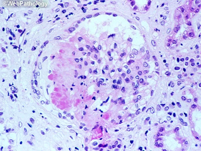
This is a light microscopy image of a formalin-fixed, paraffin-embedded tissue section stained with Hematoxylin and Eosin (H&E). The sample reveals a well-formed granulomatous lesion characterized by a surrounding cuff of lymphocytes and numerous epithelioid histiocytes with elongated, crescent-shaped nuclei. Within the granuloma, multinucleated giant cells (Langhans-type) are visible, and a central amorphous pink necrotic area (caseous necrosis) is evident. The tissue architecture demonstrates organized granulomatous inflammation with a necrotic core, consistent with infectious granulomas. The differential includes mycobacterial disease (tuberculosis) and certain fungal infections (histoplasmosis, coccioidomycosis), though non-necrotizing granulomas can occur in sarcoidosis. The staining highlights chromatin-rich nuclei and cytoplasmic eosinophilia; occasional fibroblasts and mild edema extend into the perigranulomatous region. Clinically, granulomatous tissue with caseation is highly suggestive of TB in the appropriate clinical context and warrants further diagnostic workup (acid-fast bacilli staining, fungal stains, culture, PCR). This image serves as teaching material for histopathology education, differential diagnosis of granulomatous inflammation, and correlation with radiologic and microbiologic findings in suspected infectious etiologies. Additional keywords for indexing: epithelioid macrophages, granuloma, necrosis, caseation, tuberculosis, mycobacteria, fungal infection, lymphohistiocytic rim, tissue biopsy, pathology slide, H&E, histology teaching. Clinical correlation with imaging and sputum analysis improves diagnostic yield.
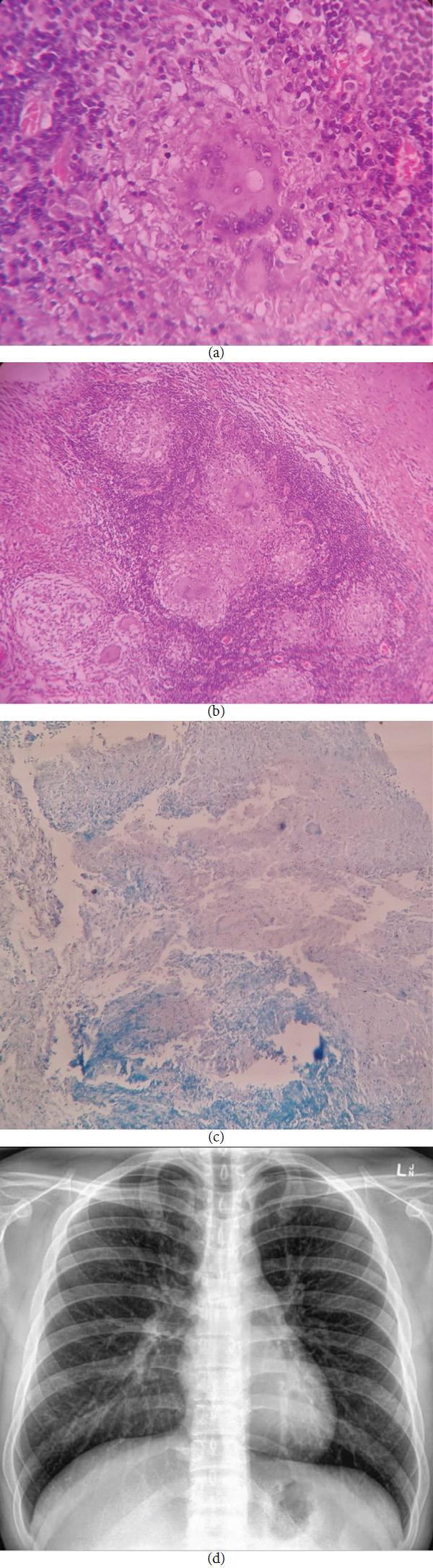
A multi-panel medical image illustrating diagnostic findings for tuberculosis. (a) High-magnification (40x) H&E stained photomicrograph showing a well-formed granuloma with central caseous necrosis, surrounded by epithelioid cells, Langhans-type multinucleated giant cells, and a peripheral rim of chronic inflammatory cells. (b) Lower-magnification (10x) H&E stained photomicrograph displaying multiple coalescing granulomatous lesions within the connective tissue. (c) Photomicrograph using Ziehl-Neelsen (ZN) stain highlighting acid-fast bacilli, visible as small pink/red rod-shaped structures against a methylene blue background, confirmatory for Mycobacterium species. (d) Posteroanterior (PA) view chest X-ray demonstrating clear lung fields with normal bronchovascular markings and no evidence of active pulmonary infiltrates, consolidation, or hilar lymphadenopathy. This visual set educates on the histopathological diagnosis of extrapulmonary tuberculosis (such as primary oral TB) where localized granulomatous inflammation is present despite a normal systemic radiological presentation.
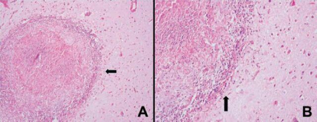
Two-panel light microscopy image of brain (glial) tissue showing necrotizing granulomatous inflammation, commonly associated with conditions like CNS tuberculosis. Image A (200x magnification) displays a well-circumscribed, circular granuloma characterized by a centrally located area of caseous necrosis, which appears as a pale, acellular eosinophilic region. The necrosis is surrounded by a dense peripheral rim of mononuclear inflammatory cells, including lymphocytes and epithelioid histiocytes. Image B (400x magnification) provides a higher-resolution view of the granuloma's border (indicated by black arrows), highlighting the transition from the necrotic core to the hypercellular inflammatory zone and the adjacent glial parenchyma. The H&E staining clearly differentiates the various zones: the central homogenous necrosis, the basophilic ring of active inflammation, and the surrounding reactive glial background. These features are diagnostic hallmarks of chronic granulomatous disease within the central nervous system.
fat necrosis pancreatic saponification histology
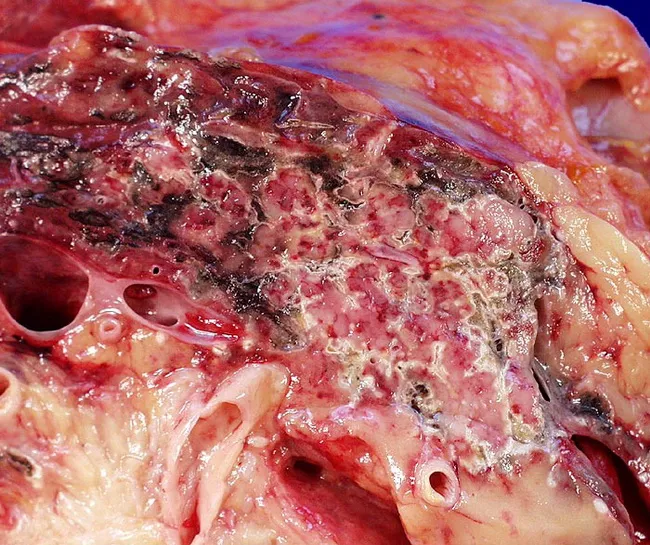
Gross pathology photograph of a resected or opened pancreatic specimen showing extensive necrosis consistent with acute necrotizing pancreatitis. The tissue demonstrates hemorrhagic, black brown necrosis of the pancreatic parenchyma with overlying peripancreatic fat appearing yellow to pale and interspersed with dark, necrotic foci. The surface is mottled with irregular areas of necrosis and hemorrhage; surrounding adipose tissue is edematous and partially necrotic, with fat saponification visible as soft, friable material. There may be scattered clearing holes from tissue removal and ducts and vascular structures are exposed. The specimen’s color variation ranges from pale pink to grayish and dark brown, indicating a spectrum from acute inflammation to liquefactive necrosis. The clinical history includes prior acute cholecystitis and cholelithiasis, consistent with gallstone pancreatitis as the etiologic context. The image underscores severe pancreatic injury with necrosis extending into peripancreatic fat, a hallmark of necrotizing pancreatitis. This finding has significant diagnostic implications, implying SMS severity, risk of systemic inflammatory response, infection, and potential need for debridement, necrosectomy, or staged intervention. In educational settings, this image illustrates the macroscopic appearance of pancreatic necrosis and peripancreatic fat necrosis in the course of gallstone-related pancreatitis. Pathology relevance includes correlative clinical and radiologic assessment. Educational value for residents and fellows.
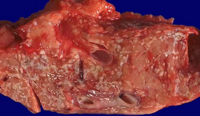
Gross pathology photograph of pancreatic tissue from a patient with acute necrotizing pancreatitis. Modality: gross pathology photography; specimen: pancreatic parenchyma with diffuse stippled necrosis and extensive peripancreatic fat necrosis. The pancreas exhibits mottled tan-brown necrotic areas intermingled with hemorrhagic streaks; focal liquefactive zones; peripancreatic adipose tissue is disrupted with edema and necrosis. The cut surface reveals areas of tissue loss with friable texture and scattered liquefactive debris; pancreatic lobules appear irregular with necrotic debris; small ducts may contain necrotic material. Peripancreatic fat shows chalky fat necrosis with saponification. Overall appearance is consistent with necrotizing pancreatitis (pancreatonecrosis). Notable features include diffuse necrosis within pancreatic parenchyma and surrounding fat, lack of a discrete capsule, and vascular congestion in affected regions. Diagnostic significance: confirms necrotizing pancreatitis and indicates severe disease burden; correlates with risk of infection, sepsis, and progression to walled-off necrosis if untreated. Differential considerations: hemorrhagic pancreatitis, sterile pancreatic necrosis, proliferative fat necrosis, pancreatitic fat necrosis (redundant). Clinical correlation: supports surgical planning for debridement or step-up therapy; prognostic implication relates to extent of necrosis and organ failure; this morphology is essential for education and research. It highlights necrotic pancreatic tissue and surrounding fat as targets for therapeutic intervention and intraoperative planning purposes.
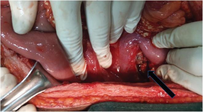
This clinical photograph shows an intraoperative view during an exploratory laparotomy, illustrating severe abdominal pathology. The image reveals loops of small and large intestines with significant inflammation. Key pathological findings include mesenteric fat saponification, visible as characteristic opaque, whitish, chalky deposits scattered across the normally yellow fatty tissue of the mesentery and omentum, indicative of enzymatic fat necrosis. A black arrow points toward the pancreatic region, which shows evidence of a necrotic transection at the junction of the body and neck, marked by darkened, devitalized tissue and loss of structural integrity. Hemorrhagic fluid is visible in the dependent portions of the peritoneal cavity. The image serves as a teaching tool for recognizing the gross appearance of acute necrotizing pancreatitis and its associated mesenteric complications in a surgical setting. The context indicates a Grade III isolated pancreatic injury with substantial hemoperitoneum.
fibrinoid necrosis vasculitis blood vessel H&E
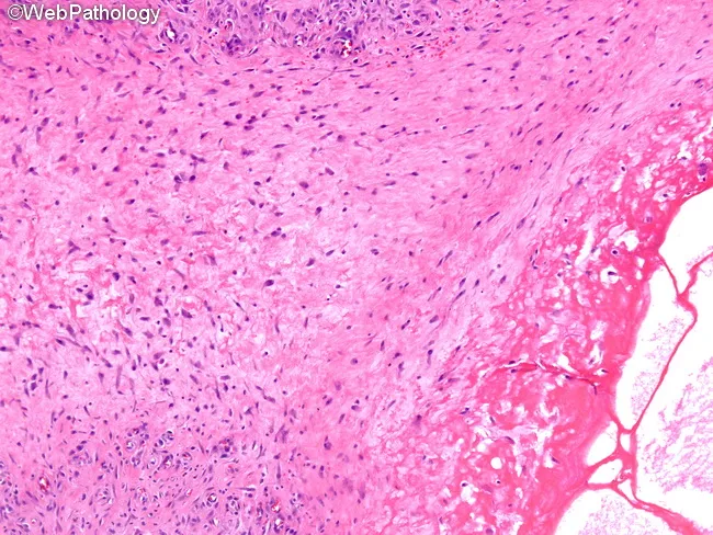
High magnification light microscopy of a formalin-fixed tissue section stained with hematoxylin and eosin (H&E) reveals a focal area of fibrinoid necrosis within the vessel wall (lower right) accompanied by cystic change. Surrounding this necrotic core is a prominent zone of vascular tissue characterized by marked fibroblastic proliferation and remodeling, with spindle-shaped cells and thickened intima forming a dense, fibrous stroma (upper left). The necrotic area displays eosinophilic, amorphous material consistent with fibrin deposition, often seen in necrotizing vasculitis. The adjacent parenchyma shows a relatively preserved architecture with scattered inflammatory cells, a pattern that may reflect an early or localized vasculitic process rather than a fully developed granulomatous lesion. Overall, the image demonstrates a cascade from acute fibrinoid injury toward progressive fibrous scarring, typical of angiitis with fibroproliferative response. The combination of fibrinoid necrosis and fibroblastic proliferation within a vascular niche has diagnostic relevance for necrotizing vasculitis syndromes (e.g., polyarteritis nodosa, granulomatosis with polyangiitis) and related connective tissue diseases. Clinically, these features are relevant for differential diagnosis, prognosis, and therapeutic decisions, as they inform decisions about immunosuppressive therapy, vascular risk assessment, and correlation with serology such as ANCA. This micrographic pattern is essential for pathology education and case documentation.
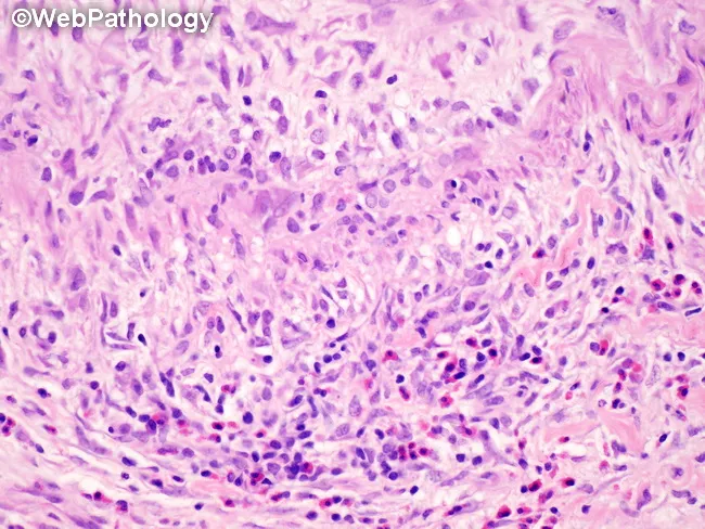
This histopathology image depicts a gallbladder wall section showing classic necrotizing vasculitis of medium-sized muscular arteries consistent with polyarteritis nodosa (PAN). The salient feature is transmural inflammation of the vessel wall with fibrinoid necrosis and disruption of the tunica media and internal elastic lamina, best seen with elastic stains but evident on routine H&E as pale, eosinophilic necrosis surrounding a dense inflammatory infiltrate. In active lesions, the infiltrate comprises neutrophils and eosinophils admixed with histiocytes; skip areas indicate discontinuous involvement with uninvolved interposed segments. The inflammatory process may spare adjacent gallbladder parenchyma, defining a bystander phenomenon rather than true parenchymal inflammation. In healing stages, the vessel shows fibrointimal proliferation and progressive luminal narrowing, reflecting irreversible remodeling. The image underscores the need to distinguish true necrotizing vasculitis from secondary vascular inflammation seen with acute or chronic cholecystitis. Clinically, PAN-related gallbladder involvement signals a systemic vasculitis that may accompany renal and gastrointestinal manifestations; diagnosis rests on histology, correlation with serology (often ANCA-negative in classic PAN), and integration with radiographic or clinical vasculitis features. This specimen exemplifies how vascular lesions can be segmental and heterogenous within a single gallbladder biopsy, influencing prognosis and therapeutic planning (steroids, immunosuppressants). Guiding patient management decisions.
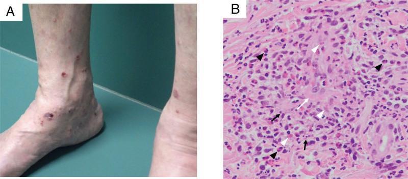
This composite image illustrates the clinical and histological features of systemic vasculitis. Panel A is a clinical photograph of the lower extremities showing multiple, discrete to confluent, erythematous to violaceous macules and papules, consistent with palpable purpura. The lesions are concentrated around the ankles and shins. Panel B is a light microscopy image of a skin biopsy stained with Hematoxylin and Eosin (H&E) at x200 magnification, demonstrating characteristic findings of leukocytoclastic vasculitis with eosinophilic involvement. Key histological features include fibrinoid necrosis of the vessel wall (white arrow), nuclear dust or leukocytoclasis (black arrows), and significant dermal extravascular leakage of red blood cells (white arrowheads). A prominent inflammatory infiltrate is visible, containing numerous eosinophils (black arrowheads) along with neutrophils and macrophages. These findings are clinically significant for diagnosing small-vessel vasculitis, such as Eosinophilic Granulomatosis with Polyangiitis (EGPA) or MPA-related eosinophilic vasculitis.
liquefactive necrosis brain abscess cerebral infarction histology
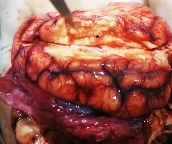
This clinical photograph shows a gross pathological specimen of a brain during necropsy. The primary finding is a focal area of liquefactive necrosis in the right cerebral hemisphere, marked by an asterisk (*). This necrotic region appears as a pale, yellowish-white, and softened area of tissue loss measuring approximately 2 cm x 0.5 cm x 1 cm. Adjacent to the necrosis, there is prominent meningeal congestion characterized by diffuse erythema and engorged vessels following the sulci and gyri. The underlying cortical tissue shows signs of hyperemia. The visual evidence supports a diagnosis of severe central nervous system inflammation or infection leading to tissue softening and degradation, typical of liquefactive processes in neural tissue. This specimen demonstrates the gross morphological presentation of cerebral infarction or inflammatory necrosis suitable for pathology education.
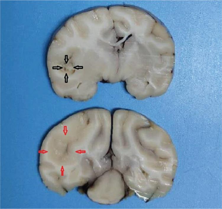
This clinical photograph displays two coronal gross pathology sections of a brain specimen, illustrating the morphological changes associated with a cerebral infarction. The sections demonstrate an area of ischemic damage localized to the territory of the middle cerebral artery (MCA). In the bottom section, red arrows delineate the boundary of the infarct lesion, which appears as a paler, fainter region with lost structural definition compared to the surrounding viable cortical and subcortical tissue. In the top section, black arrows highlight the center of the lesion, where there is clear evidence of liquefactive necrosis. This necrotic core is characterized by a more friable, darker, and fragmented texture, signifying a loss of tissue integrity. These visual markers—pallor at the boundary and central necrosis—are characteristic gross findings of subacute cerebral infarction. The specimen serves as a pathological correlation for neuroimaging findings such as high signal intensity on DWI/T2 sequences, representing the progression from initial ischemia to cellular death and structural breakdown within the brain parenchyma.
NECROSIS
DEFINITION
Necrosis = denaturation of proteins + enzymatic digestion + membrane disruption + inflammation (Robbins Pathologic Basis of Disease, p. 54)
ETIOLOGY (Causes)
CAUSES OF NECROSIS
──────────────────────────────────────────────
  1. Ischemia / Hypoxia       ← most common
     (e.g. MI, stroke, infarction)

  2. Physical agents
     (heat, cold, radiation, trauma)

  3. Chemical / toxic agents
     (CCl₄, heavy metals, drugs)

  4. Microbial toxins
     (bacteria, fungi, viruses)

  5. Immune reactions
     (hypersensitivity, vasculitis)

  6. Activated proteases
     (e.g. acute pancreatitis)

  7. Nutritional deficiencies
──────────────────────────────────────────────
PATHOGENESIS FLOWCHART
INJURIOUS STIMULUS
       │
       ▼
┌──────────────────────────────────────────────┐
│   Depletion of ATP (mainly ischemia)         │
│   ↓ Oxidative phosphorylation                │
│   ↑ Anaerobic glycolysis → ↑ lactic acid     │
│   → ↓ intracellular pH                       │
└──────────────────┬───────────────────────────┘
                   │
          ┌────────▼────────────────────────┐
          │  Na⁺-K⁺ ATPase pump fails       │
          │  → Na⁺ & water enter cell       │
          │  → CELL SWELLING (hydropic change)│
          └────────┬────────────────────────┘
                   │
       ┌───────────▼───────────────────────────┐
       │  Mitochondrial dysfunction            │
       │  → ↑ intracellular Ca²⁺              │
       │  → Cytochrome c release              │
       │  → Reactive oxygen species (ROS)     │
       └───────────┬───────────────────────────┘
                   │
       ┌───────────▼───────────────────────────┐
       │  MEMBRANE DAMAGE                      │
       │  (plasma membrane + organelle         │
       │   membranes + lysosomes)              │
       └───────────┬───────────────────────────┘
                   │
       ┌───────────▼───────────────────────────┐
       │  Lysosomal enzymes released into      │
       │  cytoplasm → digest cell contents     │
       └───────────┬───────────────────────────┘
                   │
       ┌───────────▼───────────────────────────┐
       │  Intracellular contents (DAMPs,       │
       │  ATP, uric acid) leak extracellularly │
       │  → INFLAMMATION recruited             │
       │  → Leukocytes release more proteases  │
       └───────────┬───────────────────────────┘
                   │
                   ▼
             NECROSIS (irreversible cell death)
TIMELINE (Cell vs Morphology)
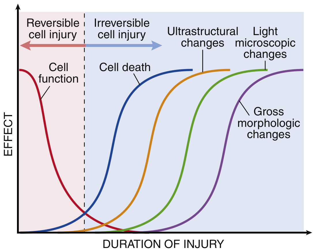
MORPHOLOGY ON H&E (Microscopy)
Cytoplasmic Changes
| Feature | Mechanism | H&E appearance |
|---|---|---|
| Increased eosinophilia | Loss of RNA (basophilia) + denatured proteins bind eosin | Pink/red cytoplasm |
| Glassy/homogeneous | Loss of glycogen particles | "Ground-glass" look |
| Vacuolated / moth-eaten | Organelles digested by enzymes | Swiss-cheese cytoplasm |
| Myelin figures | Phospholipid precipitates from membranes | Whorled intracytoplasmic inclusions |
Nuclear Changes (3 Patterns)
Normal nucleus
      │
      ├──► PYKNOSIS        ← nuclear shrinkage + dark condensed chromatin
      │
      ├──► KARYORRHEXIS    ← fragmentation of pyknotic nucleus
      │
      └──► KARYOLYSIS      ← fading of basophilia, DNA digested by DNases
                              → complete dissolution in 1-2 days
TYPES OF NECROSIS
1. COAGULATIVE NECROSIS
Ischemia → protein denaturation (including enzymes)
         → proteolysis BLOCKED
         → cell outline preserved for days/weeks
         → firm texture
         → Eventually leukocytes digest debris
2. LIQUEFACTIVE NECROSIS
Bacterial infection OR brain ischemia
   → Massive leukocyte recruitment
   → Hydrolytic enzymes released
   → COMPLETE DIGESTION of dead cells
   → Transformation to viscous liquid
   → Pus (bacteria) or soft cystic cavity (brain)
3. CASEOUS NECROSIS
TB infection → Type IV hypersensitivity
   → Macrophages form GRANULOMA
   → Central cell death + protein denaturation
   → Neither coagulative nor liquefactive
   → Friable, white "cheese-like" material
4. FAT NECROSIS
Acute pancreatitis
   → Pancreatic acinar cell injury
   → Release of activated lipases into peritoneum
   → Lipases split triglycerides in fat cells
   → Free fatty acids released
   → Fatty acids + Ca²⁺ → CALCIUM SOAPS
   → Chalky-white saponification deposits
5. FIBRINOID NECROSIS
Antigen-antibody complexes deposited
   in vessel walls (Type III hypersensitivity)
      │
      ▼
Plasma proteins (fibrin) leak into vessel wall
      │
      ▼
Bright pink, amorphous material in wall
("fibrin-like" = fibrinoid)
      │
      ▼
Vessel occlusion → ischemia downstream
6. GANGRENOUS NECROSIS
Coagulative necrosis (dry gangrene)
         +
Superimposed bacterial infection
         ↓
Liquefactive component added = WET GANGRENE
COMPARISON TABLE (Short Form for Exam)
| Type | Cause | Mechanism | H&E | Example |
|---|---|---|---|---|
| Coagulative | Ischemia | Protein denaturation blocks proteolysis | Ghost cells, eosinophilic, architecture preserved | MI, renal infarct |
| Liquefactive | Brain ischemia; bacterial infection | Enzymatic digestion dominates | Liquid pus/cavity, PMNs | Brain abscess, cerebral infarct |
| Caseous | TB, fungi | Type IV hypersensitivity, granuloma | Amorphous granular debris + granuloma | TB lung |
| Fat | Acute pancreatitis | Lipase splits triglycerides → Ca²⁺ soap | Shadow fat cells + basophilic Ca²⁺ deposits | Peripancreatic fat |
| Fibrinoid | Immune vasculitis, malignant HTN | Immune complex + fibrin in vessel wall | Bright pink amorphous vessel wall | PAN, SLE, malignant HTN |
| Gangrenous | Vascular occlusion ± infection | Coagulative ± liquefactive | Mixed | Diabetic foot, limb ischemia |
OUTCOMES OF NECROSIS
NECROTIC CELLS
      │
      ├──► Enzymatic digestion + phagocytosis → RESOLUTION (healing)
      │
      ├──► Dystrophic calcification
      │    (Ca²⁺ deposits in necrotic tissue)
      │
      ├──► Replacement by scar (fibrosis)
      │
      └──► Liquefaction → abscess or cyst
SHORT FORM SUMMARY (Memory Aid)
| Mnemonic | C - L - C - F - F - G |
|---|---|
| C | Coagulative - Ischemia - ghost cells |
| L | Liquefactive - Brain/bacteria - pus |
| C | Caseous - TB - cheese + granuloma |
| F | Fat - pancreatitis - chalk deposits |
| F | Fibrinoid - vasculitis - pink smudge |
| G | Gangrenous - limb ischemia - dry + wet |