
Trigger thumb
trigger thumb stenosing tenosynovitis A1 pulley
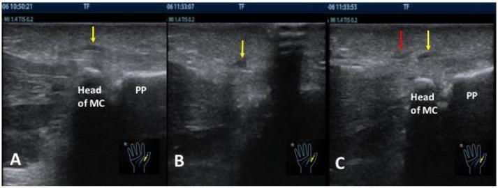
A composite of three musculoskeletal ultrasound images (A, B, and C) in the longitudinal plane of a right thumb, demonstrating the A1 pulley and adjacent anatomical structures for trigger finger release. Panel A (Pre-procedure) shows the 'Head of MC' (metacarpal) and 'PP' (proximal phalange), with a yellow arrow indicating a thickened A1 pulley, a classic sign of stenosing tenosynovitis. Panel B (Post-procedure) displays a yellow arrow pointing to hypoechoic fluid in the surrounding tissue immediately following a needle knife release procedure. Panel C (Complication) illustrates a technical failure in the 'blind group' procedure: a red arrow highlights an incorrect cutting direction or track after skin piercing, while a yellow arrow indicates that the actual thickened A1 pulley remains unaddressed at a separate location. This clinical imaging set highlights the limitations of blind needle knife release compared to guided interventions, emphasizing the risk of inaccurate targeting and tissue fluid accumulation.
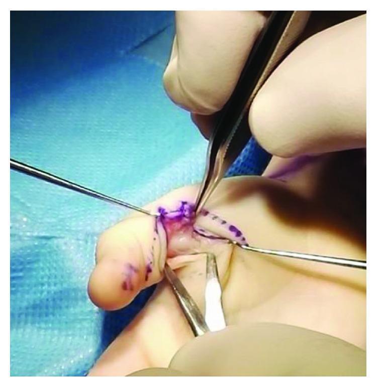
This clinical photograph captures an intraoperative view of a surgical procedure on a human digit, specifically an A1 pulley release to treat a trigger thumb. The primary anatomical focus is the flexor tendon sheath at the level of the metacarpophalangeal joint. Centrally, a 'Node of Notta'—a pathological nodular thickening of the flexor pollicis longus tendon—is exposed and being manipulated with fine surgical forceps. The surgical field is prepared with blue sterile drapes and shows purple skin markers delineating the incision path. Multiple microsurgical instruments, including adson-type forceps and a fine probe or retractor, are visible in the hands of the surgeon (gloved). The image demonstrates the classic clinical presentation of pediatric stenosing tenosynovitis where the enlarged tendon nodule becomes trapped proximal to the A1 pulley, causing mechanical locking of the joint. This visual is highly relevant for orthopedic and hand surgery education, specifically regarding the surgical management of congenital or developmental trigger digits.
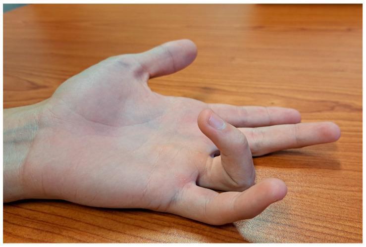
This clinical photograph shows the left hand of a 15-year-old patient in a supinated position, illustrating a presentation of a pediatric trigger finger. The ring finger (fourth digit) demonstrates a localized fixed flexion deformity at the proximal interphalangeal (PIP) joint, while the distal interphalangeal (DIP) joint remains relatively neutral. This visual captures the 'locking' or 'snapping' phenomenon characteristic of stenosing tenosynovitis, where the flexor tendon becomes trapped under the A1 pulley. In comparison, the index, middle, and small fingers are mostly extended, and the thumb is adducted with slight flexion at the metacarpophalangeal (MCP) joint. The image serves as an educational example of idiopathic trigger finger in an adolescent, a condition that may involve nodularity of the flexor digitorum superficialis (FDS) or profundus (FDP) tendons. Clinical relevance includes the diagnosis of mechanical tendon entrapment and the assessment of range of motion prior to potential surgical intervention such as an A1 pulley release.
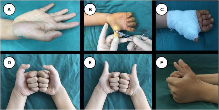
A series of six clinical photographs (A-F) documenting the surgical management and follow-up of a pediatric case of trigger thumb. Panel A shows the preoperative state with limited extension of the interphalangeal (IP) joint of the right thumb. Panel B is an intraoperative view showing the surgical A1 pulley release procedure using a small incision at the base of the thumb. Panel C illustrates immediate postoperative management with the hand immobilized in a white bandage. Panels D, E, and F provide long-term follow-up images (nearly 2 years post-surgery) demonstrating excellent functional and cosmetic outcomes. Panel D shows full flexion of the thumbs bilaterally, Panel E shows complete extension in a frontal view, and Panel F provides a lateral view of the extended thumb. These follow-up images confirm a well-healed surgical site with no visible scar formation, normal range of motion, and absence of deformity or atrophy, demonstrating successful resolution of the stenosing tenosynovitis.
"trigger thumb" OR "trigger finger" stenosing tenosynovitis treatment
Trigger Thumb
Pathophysiology
- Thickening and fibrosis of the A1 pulley
- Formation of a tendon nodule (called Notta's nodule) just proximal to the A1 pulley
- The nodule can be palpated and moves with the tendon
- In rheumatoid patients, a nodule distal to the MCP level can also cause triggering
Clinical Features
- Usually seen in individuals over 45 years of age
- Inability to extend the flexed thumb, or to flex an extended thumb
- A palpable "snap" or "triggering" at the MCP level
- Patient often reports the problem is in the interphalangeal (IP) joint - this is a classic misdirection
- Local tenderness is present but not a prominent complaint
- Pressure over the nodule accentuates the snapping
- Thumb may lock in fixed flexion in advanced cases
- Association with collagen diseases (rheumatoid arthritis), diabetes mellitus, and hypothyroidism
| Grade | Description |
|---|---|
| 0 | Normal movement |
| 1 | Uneven movement |
| 2 | Actively correctable triggering |
| 3 | Passively correctable only |
| 4 | Fixed deformity |
Pediatric Trigger Thumb
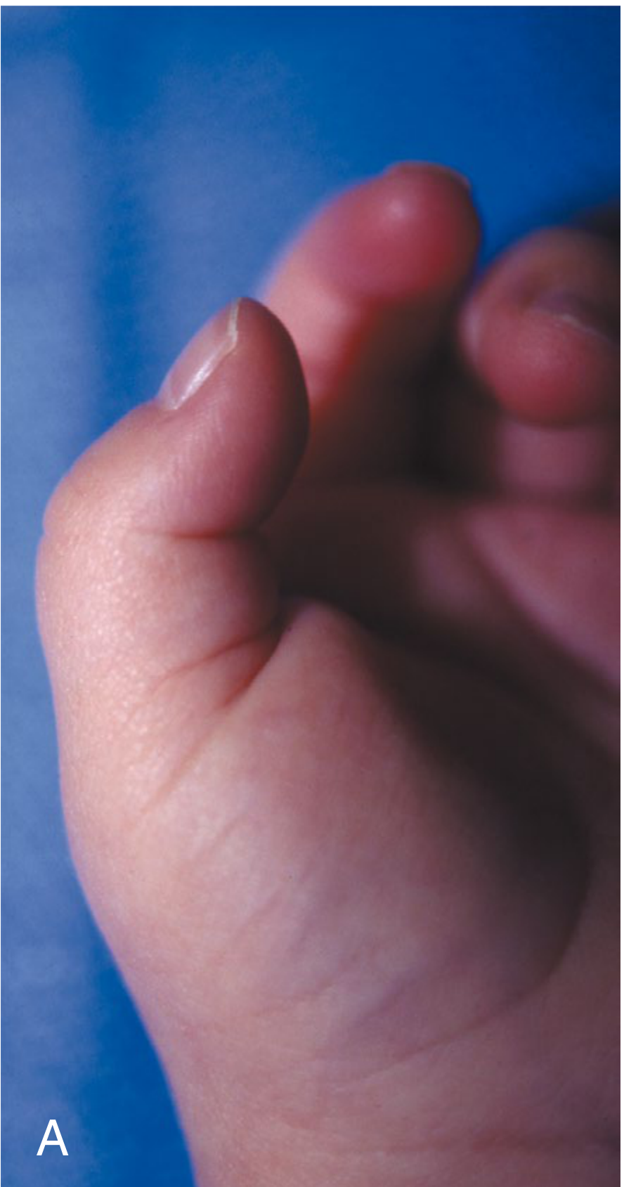
- Child typically presents in the first few years of life with a relatively fixed flexion posture of the IP joint (not the snapping/triggering complaint seen in adults)
- The clicking/snapping is usually NOT the presenting complaint
- Notta's nodule - an intratendinous nodule palpable proximal to the A1 pulley
- Must be differentiated from clasped thumb deformity (which involves primarily MCP flexion)
- Associated conditions: trisomy 13, juvenile rheumatoid arthritis, mucopolysaccharidosis
Differential Diagnosis
- Intraarticular disorders (loose bodies, degenerative joint disease, fractures)
- Common extensor tendon subluxation
- Clasped thumb deformity (pediatric)
- de Quervain's tenosynovitis (involves first dorsal compartment, not flexor)
Treatment
Nonoperative (Adults)
- Stretching, night splinting, heat/ice - first-line for short-duration, uncomplicated cases
- Corticosteroid injection - ~60% success after a single injection; repeat injections can be used for patients preferring nonoperative management; splinting after injection may improve longevity
- Diabetes caution: Corticosteroids may elevate serum glucose for ≥5 days. Patients with unstable diabetes may be better served by primary surgery. Immediate surgical release is actually the most cost-effective strategy in diabetic patients.
Surgical (Adults)
- Open A1 pulley release (gold standard): ~97% complete resolution rate
- Transverse incision in the palm at the MCP level
- Identify and protect digital nerves (the radial digital nerve of the thumb is at risk)
- Longitudinal incision through the A1 pulley under direct vision
- Release done under local anesthetic block so cessation of triggering can be confirmed intraoperatively
- Percutaneous release (needle or push knife): supported in the literature but carries risk of incomplete release and digital nerve injury, especially in the thumb and index finger where the nerve anatomy is close
- Surgery is performed in the office or operating room; both settings have equivalent complication rates for experienced surgeons
Pediatric Treatment
- Observation and splinting first: extension splinting ≥3 hours/day achieves resolution in ~67% vs. 30% with observation alone
- Surgical release (A1 pulley release): recommended at ~3 years of age if spontaneous resolution has not occurred; can be done effectively in older children too
- More extensile exposure than in adults (must visualize A1 to A3 pulley)
- Bruner incision centered over the MCP joint
- Evaluate for tendon nodularities and FDS/FDP decussation anomalies
- Consider resection of a slip of FDS to reduce tendon volume if needed
- Using A1 release + FDS slip resection: 91% success rate (Bae)
Surgical Anatomy Key Points
| Structure | Relevance |
|---|---|
| A1 pulley | Site of stenosis; released in surgery |
| Oblique pulley (thumb) | Key thumb pulley (analogous to A2 in fingers); must be preserved |
| Flexor pollicis longus | The tendon that triggers |
| Notta's nodule | Intratendinous nodule at/just proximal to A1 |
| Radial digital nerve of thumb | At risk in percutaneous release |
Complications of Surgery
- Persistent triggering (more common than true recurrence) - may catch on transverse fibers of palmar aponeurosis, usually resolves spontaneously
- Digital nerve injury (higher risk with percutaneous approach)
- Infection (higher risk in patients with preoperative hypoglycemia)
- Bow-stringing if too much of the pulley system is sacrificed