
Scabies
Reading File
Finding Sources
Finding Sources
Searching Images
scabies burrow rash skin lesions
Searching PubMed
scabies treatment
Reading File
Reading File
Scabies
Etiology and pathogenesis
Scabies is caused by the host-specific mite Sarcoptes scabiei var. hominis, which lives its entire life cycle within the epidermis. It is not a vector for systemic disease, but secondary bacterial infection with Streptococcus pyogenes or Staphylococcus aureus can occur - Dermatology 2-Volume Set, 5e.
Epidemiology
An estimated 150-200 million people are affected worldwide (~2.5% prevalence), with rates in low-income countries ranging from 4-70%. The WHO designated scabies a neglected tropical disease in 2017. Higher rates occur in tropical regions, overcrowded/institutional settings, and during natural disasters or displacement. Transmission is mainly via prolonged direct skin contact (including sexual contact), with fomite transmission possible, especially with crusted scabies - Dermatology 2-Volume Set, 5e.
Clinical presentation
- Intensely pruritic papules, vesicles, and burrows, classically favoring interdigital web spaces, wrists, ankles, axillae, nipples/areolae, waist, and groin.
- Itching is typically worse at night.
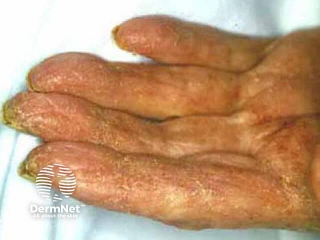
- Crusted (Norwegian) scabies: occurs in immunocompromised hosts (elderly, HIV/HTLV-1 infection, transplant recipients, those with impaired sensation/ability to scratch, e.g. leprosy or paraplegia). Presents as thick, hyperkeratotic, grayish-white plaques with sand-like scale, may involve the head/neck (usually spared in immunocompetent hosts), can be surprisingly non-pruritic despite carrying an enormous mite burden, and is highly contagious - Dermatology 2-Volume Set, 5e; Goldman-Cecil Medicine.
- Differential diagnosis includes eczema/dermatitis, contact dermatitis, psoriasis, drug eruptions, and seborrheic dermatitis (when face/scalp involved in immunosuppressed patients).
Diagnosis
Definitive diagnosis relies on dermoscopy or microscopic identification of mites, eggs, or fecal pellets (scybala) on KOH or mineral oil mounts of skin scrapings. PCR-based detection of S. scabiei DNA is also described - Dermatology 2-Volume Set, 5e.
Treatment
- First-line for classic scabies: topical permethrin 5% cream, applied twice one week apart (kills mites and eggs; repeat application needed since a single application may not kill newly hatched mites) - Harrison's Principles of Internal Medicine 22e; Red Book 2021; Textbook of Family Medicine 9e.
- Oral ivermectin (200 mcg/kg) is used for resistant, widespread, or crusted scabies, and in outbreak settings.
- Crusted scabies: difficult to treat, generally requires a combined regimen of topical permethrin plus repeated doses of oral ivermectin, with pre-application of a keratolytic agent (e.g., 6% salicylic acid) to help penetration through the thick scale - Harrison's Principles of Internal Medicine 22e; Dermatology 2-Volume Set, 5e.
- Malathion 0.5% lotion is an alternative first-line topical agent in some guidelines - Goldman-Cecil Medicine.
- All close contacts and household members should be treated simultaneously; bedding and clothing should be laundered.
- Complications: secondary bacterial superinfection, and in immunocompromised patients, potentially fatal septicemia.
Recent evidence (last 2 years)
Three relevant systematic reviews/meta-analyses were identified that may refine the above:
- A 2025 meta-analysis (PMID: 41282002) comparing oral ivermectin vs. benzyl benzoate for scabies treatment.
- A 2024 systematic review/meta-analysis on scabies treatment failure (PMID: 37625798, British Journal of Dermatology), which specifically examined failure rates with permethrin, ivermectin, malathion, and lindane - worth reviewing if treatment failure is a concern, as it may nuance the standard first-line recommendations above.
- A 2024 systematic review on risk factors for scabies transmission in hospitals/nursing homes (PMID: 38575893).
These don't overturn the textbook guidance but the treatment-failure review (37625798) is worth consulting directly if you're managing a case with poor response to standard permethrin therapy.
Let me know if you want me to go deeper into a specific aspect - diagnosis/dermoscopy, crusted scabies management, pediatric or pregnancy considerations, or outbreak control in institutional settings.
This is a shared conversation. Sign in to Orris to start your own chat.