
Generate image of middle cranial fossa and its approaches showing proper landmarks and important vital structures paasing through it in the image
Detailed anatomical diagram of the middle cranial fossa showing surgical approaches and key landmarks. The image should include: - The bony floor of the middle cranial fossa with labeled bones: greater wing of sphenoid, temporal squama, petrous part of temporal bone - Foramina and fissures clearly labeled: foramen ovale (mandibular nerve V3), foramen rotundum (maxillary nerve V2), foramen spinosum (middle meningeal artery), foramen lacerum, superior orbital fissure (CN III, IV, V1, VI), foramen lacerum - Important structures passing through: trigeminal nerve (CN V) with its divisions, middle meningeal artery and vein, greater and lesser petrosal nerves, internal carotid artery in carotid canal - Surgical approach landmarks: arcuate eminence (superior semicircular canal), tegmen tympani, petrous ridge, GSPN (greater superficial petrosal nerve) groove - Middle fossa surgical approach corridor shown with dashed lines - Meckel's cave / trigeminal impression - Hiatus of facial canal - Key anatomical relations: cochlea position, internal auditory canal (IAC) location relative to arcuate eminence - Color coding: nerves in yellow, arteries in red, veins in blue, bone in tan/beige - Clean medical illustration style with crisp labels and anatomical accuracy
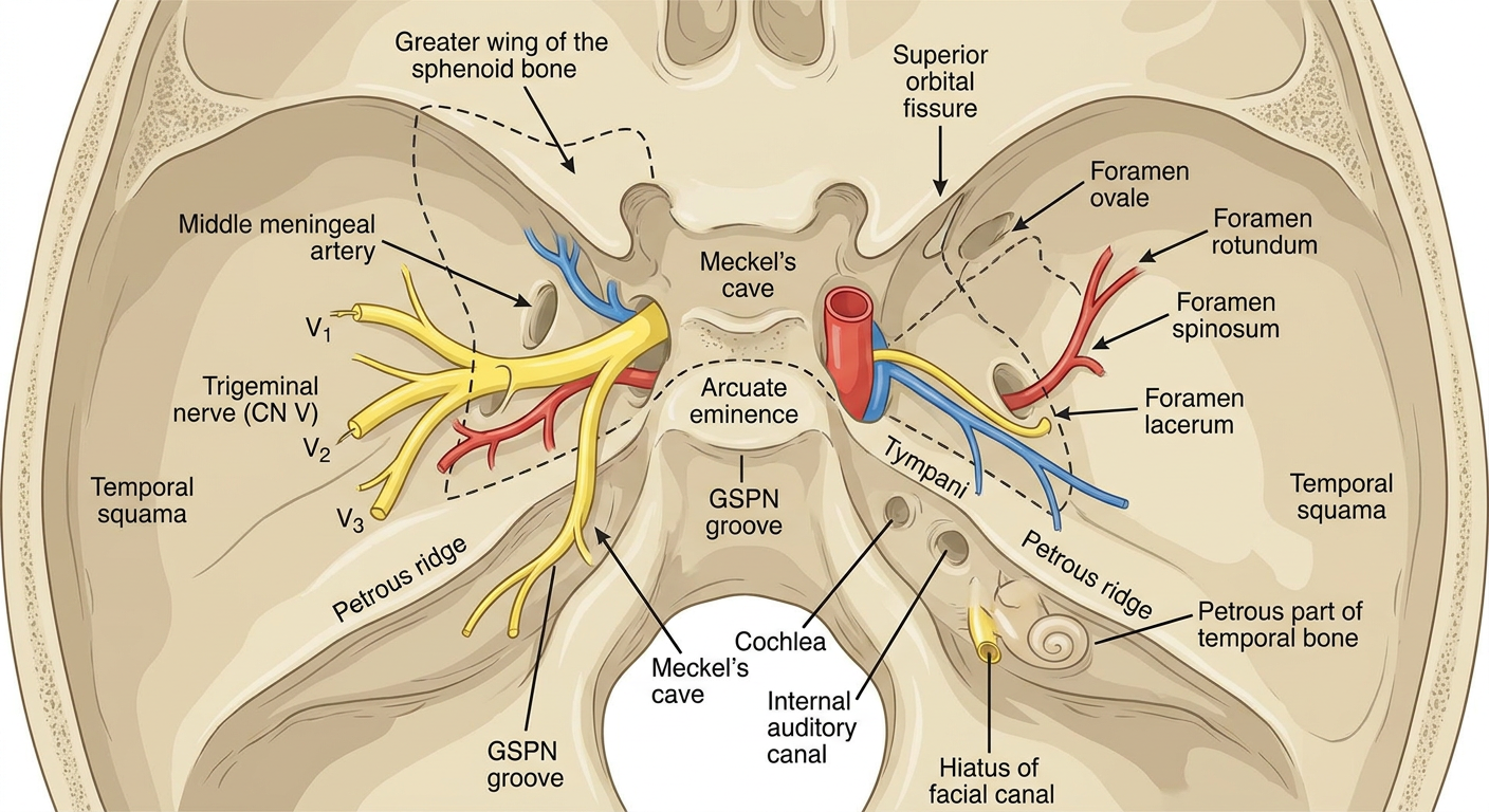
Foramina and Structures Passing Through
| Foramen / Opening | Structure(s) |
|---|---|
| Superior orbital fissure | CN III (oculomotor), CN IV (trochlear), CN V1 (ophthalmic), CN VI (abducens), ophthalmic veins |
| Foramen rotundum | CN V2 (maxillary nerve) |
| Foramen ovale | CN V3 (mandibular nerve), accessory meningeal artery |
| Foramen spinosum | Middle meningeal artery & vein |
| Foramen lacerum | Internal carotid artery (passes over it), greater petrosal nerve |
| Carotid canal | Internal carotid artery, sympathetic plexus |
| Hiatus of facial canal | Greater superficial petrosal nerve (GSPN) |
Key Surgical Landmarks (Middle Fossa Approach)
- Arcuate eminence - marks the superior semicircular canal; used to locate the IAC
- GSPN groove - leads to the geniculate ganglion and IAC
- Tegmen tympani - thin bone roof of the middle ear
- Petrous ridge - medial boundary of surgical corridor
- Meckel's cave - dural recess housing the trigeminal (Gasserian) ganglion
- Trigeminal impression - depression on the petrous apex for the trigeminal ganglion
Middle Fossa Surgical Approach (Kawase / House approach)
- Internal auditory canal (IAC) - acoustic neuroma, facial nerve tumors
- Petrous apex lesions
- Trigeminal neuralgia (Meckel's cave)
- Superior semicircular canal dehiscence repair