
body of corpus callosum vs fornix on mi brain coronal section
Reading File
The question is about neuroanatomy - specifically locating the body of corpus callosum vs the fornix on a coronal MRI brain section. This is a visual/anatomy question.
Searching sources...
Finding Sources
Finding Sources
Reading File
Reading File
Let me get the key section and images from Grainger & Allison specifically covering the limbic system/corpus callosum/fornix relationships on coronal MRI.
Reading File
Finding Sources
I found a very relevant image from Grainger & Allison (Fig. 53.7A - coronal T2 MR image showing corpus callosum, fornix, etc.) and a sagittal one showing both structures labeled. Let me fetch those images and look for a dedicated coronal section image showing the body of corpus callosum vs fornix.
Loading Image
Finding Sources
Let me find the specific coronal MRI image showing the body of corpus callosum vs fornix from the Neuroanatomy textbook (Fig 4.14B - coronal showing fornix body) and also get the Grainger & Allison coronal image.
Finding Sources
Reading File
Now let me fetch the key images - Fig 53.7 (coronal + sagittal showing corpus callosum body and fornix) and also look for the Neuroanatomy textbook coronal image (Fig 4.14B).
Loading Image
Finding Sources
Excellent! Now I have both key images. The sagittal MRI (Fig 53.7B) clearly labels the corpus callosum body (B), fornix (F), genu (G), rostrum (R), splenium (S), and massa intermedia (MI). Let me now look for a dedicated coronal MRI image that more specifically shows the spatial relationship between the body of corpus callosum and the fornix from the Neuroanatomy textbook.
Finding Sources
This is exactly what's needed - Gray's Anatomy for Students has a labeled diagram with corpus callosum (#1) and body of fornix (#3). Let me get that image.
Reading File
This is perfect - Gray's Anatomy for Students has eFig. 9.22 "Coronal Section of the Brain Through the Diencephalon" with numbered labels including corpus callosum (#1) and body of fornix (#3). Let me fetch that image.
Loading Image
This is exactly the image needed. Now I have everything to give a complete, well-illustrated answer. Let me also get the full label list for eFig. 9.22.
Reading File
Now I have all the information needed. Here is the complete answer:
Body of Corpus Callosum vs Fornix - Coronal MRI Section
On a coronal MRI through the brain at the level of the diencephalon, these two structures are close to each other in the midline but occupy distinct positions relative to the lateral ventricle. The key distinction is their vertical relationship:
Positional Relationship
| Structure | Position on Coronal Section |
|---|---|
| Body of Corpus Callosum | Superior - forms the roof of the body of the lateral ventricle; appears as a broad, horizontal white matter band arching across the midline at the top of the ventricle |
| Body of Fornix | Inferior - hangs below the corpus callosum, just beneath the septum pellucidum, appearing as a small rounded/oval white matter bundle at the midline |
How to Tell Them Apart on Coronal MRI
-
Corpus callosum body - broad, thick, flat band of white matter that forms the ceiling of the lateral ventricle. Appears low signal (dark) on T2, high signal on T1. Wider and more prominent.
-
Body of fornix - a smaller, more discrete bundle that runs just inferior to the corpus callosum and superior to the third ventricle. The two columns of the fornix hang side by side, separated by the thin septum pellucidum above and the third ventricle below. The fornix is suspended from the undersurface of the corpus callosum by the septum pellucidum.
Memory Tip
"Callosum is the Ceiling, Fornix hangs below" - on coronal sections, corpus callosum (#1) is the roof, septum pellucidum (#2) descends from it, and the body of the fornix (#3) is attached inferiorly.
Coronal Brain Section (Gray's Anatomy for Students, eFig. 9.22)
The image below is a labeled coronal section through the diencephalon. Key labels:
- 1 = Corpus callosum (the broad superior band)
- 2 = Septum pellucidum (thin midline sheet below it)
- 3 = Body of fornix (the small bundle just below the septum pellucidum)
- 5 = Body of lateral ventricle (the cavity between them and the thalamus)
- 6 = Thalamus
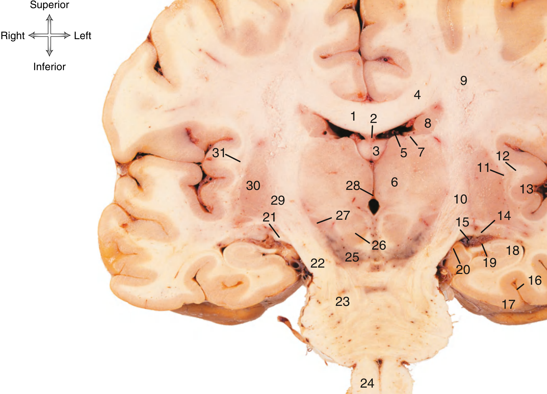
Gray's Anatomy for Students, eFig. 9.22 - Coronal Section Through the Diencephalon
MRI Coronal + Sagittal Reference (Grainger & Allison Radiology, Fig. 53.7)
The image below shows a coronal T2 MRI (A) and a sagittal T2 MRI (B). In the sagittal view, the corpus callosum body is labeled B, and the fornix is labeled F - note how the fornix runs inferior and parallel to the corpus callosum. The massa intermedia (MI) of the thalamus is also shown within the third ventricle.
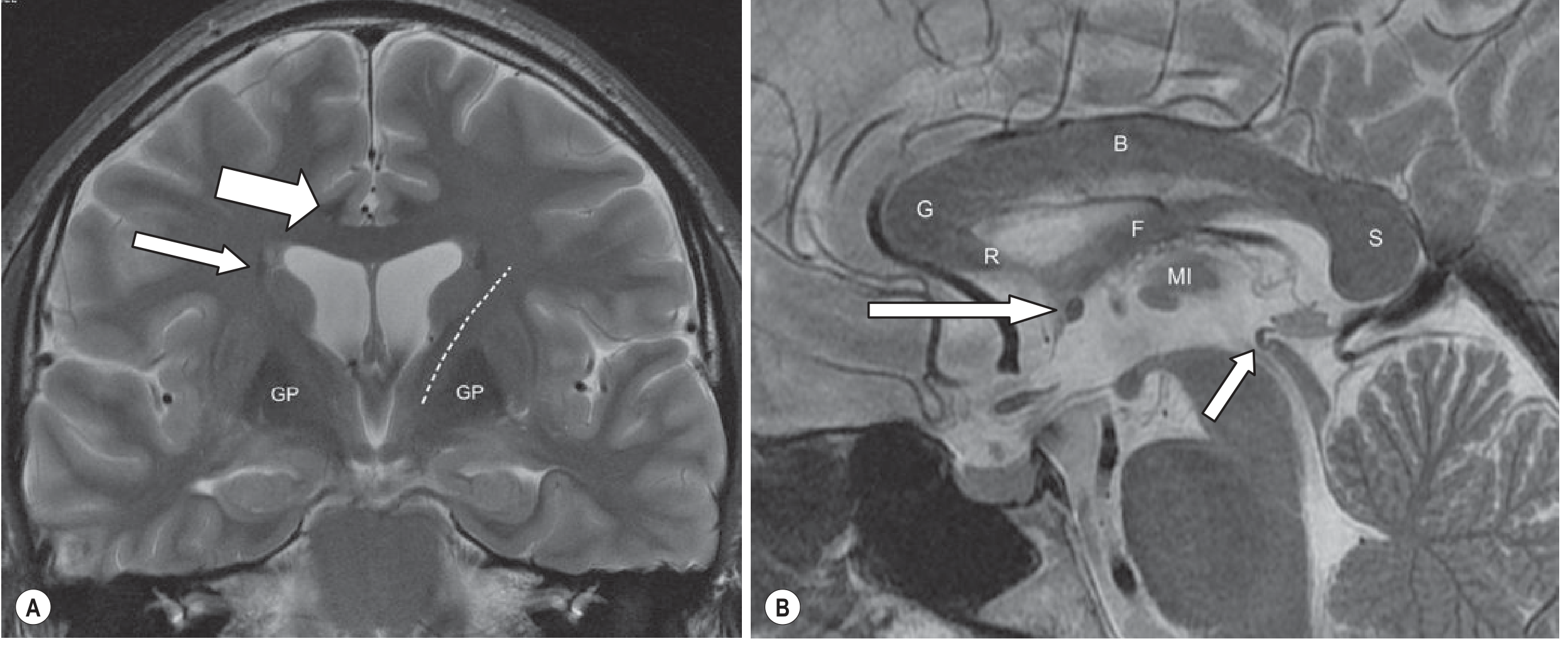
Grainger & Allison's Diagnostic Radiology, Fig. 53.7 - The body (B), fornix (F), and massa intermedia (MI) are labeled on the sagittal T2 image
Summary for Exam
On a coronal MRI:
-
The body of corpus callosum = superior midline structure, the broad white roof of the lateral ventricles
-
The body of fornix = inferior midline structure, a small rounded bundle just below the corpus callosum and septum pellucidum, superior to the third ventricle
-
The septum pellucidum lies between them, separating left from right lateral ventricle
-
The massa intermedia (interthalamic adhesion) is found within the third ventricle, just below the fornix at this level
-
Gray's Anatomy for Students, eFig. 9.22
-
Grainger & Allison's Diagnostic Radiology, Fig. 53.7
This is a shared conversation. Sign in to Orris to start your own chat.