
Joseph priestly , humphy davy, horacewells, Sir ivan whiteside magill, johnsnow, james young simpson, Crawford long, WTG Morton, Ralph waters , Sir Robert reynods macintosh, August karl gustav bier scientist in Anaesthesia - make a short notes with pictures in MD anaesthesia
Joseph Priestley oxygen discovery scientist portrait
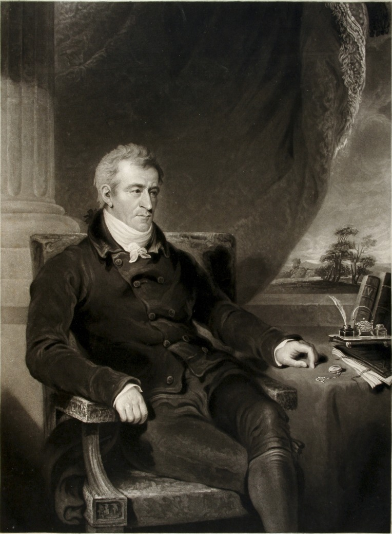
**Imaging Modality:** Historical medical portrait; mezzotint engraving. **Subject:** Joseph Constantine Carpue (1764–1846), a prominent English surgeon and anatomist. **Anatomical Focus:** Full-body seated portrait with emphasis on facial features and hands. **Clinical and Historical Context:** Carpue is a foundational figure in the history of plastic surgery, specifically renowned for performing the first successful "Indian method" rhinoplasty in the Western world (1814). He was also a noted teacher of anatomy and a pioneer in the medical application of electricity (galvanism). **Visual Features:** The subject is depicted in 19th-century professional attire, seated in a study environment. Key symbolic elements include a quill and inkwell, medical or anatomical texts, and a pocket watch, signifying scientific observation and scholarly contribution. The background features a classical column and an outdoor vista, typical of Georgian-era professional portraiture. **Key Diagnostic/Search Terms:** History of medicine, reconstructive surgery, rhinoplasty, British anatomy, 19th-century surgery, medical portraiture, Joseph Constantine Carpue.
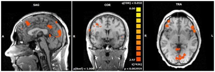
A functional MRI (fMRI) statistical map overlaid on a T1-weighted structural brain template, demonstrating Blood Oxygen Level Dependent (BOLD) activity in healthy subjects during a voluntary selection task. The image displays three standard planes: Sagittal (SAG), Coronal (COR), and Transverse/Axial (TRA). Significant activations are represented by a heat map ranging from orange to yellow, corresponding to t-values from 2.97 to 8.00 at a False Discovery Rate (q(FDR) < 0.05). In the sagittal view, increased BOLD responses are visible in the medial and lateral frontal regions, as well as the posterior cortex. The coronal and transverse views confirm bilateral but asymmetrical activation clusters, primarily located in the superior and middle frontal gyri (BA 6/8/9/46) and the dorsolateral prefrontal cortex. Distinct posterior activity is localized to the cuneus (BA 7/18/19) and the medial posterior regions. This clinical imaging set illustrates the functional neuroanatomy associated with the N2 event-related potential component, highlighting the cognitive control network involved in response selection.
Humphry Davy nitrous oxide laughing gas discovery
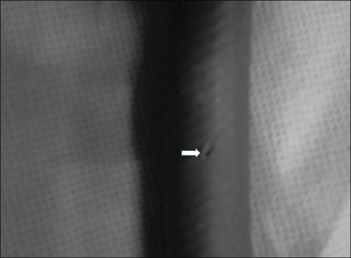
**Imaging Modality:** Clinical photograph (close-up) **Anatomical/Equipment Region:** Medical gas delivery system; specifically, a nitrous oxide pipeline/hose. **Observed Pathology/Abnormality:** Structural failure of a high-pressure medical gas pipeline characterized by localized dilation (aneurysmal bulging) and a discrete perforation. **Characteristic Visual Features:** * **Dilation:** The image shows a segment of a dark, flexible pipeline with a visible focal expansion or bulging, indicating a loss of structural integrity of the wall. * **Perforation:** A white arrow points directly to a small, longitudinal slit-like hole or rupture located within the center of the dilated area. * **Surface Texture:** The pipeline exhibits a cross-hatched or braided reinforced texture typical of medical-grade high-pressure hoses. **Clinical Context:** This represents a critical equipment failure in an anesthesia or perioperative setting. Such defects in a nitrous oxide pipeline can lead to gas leaks, loss of delivery pressure, and potential environmental exposure to anesthetic gases. **Key Diagnostic Features:** Focal wall thinning and rupture in a pressurized conduit, distinguished by the physical deformation (bulge) preceding the breach.
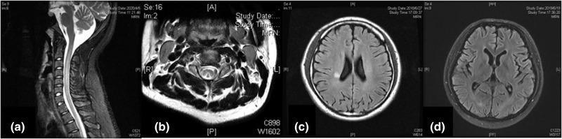
A multi-panel figure displaying diagnostic MRI scans related to central nervous system complications of nitrous oxide abuse, primarily illustrating subacute combined degeneration (SCD) of the spinal cord and cerebral changes. Panel (a) is a sagittal T2-weighted MRI of the cervical spine showing a continuous linear hyperintense signal within the posterior aspect of the spinal cord. Panel (b) shows an axial T2-weighted section of the cervical spinal cord demonstrating the classic 'inverted V sign' (also known as the rabbit ear sign), representing symmetric hyperintensity in the posterior columns (funiculus gracilis and cuneatus). Panel (c) is an axial T2-FLAIR brain MRI showing a focal hyperintense signal in the right corona radiata. Panel (d) is an axial T2-FLAIR brain MRI illustrating generalized cerebral atrophy, characterized by widened cortical sulci and enlarged lateral ventricles. These findings are characteristic of vitamin B12 deficiency-induced myelopathy and encephalopathy, often seen in the clinical context of chronic nitrous oxide exposure.
Horace Wells dentist nitrous oxide anaesthesia history
![Recommendation Table 47. Recommendations for volatile and intravenous anaesthetics, opioids and neuromuscular blocking agents
<table><thead><tr><th>Recommendations</th><th>Class<sup>a</sup></th><th>Level<sup>b</sup></th><th>Ref<sup>c</sup></th></tr></thead><tbody><tr><td>Oxygenator exhaust concentrations of volatile anaesthetic agents should be considered for monitoring during CPB.</td><td>IIa</td><td>B</td><td>[157, 621, 622]</td></tr><tr><td>It is recommended that approved equipment be used for delivering volatile anaesthetics during CPB.</td><td>I</td><td>C</td><td>[618, 619]</td></tr><tr><td>The administration of nitrous oxide immediately before and after CPB is not recommended.</td><td>III</td><td>B</td><td>[623, 624]</td></tr><tr><td>Volatile anaesthetic agents may be considered for the maintenance of anaesthesia during CPB.</td><td>IIb</td><td>B</td><td>[627-633]</td></tr><tr><td>During the maintenance phase of CPB, it should be considered to administer intravenous anaesthetics and opioids, except remifentanil, at a dose at least as high as prior to CPB.</td><td>IIa</td><td>B</td><td>[640, 642-646, 652-654]</td></tr><tr><td>Intravenous fentanyl administration by intermittent bolus or infusion may be considered.</td><td>IIb</td><td>C</td><td>[652, 653]</td></tr><tr><td>After the initiation of CPB and induction of hypothermia, reducing the dose of remifentanil after 20-30 min by 30% at 32°C, and immediately by 60% with moderate to deep hypothermia below 28°C, may be considered</td><td>IIb</td><td>B</td><td>[654]</td></tr><tr><td>The Minto model for remifentanil may be considered during CPB with normothermia or mild hypothermia, with the expectation that it may underestimate blood levels.</td><td>IIb</td><td>B</td><td>[662, 663]</td></tr></tbody></table>](https://cdn.orris.care/cdss_images/GLGCA_4542999_1766781081181_a906d53c-0c57-47e4-a317-cd50da18f86c_035cf636-4923-4c5a-a531-c25d9af6382a.png)
Recommendation Table 47. Recommendations for volatile and intravenous anaesthetics, opioids and neuromuscular blocking agents <table><thead><tr><th>Recommendations</th><th>Class<sup>a</sup></th><th>Level<sup>b</sup></th><th>Ref<sup>c</sup></th></tr></thead><tbody><tr><td>Oxygenator exhaust concentrations of volatile anaesthetic agents should be considered for monitoring during CPB.</td><td>IIa</td><td>B</td><td>[157, 621, 622]</td></tr><tr><td>It is recommended that approved equipment be used for delivering volatile anaesthetics during CPB.</td><td>I</td><td>C</td><td>[618, 619]</td></tr><tr><td>The administration of nitrous oxide immediately before and after CPB is not recommended.</td><td>III</td><td>B</td><td>[623, 624]</td></tr><tr><td>Volatile anaesthetic agents may be considered for the maintenance of anaesthesia during CPB.</td><td>IIb</td><td>B</td><td>[627-633]</td></tr><tr><td>During the maintenance phase of CPB, it should be considered to administer intravenous anaesthetics and opioids, except remifentanil, at a dose at least as high as prior to CPB.</td><td>IIa</td><td>B</td><td>[640, 642-646, 652-654]</td></tr><tr><td>Intravenous fentanyl administration by intermittent bolus or infusion may be considered.</td><td>IIb</td><td>C</td><td>[652, 653]</td></tr><tr><td>After the initiation of CPB and induction of hypothermia, reducing the dose of remifentanil after 20-30 min by 30% at 32°C, and immediately by 60% with moderate to deep hypothermia below 28°C, may be considered</td><td>IIb</td><td>B</td><td>[654]</td></tr><tr><td>The Minto model for remifentanil may be considered during CPB with normothermia or mild hypothermia, with the expectation that it may underestimate blood levels.</td><td>IIb</td><td>B</td><td>[662, 663]</td></tr></tbody></table>
A multi-panel figure displaying diagnostic MRI scans related to central nervous system complications of nitrous oxide abuse, primarily illustrating subacute combined degeneration (SCD) of the spinal cord and cerebral changes. Panel (a) is a sagittal T2-weighted MRI of the cervical spine showing a continuous linear hyperintense signal within the posterior aspect of the spinal cord. Panel (b) shows an axial T2-weighted section of the cervical spinal cord demonstrating the classic 'inverted V sign' (also known as the rabbit ear sign), representing symmetric hyperintensity in the posterior columns (funiculus gracilis and cuneatus). Panel (c) is an axial T2-FLAIR brain MRI showing a focal hyperintense signal in the right corona radiata. Panel (d) is an axial T2-FLAIR brain MRI illustrating generalized cerebral atrophy, characterized by widened cortical sulci and enlarged lateral ventricles. These findings are characteristic of vitamin B12 deficiency-induced myelopathy and encephalopathy, often seen in the clinical context of chronic nitrous oxide exposure.
Crawford Long ether anaesthesia first use history
A series of weight-bearing and non-weight-bearing radiographs showing the surgical management of Crawford type IV congenital pseudarthrosis of the tibia (CPT) in a pediatric patient. Panels (a) and (b) represent the preoperative state, showing significant anterolateral bowing and mid-diaphyseal pseudarthrosis of the tibia with an intact fibula. Panels (c) and (d) are postoperative images at 2 months, demonstrating an Ilizarov circular external fixator applied for stability and compression; the frame consists of rings connected by threaded rods and tensioned Kirschner wires. An intramedullary rod is also visible. Panels (e) and (f) show the long-term follow-up at 5.4 years post-operation, highlighting successful solid bony union, restoration of anatomical alignment, and a persistent intramedullary rod. The images demonstrate the absence of ankle valgus or significant limb-length discrepancy (LLD), marking effective treatment of pediatric tibial dysplasia through combined internal fixation and circular external distraction/compression osteogenesis.
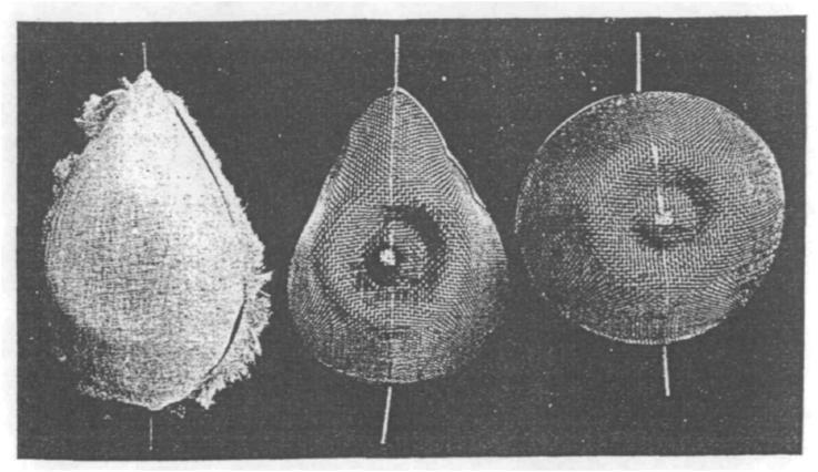
This historical clinical image, originally published in 1923, depicts three variations of ether masks used in early 20th-century anesthesia. The masks are shown in a comparison layout, illustrating the evolution of anesthetic equipment. The left-most mask has a pear-like, teardrop shape with a visible fibrous, lint-like texture along its lateral margins, suggesting a primitive design for holding liquid ether. The middle mask demonstrates an 'improved' teardrop design attributed to S.R. Wilson, featuring a more structured wire mesh or woven lattice with concentric rings and a central depression to facilitate even vapor distribution. The right-most mask is a circular variant with a similar fine-mesh construction and a central indentation. Each device is mounted on a vertical handling rod. These masks represent the transition in clinical practice from simple gauze-and-frame methods to more engineered designs intended to optimize the concentration and delivery of volatile anesthetic agents like ether and chloroform. This visual is significant for medical history and the study of the development of the specialty of anesthesiology.
William Morton ether anaesthesia Massachusetts General Hospital 1846

<table> <tr> <th>Name</th> <th>Employment</th> <th>Representation in Peer Review Process</th> </tr> <tr> <td>G. William Dec</td> <td>Massachusetts General Hospital—Chief Emeritus, Cardiology Division and Roman W. DeSanctis Professor of Medicine</td> <td>Content Reviewer, Former ACC Competency Management Committee Member</td> </tr> <tr> <td>Douglas E. Drachman</td> <td>Massachusetts General Hospital, Division of Cardiology—Heart Center Director of Education</td> <td>Content Reviewer, Interventional Cardiology Program Director</td> </tr> <tr> <td>Bailey Ann Estes</td> <td>Hendrick Medical Center—Cardiac Catheterization Laboratory Nurse and Research Coordinator</td> <td>Content Reviewer, ACC Cardiovascular Team Section Leadership Council</td> </tr> <tr> <td>Kendra J. Grubb</td> <td>Emory University—Associate Professor of Surgery and Medicine; Surgical Director Structural Heart and Valve Center</td> <td>Content Reviewer, ACC Cardiac Surgery Team Section Leadership Council</td> </tr> <tr> <td>Rebecca T. Hahn</td> <td>Columbia Structural Heart and Valve Center—Chief Scientific Officer of the Echocardiography Core Lab at the Cardiovascular Research Foundation and Director of Interventional Echocardiography; Columbia University Irving Medical Center—Professor of Medicine</td> <td>Content Reviewer, ACC Competency Management Committee</td> </tr> <tr> <td>Uzoma Ibebuogu</td> <td>Methodist University Hospital—Director of Structural Heart Disease Intervention; University of Tennessee Health Sciences Center, Division of Cardiovascular Diseases—Associate Professor of Medicine</td> <td>Content Reviewer, ACC Interventional Cardiology Section Leadership Council</td> </tr> <tr> <td>Ahesqal Islam</td> <td>University of Massachusetts Chan Medical School-Baystate—Program Director, Interventional Cardiology Fellowship</td> <td>Content Reviewer, Interventional Cardiology Program Director</td> </tr> <tr> <td>Henry S. Jennings III</td> <td>Vanderbilt University Medical Center, Vanderbilt Heart & Vascular Institute/Interventional Cardiology Section—Assistant Professor of Medicine</td> <td>Content Reviewer, ACC Cardiovascular Imaging Section Leadership Council</td> </tr> <tr> <td>Tara L. Jones</td> <td>University of Utah—Assistant Professor of Medicine, Interventional Cardiology</td> <td>Content Reviewer, Individual Contributor</td> </tr> <tr> <td>Sabeeda Kadavath</td> <td>Vanderbilt University—Structural Heart Disease Fellow</td> <td>Content Reviewer, ACC Interventional Cardiology Section Leadership Council</td> </tr> <tr> <td>Clifford J. Kavinsky</td> <td>Rush University Medical Center—Professor of Medicine and Pediatrics, Chief, Section of Structural and Interventional Cardiology, Director, Rush Center for Congenital and Structural Heart Disease, Director, Interventional Fellowship</td> <td>Content Reviewer, ACC Interventional Cardiology Section Leadership Council</td> </tr> <tr> <td>Sadiya S. Khan</td> <td>Northwestern University Feinberg School of Medicine—Assistant Professor of Medicine (Cardiology) and Preventive Medicine (Epidemiology)</td> <td>Content Reviewer, Former ACC Competency Management Committee Member</td> </tr> <tr> <td>Rami Khouzam</td> <td>University of Tennessee Health Science Center—Professor of Medicine, Vice Chair, Internal Medicine Department, Program Director, Interventional Cardiology, Associate Program Director, Cardiology Fellowship; Methodist University Hospital and Methodist Le Bonheur Healthcare System—Director, Cardiac Catheterization Laboratory</td> <td>Content Reviewer, Interventional Cardiology Program Director</td> </tr> <tr> <td>Viet Le</td> <td>Intermountain Healthcare—Cardiology Research PA</td> <td>Content Reviewer, ACC Cardiovascular Team Section Leadership Council</td> </tr> <tr> <td>Alexander Lee</td> <td>Northwell Health - Long Island Jewish Medical Center—Program Director, Interventional Cardiology Fellowship</td> <td>Content Reviewer, Interventional Cardiology Program Director</td> </tr> <tr> <td>Stamatios Lerakis</td> <td>Mount Sinai Hospital—Director of Noninvasive Cardiology; Icahn School of Medicine at Mount Sinai—Director of Imaging for Structural and Valve Interventions</td> <td>Content Reviewer, ACC Cardiovascular Imaging Section Leadership Council</td> </tr> <tr> <td>Ryan Mallory</td> <td>Indiana University—Cardiology Fellow in Training</td> <td>Content Reviewer, ACC Fellows in Training Section Leadership Council</td> </tr> <tr> <td>John Moulow</td> <td>Methodist Cardiology Clinic of San Antonio—Cardiologist</td> <td>Content Reviewer, ACC Geriatric Cardiology Section Leadership Council</td> </tr> <tr> <td>Abhiram Prasad</td> <td>Mayo Clinic, Department of Cardiovascular Diseases—Consultant, Program Director, Interventional Cardiology Fellowship; Mayo Clinic College of Medicine—Professor of Medicine</td> <td>Content Reviewer—Interventional Cardiology Program Director</td> </tr> <tr> <td>Tanveer Rab</td> <td>Emory University—Professor of Medicine, Interventional Cardiology</td> <td>Content Reviewer, ACC Interventional Cardiology Section Leadership Council</td> </tr> <tr> <td>Michael Ragosta</td> <td>University of Virginia Health System—Professor of Medicine, Director, Cardiac Catheterization Laboratories</td> <td>Content Reviewer, Interventional Cardiology Program Director</td> </tr> <tr> <td>Bharath Rajagopalan</td> <td>Prairie Heart Institute—Cardiac Electrophysiologist</td> <td>Content Reviewer, ACC Electrophysiology Section Leadership Council</td> </tr> <tr> <td>Robert F. Riley</td> <td>Overlake Medical Center—Director, Complex Coronary Therapeutics Program</td> <td>Content Reviewer, ACC Interventional Cardiology Section Leadership Council</td> </tr> <tr> <td>Shubha Deep Roy</td> <td>University of Iowa Hospitals and Clinics—Interventional Cardiology Fellow, Division of Cardiovascular Medicine, Department of Internal Medicine, University of Iowa Hospitals and Clinics</td> <td>Content Reviewer, ACC Fellows in Training Section Leadership Council</td> </tr> <tr> <td>Matthew Sherwood</td> <td>Inova Heart and Vascular Institute—Co-Director of Structural Heart Program, Co-Director of Cardiac Catheterization Laboratory</td> <td>Content Reviewer, ACC Interventional Section Leadership Council</td> </tr> <tr> <td>Adhir Shroff</td> <td>University of Illinois College of Medicine at Chicago—Interventional Cardiology Program Director; Professor of Medicine</td> <td>Content Reviewer, Interventional Cardiology Program Director</td> </tr> <tr> <td>Scott Shurmur</td> <td>Texas Tech University Health Sciences Center—Chairman, Internal Medicine; Division Chief, Cardiology; Interventional Cardiology Program Director; Professor</td> <td>Content Reviewer, Interventional Cardiology Program Director</td> </tr> <tr> <td>Michael A. Solomon</td> <td>National Institutes of Health Clinical Center—Senior Research Physician</td> <td>Content Reviewer, ACC Competency Management Committee Member</td> </tr> <tr> <td>Anwar Tandar</td> <td>University of Utah School of Medicine—Director of Structural Cardiac Intervention; Associate Professor of Medicine</td> <td>Content Reviewer, Interventional Cardiology Program Director</td> </tr> </table>

<table><tr><td>Theofanie Mela, MD</td><td>Massachusetts General Hospital,<br>Boston,<br>Massachusetts</td><td>1: Abbott<br>1: Biotronik<br>1: Medtronic</td><td>None</td><td>None</td><td>None</td><td>None</td><td>None</td><td>None</td><td>None</td></tr></table>
James Young Simpson chloroform anaesthesia obstetrics history
This medical figure illustrates the application of the Single Plane Simpson's method (disk summation) for calculating left ventricular (LV) volume from ultrasound imaging. Image (a) is a 2D echocardiographic ultrasound frame in a grayscale sectoral view. The left ventricle is highlighted by a yellow solid contour tracing the endocardial border. Within this contour, a series of parallel red horizontal lines represent the individual axes used to divide the LV cavity into discrete cylindrical disks. Image (b) presents a corresponding 3D volumetric model rendered in solid red. This model visualizes the LV as a stack of short cylinders, creating a truncated conical shape that tapers from the base toward the apex. The horizontal segments in the 3D model demonstrate the additive geometry used to estimate total volume. This visualization is used in cardiology and obstetrics for assessing cardiac function parameters such as stroke volume (SV) and ejection fraction (EF) by modeling the complex geometry of the heart more accurately than simpler elliptical assumptions.
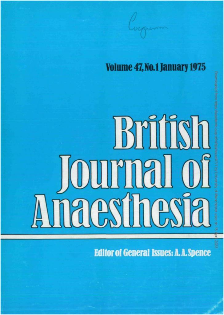
Educational image of a historical medical journal cover. The visual features a vibrant cyan-blue background, identifying this as the January 1975 issue (Volume 47, No. 1) of the 'British Journal of Anaesthesia'. The title is displayed in prominent, white, serif typeface with black outlines. A horizontal white line separates the main title from the lower section, which identifies A.A. Spence as the 'Editor of General Issues'. This specific design represents a significant transition in the journal's publication history when Macmillan Journals Limited took over production, introducing a color-coded system for different volumes and moving the table of contents to the back cover. The image serves as a visual record of the evolution of medical publishing within the specialty of anesthesiology.
John Snow chloroform anaesthesia epidemiology history pioneer
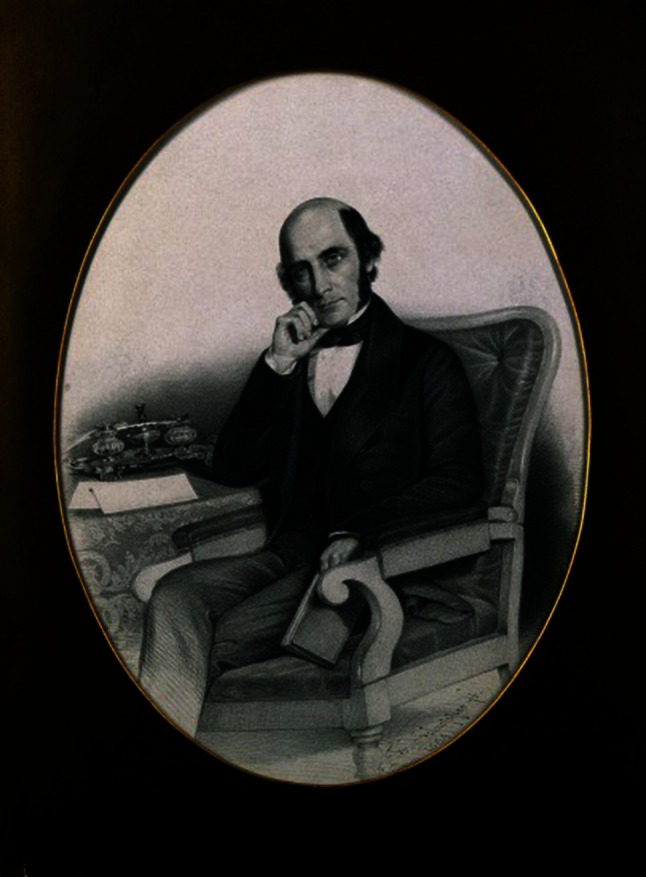
**Modality:** Historical medical portrait (lithograph/stipple engraving). **Subject:** Portrait of William John Little (1810–1894), a pioneer in orthopedic surgery and the formal identification of cerebral palsy (historically referred to as "Little’s Disease"). **Composition and Details:** The image is a mid-19th-century oval-framed portrait depicting a male figure in a seated, reflective pose. He is dressed in formal Victorian-era professional attire, including a dark frock coat, waistcoat, and high-collared shirt with a necktie. **Relevant Clinical and Historical Context:** * **Anatomical/Clinical Focus:** Little is renowned for his work on congenital and acquired deformities, specifically clubfoot (talipes equinovarus) and the neurological sequelae of neonatal asphyxia (spastic diplegia). * **Symbolic Elements:** The subject is seated at a desk featuring writing implements (inkwell and paper), signifying his role as a medical scholar and author of seminal texts such as *On the Deformities of the Human Frame*. **Database Indexing Keywords:** Medical History, Orthopedic Surgery, Cerebral Palsy, Little’s Disease, 19th-century Physician, Neurological History, William John Little.
Educational image of a historical medical journal cover. The visual features a vibrant cyan-blue background, identifying this as the January 1975 issue (Volume 47, No. 1) of the 'British Journal of Anaesthesia'. The title is displayed in prominent, white, serif typeface with black outlines. A horizontal white line separates the main title from the lower section, which identifies A.A. Spence as the 'Editor of General Issues'. This specific design represents a significant transition in the journal's publication history when Macmillan Journals Limited took over production, introducing a color-coded system for different volumes and moving the table of contents to the back cover. The image serves as a visual record of the evolution of medical publishing within the specialty of anesthesiology.
Ralph Waters anaesthesia CO2 absorption soda lime circle system
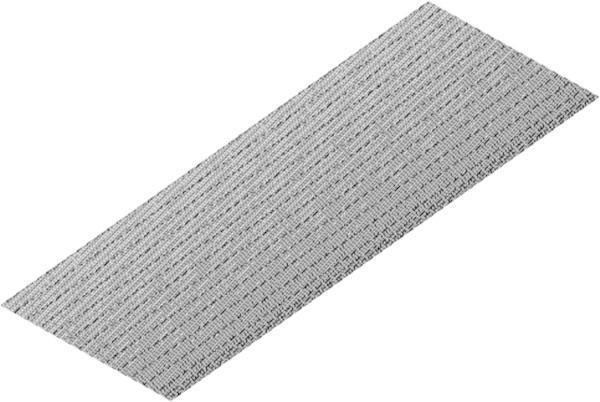
This diagnostic and engineering schematic illustrates a 1-degree axial scrubber segment utilized in Closed Circuit Rebreather (CCR) systems for CO2 absorption. The image depicts a computational fluid dynamics (CFD) model of the scrubber's internal mesh structure, specifically showing a structured hexahedron (hex) grid pattern with an edge sizing of 400. The visualization displays a highly uniform and dense mesh composed of repeating rectangular elements aligned along the longitudinal axis of the canister slice. This modeling technique is used in medical and safety research to simulate exothermic chemical reactions between CO2 and soda lime, as well as to analyze laminar flow and heat transfer within the porous medium of the scrubber. The high-density grid is critical for ensuring numerical convergence and accurate temperature prediction during the simulation of the respiratory gas purification process.
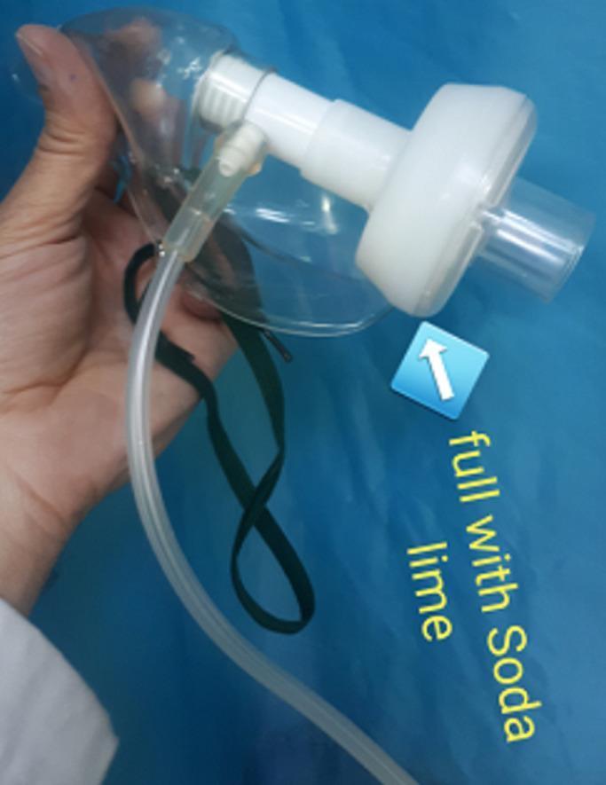
A clinical photograph displaying a modified respiratory face mask assembly designed for non-invasive ventilation. The apparatus consists of a clear plastic oxygen mask with dark elastic head straps and a flexible oxygen supply tube. Central to the modification is a white, rigid plastic cylindrical housing connected directly to the mask's main port. This housing is annotated as being 'full with Soda lime', acting as a carbon dioxide (CO2) absorbent filter within the expiratory circuit. This clinical setup is intended to mitigate hypercapnia in patients, such as those with severe COVID-19, by reducing the re-breathing of CO2. The image demonstrates a specialized adaptation of standard medical equipment for advanced respiratory support, focusing on preventing the need for invasive tracheal intubation through effective dead-space gas management.
spinal anaesthesia history regional block technique
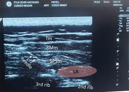
This diagnostic ultrasound image demonstrates a Serratus Posterior Superior Intercostal Plane (SPSIP) block, an ultrasound-guided regional anaesthesia technique. The scan, captured in the sagittal plane along the medial border of the scapula, shows the layered musculature of the upper back. From superficial to deep, the labeled structures include the trapezius muscle (Tm), rhomboid major muscle (RMm), and the serratus posterior superior muscle (SPSm). Deep to these muscles, the hyperechoic surfaces of the 2nd and 3rd ribs are visible with characteristic acoustic shadowing. A needle is visualized approaching the fascial plane. A hypoechoic (dark) oval region labeled 'LA' indicates the successful infiltration and spread of local anaesthetic between the serratus posterior superior muscle and the underlying intercostal space/ribs. This image serves as a clinical guide for identifying target anatomical planes for postoperative analgesia in thoracic or shoulder surgeries.
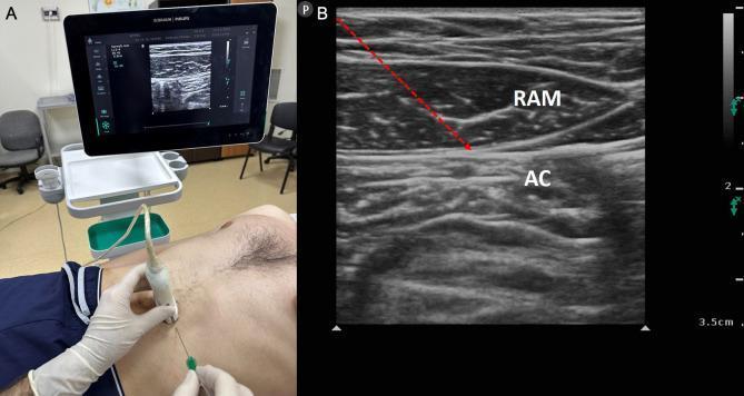
This dual-panel image demonstrates the clinical and sonographic components of a rectus sheath block (RSB), a regional anesthesia technique targeting T7-T12 spinal nerve endings. Panel A is a clinical photograph showing a patient in the supine position for a mid-abdominal intervention. A clinician, wearing sterile gloves, is performing the procedure using a high-frequency linear ultrasound probe placed in a transverse orientation over the rectus abdominis muscle. A block needle is visible being inserted in-plane toward the target site. An ultrasound monitor in the background displays the real-time procedural imaging. Panel B is a corresponding musculoskeletal ultrasound image (sonogram) labeling the relevant anatomy. The rectus abdominis muscle (RAM) is identified as a hypoechoic, spindle-shaped structure with internal fibrillar striations. Deep to the RAM is the abdominal cavity (AC), appearing heterogeneous and hyperechoic due to bowel interfaces. A red dashed arrow indicates the needle's trajectory, targeting the potential space between the posterior aspect of the rectus muscle and the posterior rectus sheath to deliver local anesthetic.
pioneers of anaesthesia history Priestley Davy Wells Morton Simpson Long Magill Snow Macintosh Waters Bier contributions
https://aneskey.com/the-history-of-anesthesia-3
Macintosh laryngoscope blade intubation anaesthesia airway equipment
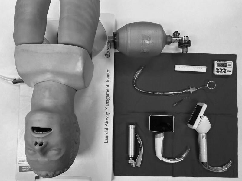
A high-angle clinical photograph displays standard and advanced airway management equipment for endotracheal intubation, positioned alongside a Laerdal Airway Management Trainer mannequin. The equipment includes three types of laryngoscopes: a conventional Macintosh-style laryngoscope with a metal handle and curved blade, and two video laryngoscopes featuring integrated digital screens and specialized curved blades for enhanced glottic visualization. Also present is a Bag Valve Mask (BVM) for manual ventilation. Centrally located is a clear cuffed endotracheal tube (ETT) with an internal stylet bent into a 'hockey-stick' configuration to facilitate insertion; the ETT includes a visible pilot balloon, connector, and depth markings. Ancillary procedural tools include a ruler for measurement and a digital timer for monitoring intubation time. This setup is typical for anesthesia training, emergency medicine simulation, or comparative studies between direct and video laryngoscopy techniques.
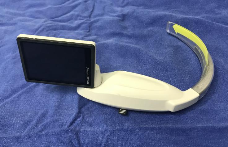
This clinical photograph displays a McGrath MAC video laryngoscope, an essential airway management tool used for endotracheal intubation. The device consists of a white, ergonomically contoured handle and a built-in rectangular LCD screen with a black bezel labeled 'McGrath® MAC'. Attached to the handle is a curved, Macintosh-style blade. The blade assembly features a metallic core encased in a clear, disposable transparent plastic sheath, which is characterized by a prominent yellow directional arrow pointing toward the distal tip. The device is shown resting on a blue surgical drape, highlighting its role in anesthesiology and emergency medicine. This visual is designed to educate medical professionals on the physical components and assembly of video laryngoscopy equipment used to improve laryngeal visualization during difficult airway scenarios.
Pioneers of Anaesthesia - Short Notes for MD Anaesthesia
A chronological overview of the 11 founding figures of modern anaesthesia, covering their life, key contributions, and legacy.
1. Joseph Priestley (1733-1804)
| Feature | Detail |
|---|---|
| Nationality | British (English) |
| Profession | Clergyman, natural philosopher, chemist |
| Key Discovery | Nitrous oxide (1773), Oxygen (co-discovered 1774), also isolated NO, HCl, NH3, CO |
- Prepared nitrous oxide (N2O) for the first time in 1773 by heating ammonium nitrate
- Discovered oxygen independently in 1774 (alongside Carl Wilhelm Scheele and Antoine Lavoisier)
- Published his findings in Experiments and Observations on Different Kinds of Air (6 volumes, from 1775)
- Had no formal scientific training yet described the properties of multiple gases systematically
- Did not use N2O clinically - that step came later with Humphry Davy
2. Sir Humphry Davy (1778-1829)
| Feature | Detail |
|---|---|
| Nationality | British (English) |
| Profession | Chemist, inventor |
| Key Work | First to suggest N2O for pain relief (1800) |
- In 1795, building on Priestley's work, Davy isolated, contained, and inhaled nitrous oxide
- Coined the term "laughing gas"
- Published Researches, Chemical and Philosophical, Chiefly Concerning Nitrous Oxide (1800)
- Wrote: "As nitrous oxide in its extensive operation appears capable of destroying physical pain, it may probably be used with advantage during surgical operations" - the first published suggestion of inhalational anaesthesia
- Despite this insight, N2O was not used clinically for another 44 years
- Davy was more focused on recreational/social use with the "laughing gas parties" popular among artists and intellectuals of the era (Coleridge, Wordsworth were attendees)
3. Horace Wells (1815-1848)
| Feature | Detail |
|---|---|
| Nationality | American |
| Profession | Dentist |
| Key Contribution | First clinical use of N2O for dental pain (1844) |
- Witnessed a "laughing gas frolic" in December 1844 where a participant injured himself but felt no pain
- Concluded N2O could be used for painless tooth extraction
- Tested it on himself (had a tooth pulled the next day with N2O - described it as "no more pain than a pinprick")
- Organized a public demonstration at Massachusetts General Hospital (MGH) in January 1845 - this failed when the patient cried out (likely under-dosed), and Wells was hissed out of the theater
- Disillusioned, Wells later experimented with chloroform, became addicted, and self-administered it while in prison (arrested for assaulting people while intoxicated with chloroform)
- Died in 1848 by self-inflicted chloroform poisoning while in custody
4. Crawford Williamson Long (1815-1878)
| Feature | Detail |
|---|---|
| Nationality | American |
| Profession | Physician/Surgeon, rural Georgia |
| Key Contribution | First documented use of ether anaesthesia (March 30, 1842) |
- On March 30, 1842, in Jefferson, Georgia, Long administered diethyl ether to James Venable and removed a neck cyst - this is the first recorded use of surgical ether anaesthesia
- Was familiar with "ether frolics" (recreational ether parties) common in the American South at the time
- Used ether in 8 more surgical cases before Morton's famous 1846 demonstration
- Did not publish his findings until 1849 (prompted by Morton's priority claim) - this is why he lost credit historically
- Continued practicing medicine and administering anaesthesia until his death in 1878 - he died while administering ether to a woman in labor
5. William Thomas Green Morton (1819-1868)
| Feature | Detail |
|---|---|
| Nationality | American |
| Profession | Dentist, then medical student |
| Key Contribution | First public demonstration of ether anaesthesia (October 16, 1846) |
- On October 16, 1846 (now known as "Ether Day"), Morton administered diethyl ether to Edward Gilbert Abbott at MGH
- Surgeon John Collins Warren then removed a jaw tumor and declared: "Gentlemen, this is no humbug"
- October 16 is now celebrated as World Anaesthesia Day
- The operating theater at MGH where this occurred is called the "Ether Dome"
- Morton tried to patent ether under the name "Letheon" (a colored, scented form), which created controversy
- Became embroiled in the bitter "Ether Controversy" with Wells, Long, and chemist Charles Jackson over priority
- Died in 1868 at age 48 of a probable stroke while traveling to New York to contest an article crediting Jackson
6. James Young Simpson (1811-1870)
| Feature | Detail |
|---|---|
| Nationality | Scottish |
| Profession | Obstetrician, Professor at Edinburgh |
| Key Contribution | Introduced chloroform as anaesthetic agent (1847) |
- Aware of ether's disadvantages (pungent smell, slow induction, nausea), Simpson systematically tested new volatile agents
- On November 4, 1847, Simpson and his assistants inhaled chloroform at a dinner party - they awoke from under the table having been rendered unconscious
- Simpson introduced chloroform for obstetric pain relief - first used in a difficult breech delivery (a boy was named "Anaesthesia" in gratitude)
- Championed obstetric anaesthesia against fierce religious opposition (the "Biblical argument" - women must suffer in childbirth per Genesis)
- His famous rebuttal: "And the Lord God caused a deep sleep to fall upon Adam...and He took one of his ribs" (Genesis 2:21) - arguing God himself performed the first anaesthetic
- Administered chloroform to Queen Victoria during the births of Prince Leopold (1853) and Princess Beatrice (1857) - this gave anaesthesia a "royal seal of approval" and silenced critics
- Invented the Simpson's forceps (obstetric forceps)
- The Simpson lithotrite (for urinary stones)
7. John Snow (1813-1858)
| Feature | Detail |
|---|---|
| Nationality | British (English) |
| Profession | Physician - considered the world's first professional anaesthetist AND father of epidemiology |
| Key Contributions | Standardized ether/chloroform delivery; mapped the 1854 Broad Street cholera outbreak |
- After ether was introduced to the UK in 1846, Snow immediately began systematizing its use
- Developed a calibrated ether inhaler that delivered controlled, measured concentrations (opposed to the haphazard practice of the day)
- Wrote On the Inhalation of the Vapour of Ether (1847) - the first scientific monograph on anaesthesia
- Described 5 stages of ether anaesthesia (precursor to Guedel's signs)
- When chloroform replaced ether, he adapted and wrote On Chloroform and Other Anaesthetics (1858, published posthumously)
- Administered chloroform to Queen Victoria for the births of Prince Leopold (1853) and Princess Beatrice (1857)
- Insisted on monitoring, controlled dosage, and pulse observation - principles of modern anaesthetic practice
- During the 1854 Soho cholera outbreak, mapped cases to a single water pump on Broad Street - convincingly demonstrated water-borne transmission of cholera (predating Pasteur's germ theory)
- The Broad Street pump handle was removed; the epidemic subsided
8. Sir Ivan Whiteside Magill (1888-1986)
| Feature | Detail |
|---|---|
| Nationality | British-Irish |
| Profession | Anaesthetist |
| Key Contribution | Pioneered endotracheal intubation and awake nasal intubation; invented Magill forceps |
- In 1919 at Queen Mary's Hospital, Sidcup (working with Harold Gillies on facial reconstruction for WWI casualties), Magill (alongside Stanley Rowbotham) developed techniques for blind nasal intubation to keep the airway clear while the surgeon operated on the face
- Developed the Magill tube - a plain rubber endotracheal tube with a curve suited to nasal passage
- Invented the Magill forceps - angled forceps used to guide a nasotracheal tube under direct laryngoscopy - still in routine use today
- Described the technique of awake blind nasal intubation - the gold standard for difficult airways before fibreoptic scopes
- Developed the Magill attachment - a breathing circuit T-piece modification for spontaneously breathing patients (Mapleson A circuit is also known as the Magill circuit)
- Elected Fellow of the Royal College of Surgeons; became the first president of the Association of Anaesthetists of Great Britain and Ireland (AAGBI) in 1932
- Knighted in 1960
9. Ralph Milton Waters (1883-1979)
| Feature | Detail |
|---|---|
| Nationality | American |
| Profession | Anaesthesiologist |
| Key Contributions | To-and-fro CO2 absorption system; first academic anaesthesia department; cyclopropane |
- Developed the "to-and-fro" CO2 absorption system using soda lime (1924) - this allowed anaesthetic gases to be recycled and rebreathed, reducing waste and cost, and enabling low-flow anaesthesia
- The to-and-fro canister directly preceded the development of the modern circle system (developed by Brian Sword in 1926, building on Waters' work)
- Introduced cyclopropane anaesthesia into clinical practice (1934)
- In 1927, established the first academic department of anaesthesia in the world at the University of Wisconsin, Madison - this transformed anaesthesia from a nursing/technical task into a medical specialty
- Trained numerous anaesthesiologists who went on to lead departments across the USA and world - the "Wisconsin school" shaped modern academic anaesthesia
- Introduced the concept of anaesthesia as a medical specialty requiring academic training and research
10. Sir Robert Reynolds Macintosh (1897-1989)
| Feature | Detail |
|---|---|
| Nationality | New Zealand-born, British by adoption |
| Profession | Anaesthesiologist, Oxford |
| Key Contribution | Invented the Macintosh laryngoscope blade (1943) |
- In 1937, appointed as the world's first Professor of Anaesthetics at Oxford University - a landmark in the recognition of anaesthesia as a full academic discipline
- Invented the curved (Macintosh) laryngoscope blade in 1943 - designed to fit into the vallecula and lift the epiglottis indirectly, providing better visualization while being safer and more comfortable than straight blades
- The Macintosh blade remains the most widely used laryngoscope blade in the world
- Wrote Essentials of General Anaesthesia (1940) and Physics for the Anaesthetist (co-authored with Mushin) - textbooks that shaped generations of trainees
- During World War II, served as anaesthesia consultant to the RAF and British forces in Spain (Spanish Civil War) - gained extraordinary clinical experience
- Was known for emphasizing safety, simplicity, and careful technique
11. August Karl Gustav Bier (1861-1949)
| Feature | Detail |
|---|---|
| Nationality | German |
| Profession | Surgeon |
| Key Contributions | Invented spinal anaesthesia (1898); invented intravenous regional anaesthesia / Bier's block (1908) |
- On August 16, 1898, Bier performed the first spinal anaesthesia in a human - injecting cocaine intrathecally to provide surgical anaesthesia of the lower body in a patient undergoing ankle surgery
- Subsequently, his assistant Dr. Hildebrandt injected cocaine into Bier's own spine - and Bier injected cocaine into Hildebrandt's spine - making this one of medicine's most famous self-experimentation episodes (they later celebrated with wine, cigars, and a large meal despite postdural puncture headaches)
- Published his results in Deutsche Zeitschrift für Chirurgie (1899) - the foundational paper on spinal anaesthesia
- In 1908, Bier pioneered intravenous regional anaesthesia - now universally known as "Bier's block" - injecting local anaesthetic into a limb isolated by a tourniquet for surgical procedures
- Bier's block is still used today for upper limb surgery (distal forearm/wrist/hand operations)
- Also contributed to the understanding of Bier's spots (physiological ischemic blotching)
Summary Table for Quick Revision
| Scientist | Year | Key Contribution | Agent/Device |
|---|---|---|---|
| Joseph Priestley | 1773 | Discovered N2O and O2 | N2O, O2 |
| Humphry Davy | 1800 | First suggested N2O for surgery | N2O |
| Horace Wells | 1844 | First clinical use of N2O (dental) | N2O |
| Crawford Long | 1842 | First surgical ether use (unpublished) | Diethyl ether |
| W.T.G. Morton | 1846 | First PUBLIC demonstration of ether | Diethyl ether (Letheon) |
| James Y. Simpson | 1847 | Introduced chloroform; obstetric anaesthesia | Chloroform |
| John Snow | 1847-58 | First scientific anaesthetist; systematic dosing | Ether & Chloroform |
| Ivan Magill | 1919 | Nasal intubation; Magill forceps & circuit | Airway devices |
| Ralph Waters | 1924-27 | CO2 absorption; first academic department | Soda lime; cyclopropane |
| Robert Macintosh | 1937-43 | First professor; curved laryngoscope blade | Macintosh blade |
| August Bier | 1898-1908 | Spinal & IV regional anaesthesia | Cocaine; Bier's block |
Memory Aids for MD Examination
Priestley → Davy → Long → Wells → Morton → Simpson → Snow → Magill → Waters → Macintosh → Bier
- World Anaesthesia Day = October 16 (Morton's Ether Demo, 1846)
- First professor of anaesthesia = Macintosh (Oxford, 1937)
- First academic anaesthesia dept = Waters (Wisconsin, 1927)
- Magill circuit = Mapleson A = most efficient for spontaneous ventilation
- Bier's block = IVRA, tourniquet, prilocaine (modern agent)
- Snow administered chloroform to Queen Victoria = popularized obstetric anaesthesia