
Hi
Hey! How can I help you today?
Na medical log book elutha number Tamil Nadu mgr university b sc accident and emergency Care technician Give plan
Asking for Preferences
எந்த மொழியில் plan வேண்டும்? · Medical log book எந்த வகையில் எழுத வேண்டும்?
Searching the Web
Tamil Nadu Dr MGR University B.Sc Accident Emergency Care Technician log book format syllabus
Here is a complete plan to write your Medical Log Book for B.Sc Accident & Emergency Care Technician - Tamil Nadu Dr. MGR University:
Medical Log Book - Complete Writing Plan
B.Sc Accident & Emergency Care Technology | TN Dr. MGR University
SECTION 1: COVER PAGE
Write the following details:
- Name of Student
- Register Number
- College Name & Address
- Year & Semester
- Academic Year (e.g., 2024-2025)
- Guide / Clinical Supervisor Name
- Hospital / Clinical Posting Venue
SECTION 2: CERTIFICATE PAGE
A short declaration signed by:
- Clinical Supervisor / Faculty Guide
- Head of Department
- College Principal
Format:
"This is to certify that this log book is the bonafide record of clinical work done by [Student Name], Register No. [XXXXX], during the academic year [YYYY-YYYY]."
SECTION 3: DAILY DUTY LOG
Create a table with these columns:
| S.No | Date | Posting Dept. | Time In | Time Out | Duty Done | Supervisor Sign |
|---|
- Fill every clinical posting day
- Departments include: Emergency, Casualty, ICU, OT, Ambulance Duty, OPD, Trauma Unit
SECTION 4: CLINICAL CASE LOG (Most Important Section)
For every patient case you handle or observe, write:
Case Format:
- Case No & Date
- Patient Details - Age, Sex, IP/OP Number
- Chief Complaint - What brought them to Emergency
- History of Present Illness
- Vitals on Arrival
- BP, Pulse, RR, SpO2, Temperature, GCS Score
- Triage Category (Red / Yellow / Green / Black)
- Assessment / Diagnosis
- Emergency Interventions Done
- IV Access, O2 therapy, CPR, Airway management, Wound dressing, Splinting, etc.
- Outcome - Admitted / Discharged / Referred / Expired
- Supervisor Signature
Target: Minimum 50-100 cases across 3 years
SECTION 5: SKILLS & PROCEDURES LOG
List every skill/procedure you performed or assisted with:
| S.No | Date | Skill / Procedure | Done by / Assisted | Supervisor Sign |
|---|
Key skills to include:
- Airway Management - Oral airway, Bag-Valve-Mask, Intubation assist
- BLS / CPR - Basic Life Support
- ACLS - Advanced Cardiac Life Support
- IV Cannulation - Peripheral IV access
- Wound Management - Dressing, suture assist
- Splinting & Immobilization
- 12-Lead ECG recording
- Oxygen therapy - Nasal cannula, Face mask, Non-rebreather mask
- Vital Signs Monitoring
- Triage Assessment
- Trauma Assessment - ABCDE approach
- Spinal Precautions / Cervical collar application
- Nebulization
- NG Tube / Urinary Catheter assist
- Ambulance / Pre-hospital care
- Patient Transfer / MICU transport
SECTION 6: EMERGENCY CATEGORIES LOG
Record cases under these headings:
- Trauma Emergencies - RTA, Fall, Assault
- Cardiovascular Emergencies - Chest pain, MI, Cardiac Arrest
- Respiratory Emergencies - Asthma, COPD, Pneumothorax
- Neurological Emergencies - Stroke, Seizure, Altered Sensorium
- Gastro-Intestinal Emergencies - GI Bleed, Acute Abdomen
- Nephro/Urology Emergencies - Renal failure, Poisoning
- Obstetric Emergencies - PPH, Eclampsia
- Paediatric Emergencies
- Environmental Emergencies - Snake bite, Burns, Drowning, Heat stroke
- Mental Health Emergencies
- Mass Casualty / Disaster Management
For each category, mention at least 2-5 case summaries.
SECTION 7: RESUSCITATION LOG
Separate record for Resuscitation cases:
| Date | Case | Type | Actions Taken | Outcome | Sign |
|---|
- CPR performed
- Defibrillation assisted
- Emergency drug administration assisted (Adrenaline, Atropine, etc.)
SECTION 8: AMBULANCE / PRE-HOSPITAL LOG
If ambulance postings are done:
| Date | Call Type | Scene Description | Care Given | Transport Outcome | Sign |
|---|
SECTION 9: DISASTER MANAGEMENT / MOCK DRILL LOG
Record any:
- Mass Casualty Incident (MCI) drills
- Hospital disaster drills
- NDRF / Civil exercises observed
Include: Date, Type of drill, Your role, Skills demonstrated
SECTION 10: SPECIAL POSTINGS LOG
Record postings in:
- ICU / MICU
- Operation Theatre
- Trauma Bay / Resuscitation Room
- Cardiac Cath Lab (observation)
- Radiology (Emergency X-ray, FAST scan)
SECTION 11: ACADEMIC ACTIVITIES LOG
| S.No | Date | Activity Type | Topic | Conducted by | Remarks |
|---|
Activities:
- Case presentations
- Journal clubs
- Seminars attended
- CME / workshops attended (BLS, ACLS, PALS certification)
- Skill lab sessions
SECTION 12: INTERNSHIP POSTING SCHEDULE (Final Year)
As per MGR University guidelines, the 4th year internship includes postings in:
| Posting | Duration |
|---|---|
| Emergency Department | 3 months |
| Trauma & Casualty | 2 months |
| ICU / Critical Care | 1 month |
| Ambulance / EMS | 1 month |
| OT / Anaesthesia | 1 month |
| General Wards | 1 month |
| Community / Disaster | 1 month |
Record attendance and supervisor signatures for each posting.
SECTION 13: SELF-ASSESSMENT / REFLECTION
At end of each rotation, write a half page reflection:
- What you learned
- Challenging cases
- Skills improved
- Areas to improve
SECTION 14: GUIDE / SUPERVISOR REMARKS
Leave blank pages at end of each section for your supervisor to write periodic remarks and sign.
WRITING TIPS
- Write neatly in blue or black ink only
- Get supervisor signature on every page or table
- No patient names - use IP/OP numbers only (patient privacy)
- Use standard medical abbreviations (BP, HR, RR, GCS, SpO2)
- Submit log book for internal marks - it carries 10 marks as per MGR University
- Keep log book updated weekly - do not leave it for last minute
- Take a photocopy before submitting to college
Internal Assessment Marks: Log Book / Project = 10 marks (MGR University regulation - must score minimum 50% in IA to appear for university exam)
This full plan covers every section you need. Start from the cover page and fill section by section as your clinical posting progresses. Let me know if you want a specific case format, sample case write-up, or a downloadable template created for you!
Blood and blood products Details With emergency medicine
Blood and blood products are biological substances derived from human donor blood used to treat various conditions. In emergency medicine, they are given when patients have severe bleeding, shock, or clotting problems. The main products include whole blood, packed red cells, platelets, fresh frozen plasma, and cryoprecipitate.
Searching sources...
Reading File
Finding Sources
Finding Sources
Reading File
Finding Sources
Reading File
Reading File
Reading File
Here is a complete, detailed write-up on Blood and Blood Products in Emergency Medicine - fully suitable for your log book.
Blood and Blood Products in Emergency Medicine
1. INTRODUCTION
Blood is a connective tissue composed of plasma and cellular components. In emergency medicine, blood and blood products are used to:
- Restore oxygen-carrying capacity (in hemorrhage/anemia)
- Correct coagulation defects (in bleeding disorders)
- Replace volume (in shock)
- Treat thrombocytopenia (in massive bleeding)
Modern transfusion practice uses component therapy - giving the specific component the patient needs, rather than whole blood.
2. COMPONENTS OF BLOOD
| Component | Normal Value |
|---|---|
| Red Blood Cells (RBCs) | 4.5-5.5 million/µL |
| White Blood Cells (WBCs) | 4,000-11,000/µL |
| Platelets | 1,50,000-4,00,000/µL |
| Plasma | 55% of blood volume |
| Total Blood Volume | ~5 litres (adult) |
3. BLOOD PRODUCTS AND THEIR DETAILS
A. WHOLE BLOOD
| Feature | Details |
|---|---|
| Volume | 450-500 mL per unit |
| Contents | RBCs + WBCs + Platelets + Plasma + Clotting factors |
| Storage | 2-6°C, shelf life 35 days |
| Indication | Massive haemorrhage, trauma resuscitation, exchange transfusion in neonates |
| Blood Group | Must be ABO and Rh compatible |
Emergency Use: Military studies showed whole blood improves mortality in massive trauma. Some Level I trauma centres now use Low-Titre Group O Whole Blood (LTOWB) as universal emergency product.
B. PACKED RED BLOOD CELLS (PRBC)
| Feature | Details |
|---|---|
| Volume | 250-350 mL per unit |
| Haematocrit | 55-80% |
| Storage | 2-6°C, shelf life 35-42 days |
| Raises Hb by | ~1 g/dL per unit (adult) |
| Indication | Anaemia with Hb <7 g/dL (or <8 g/dL in cardiac patients), active bleeding, haemorrhagic shock |
Emergency Transfusion Triggers (Guidelines):
- Hb < 7 g/dL - stable ICU patients
- Hb < 8 g/dL - cardiovascular disease or active bleeding
- Hb 7-9 g/dL - severe trauma patients
In Emergency (No Time for Cross-match):
- Use Group O Negative (O-) - universal donor RBCs
- O- is preferred for females of childbearing age and pregnant women
- O+ can be used for adult males in life-threatening emergencies
C. PLATELETS
| Feature | Details |
|---|---|
| Volume | 50-70 mL (single donor) / 300 mL (apheresis) |
| Storage | 20-24°C with agitation, shelf life 5-7 days |
| Raises Platelet count | ~30,000-50,000/µL per pool |
| Indication | Platelet count <10,000/µL (prophylactic), <50,000/µL with active bleeding or before surgery, <100,000/µL in massive transfusion |
Emergency Use:
- Active haemorrhage with thrombocytopenia
- Massive transfusion protocol (MTP)
- Post-cardiopulmonary bypass bleeding
- Dengue haemorrhagic fever with severe thrombocytopenia
Note: ABO compatible preferred but not mandatory. Rh-negative patients receiving Rh-positive platelets should receive anti-D immunoglobulin.
D. FRESH FROZEN PLASMA (FFP)
| Feature | Details |
|---|---|
| Volume | 200-250 mL per unit |
| Contents | All clotting factors, fibrinogen, albumin, immunoglobulins |
| Storage | -18°C or below, shelf life 12 months; once thawed use within 24 hours |
| Dose | 10-20 mL/kg to achieve >30% factor activity |
| Indication | Coagulopathy, liver disease with bleeding, DIC, warfarin reversal (when PCC not available), TTP, massive transfusion |
Emergency Indications for FFP:
- Massive transfusion (give with PRBCs and platelets in 1:1:1 ratio)
- DIC (Disseminated Intravascular Coagulation)
- Liver failure with bleeding
- Reversal of warfarin when Prothrombin Complex Concentrate (PCC) is not available
- Factor deficiencies (V, XI) where concentrates are unavailable
- Coagulopathy: PT/INR > 1.5 times control
Contraindication: Do NOT use FFP prophylactically without clinical evidence of bleeding.
E. CRYOPRECIPITATE
| Feature | Details |
|---|---|
| Volume | 10-20 mL per unit |
| Contents | Factor VIII, Factor XIII, von Willebrand factor (vWF), Fibrinogen, Fibronectin |
| Storage | -18°C, shelf life 12 months |
| Dose | 1 unit per 5-10 kg body weight; target fibrinogen >1.5 g/L |
| Indication | Haemophilia A (if Factor VIII concentrate not available), von Willebrand disease, hypofibrinogenaemia, DIC, massive transfusion |
Emergency Use: Used when fibrinogen falls below critical level (<1.5 g/L) in massive haemorrhage or DIC.
F. FIBRINOGEN CONCENTRATE
- More concentrated than cryoprecipitate
- Used in severe hypofibrinogenaemia (fibrinogen <1.5 g/L)
- Faster to prepare than cryoprecipitate in emergency
- Dose: 2-4 grams IV
G. PROTHROMBIN COMPLEX CONCENTRATE (PCC)
- Contains Factors II, VII, IX, X (and Proteins C & S)
- Emergency use: Rapid reversal of warfarin/Vitamin K antagonists, life-threatening bleeding
- Acts faster than FFP
- Dose: Based on INR and body weight
- Does NOT require thawing - ready to use immediately
H. ALBUMIN
| Feature | Details |
|---|---|
| Type | 5% (iso-oncotic) and 25% (hyper-oncotic) |
| Indication | Hypoalbuminaemia, liver cirrhosis (SBP, large volume paracentesis), nephrotic syndrome, burns |
| Emergency Use | Volume replacement in septic shock, burns >30% BSA |
I. ANTI-D IMMUNOGLOBULIN
- Given to Rh-negative mothers or patients who receive Rh-positive blood products
- Prevents anti-D alloimmunization
- Important in emergency transfusion of Rh- females
4. COMPATIBILITY TESTING IN EMERGENCY
| Test | Time Required | Notes |
|---|---|---|
| Full Cross-match (type & screen) | 45-60 minutes | Standard, safest |
| Type-specific but uncross-matched | 10-15 minutes | Acceptable in emergency |
| Group O Negative (uncross-matched) | Immediate | Life-threatening emergency only |
| Electronic cross-match | 5-10 minutes | If no antibodies on record |
Key Rule:
- O Negative RBCs = Universal donor RBCs (emergency use)
- AB Positive Plasma = Universal donor plasma (no anti-A or anti-B antibodies)
5. MASSIVE TRANSFUSION PROTOCOL (MTP)
Definition: Transfusion of >10 units PRBCs in 24 hours OR >4 units in 1 hour
Trigger Criteria for MTP:
- Systolic BP < 90 mmHg
- Heart Rate > 120 bpm
- Mechanism suggesting major haemorrhage (RTA, stab, gunshot, ruptured AAA)
- Haemoglobin < 7 g/dL with ongoing bleeding
MTP Ratio - PROPPR Trial Evidence:
PRBCs : FFP : Platelets = 1 : 1 : 1
This ratio:
- Prevents dilutional coagulopathy
- Reduces crystalloid use
- Reduces acidosis
- Improves 24-hour and 30-day survival
MTP Steps in Emergency:
- Activate MTP - inform blood bank immediately
- Start with 6 units PRBCs + 6 units FFP + 1 apheresis platelet
- Check labs every 30-60 minutes (Hb, PT, aPTT, platelet count, fibrinogen)
- Add cryoprecipitate if fibrinogen < 1.5 g/L
- Consider Tranexamic Acid (TXA)
- Aim for: Hb > 7, Platelets > 50,000, Fibrinogen > 1.5 g/L, INR < 1.5
6. TRANEXAMIC ACID (TXA) - Emergency Adjunct
- Drug class: Antifibrinolytic
- Mechanism: Inhibits plasminogen activation, prevents clot breakdown
- Dose: 1 gram IV over 10 minutes, then 1 gram IV over 8 hours
- Time limit: Must be given within 3 hours of injury (CRASH-2 trial)
- Indications: Trauma with haemorrhage, post-partum haemorrhage, TBI with haemorrhage
- Contraindication: Do NOT give after 3 hours - may increase mortality
7. DAMAGE CONTROL RESUSCITATION (DCR) - Emergency Principle
Core principles:
- Direct pressure / tourniquet to control external bleeding
- Permissive hypotension - target SBP 70-90 mmHg until surgical haemorrhage control (avoids "popping the clot")
- Minimal crystalloid - avoid normal saline (causes hyperchloraemic acidosis)
- Early blood product transfusion in 1:1:1 ratio
- Early operative control - transfer to OT or angiography for definitive haemostasis
- TXA within 3 hours
- Warm blood products - prevent hypothermia (use blood warmers)
- Correct the Lethal Triad:
- Hypothermia (<35°C)
- Acidosis (pH <7.35)
- Coagulopathy
8. TRANSFUSION REACTIONS
| Reaction | Timing | Features | Management |
|---|---|---|---|
| Acute Haemolytic (AHTR) | During/immediately after | Fever, chills, back pain, hypotension, haemoglobinuria, renal failure | STOP transfusion immediately; IV fluids; monitor urine output |
| Febrile Non-Haemolytic (FNHTR) | During transfusion | Fever +1°C, chills without haemolysis | Stop/slow transfusion; paracetamol; can restart if mild |
| Allergic / Urticarial | During transfusion | Urticaria, itching | Stop; antihistamine; restart if mild |
| Anaphylaxis | Within minutes | Bronchospasm, hypotension, stridor | STOP; Adrenaline 0.5 mg IM; fluids; steroids |
| TRALI (Transfusion-Related Acute Lung Injury) | Within 6 hours | Acute hypoxia, bilateral infiltrates, non-cardiogenic pulmonary oedema | STOP; O2; supportive; most common with FFP |
| TACO (Transfusion-Associated Circulatory Overload) | During/after | Hypertension, pulmonary oedema, raised JVP | Slow/stop; diuretics; sit upright |
| Delayed Haemolytic | 3-10 days later | Falling Hb, jaundice, mild fever | Supportive |
| Hyperkalaemia | During rapid transfusion | Cardiac arrhythmia | ECG monitoring; calcium gluconate |
9. SPECIAL SITUATIONS IN EMERGENCY
| Situation | Blood Product Choice |
|---|---|
| Haemorrhagic Shock (Trauma) | O- PRBCs + AB FFP immediately; activate MTP |
| DIC | FFP + Cryoprecipitate + Platelets |
| Haemophilia A | Factor VIII concentrate; if unavailable: Cryoprecipitate |
| Haemophilia B | Factor IX concentrate; if unavailable: FFP |
| Warfarin Overdose | PCC (first choice) + Vitamin K; FFP if PCC unavailable |
| TTP | Plasma exchange with FFP; do NOT give platelets |
| Dengue with severe thrombocytopenia | Platelet transfusion if <10,000 or active bleeding |
| Post-partum Haemorrhage (PPH) | PRBCs + FFP + Cryoprecipitate + TXA |
| Sickle Cell Crisis | Exchange transfusion or simple transfusion |
10. BLOOD GROUPS - QUICK REFERENCE
| Blood Group | Can Receive RBCs from | Can Donate RBCs to |
|---|---|---|
| A | A, O | A, AB |
| B | B, O | B, AB |
| AB | A, B, AB, O (universal recipient) | AB |
| O | O only | A, B, AB, O (universal donor) |
- O Negative = Universal RBC Donor
- AB Positive = Universal Plasma Donor
11. SUMMARY TABLE - BLOOD PRODUCTS AT A GLANCE
| Product | Volume | Storage Temp | Shelf Life | Main Emergency Use |
|---|---|---|---|---|
| Whole Blood | 450 mL | 2-6°C | 35 days | Massive trauma |
| PRBC | 250-350 mL | 2-6°C | 35-42 days | Haemorrhage, anaemia |
| Platelets | 50-300 mL | 20-24°C | 5-7 days | Thrombocytopenia, MTP |
| FFP | 200-250 mL | -18°C | 12 months | Coagulopathy, MTP |
| Cryoprecipitate | 10-20 mL | -18°C | 12 months | DIC, hypofibrinogenaemia |
| PCC | Powder (reconstitute) | Room temp | 2-3 years | Warfarin reversal |
| Albumin 5%/25% | 250/100 mL | Room temp | 3-5 years | Burns, septic shock |
Sources: Barash's Clinical Anesthesia 9e; Current Surgical Therapy 14e
This is a complete, exam-ready log book entry. You can copy this section under your "Blood and Blood Products" topic in your log book. Let me know if you want a case study on haemorrhagic shock or any specific section expanded further!
ABG Details One by one 4 ang result um
ABG (Arterial Blood Gas) is a test done on arterial blood to measure pH, PaCO2, PaO2, HCO3, and oxygen saturation. It tells us about the acid-base balance and respiratory status of a patient. The 4 main results are: Respiratory Acidosis, Respiratory Alkalosis, Metabolic Acidosis, and Metabolic Alkalosis.
Searching sources...
Finding Sources
Finding Sources
Reading File
Reading File
Loading Image
Reading File
Here is the complete, detailed write-up on ABG - Arterial Blood Gas, one by one, with all 4 results explained fully for your log book.
ARTERIAL BLOOD GAS (ABG) - Complete Details
WHAT IS ABG?
Arterial Blood Gas (ABG) is a blood test taken from an artery (usually radial artery at wrist) that measures:
- How well the lungs are working (oxygenation and ventilation)
- Acid-base balance of the body
NORMAL ABG VALUES (Must Memorize)
| Parameter | Full Form | Normal Value |
|---|---|---|
| pH | Potential of Hydrogen | 7.35 - 7.45 |
| PaCO2 | Partial pressure of CO2 in arterial blood | 35 - 45 mmHg |
| PaO2 | Partial pressure of O2 in arterial blood | 80 - 100 mmHg |
| HCO3- | Bicarbonate (Metabolic indicator) | 22 - 26 mEq/L |
| SaO2 | Arterial Oxygen Saturation | 95 - 100% |
| Base Excess (BE) | Buffer base deviation | -2 to +2 mEq/L |
Quick Memory Trick:
- pH = Lungs + Kidneys balance
- PaCO2 = Lungs control this (respiratory indicator)
- HCO3- = Kidneys control this (metabolic indicator)
HOW TO INTERPRET ABG - STEP BY STEP
Step 1: Look at pH - Is it acidosis or alkalosis?
- pH < 7.35 = Acidosis
- pH > 7.45 = Alkalosis
Step 2: Look at PaCO2 and HCO3 - Is it respiratory or metabolic?
- PaCO2 > 45 = Respiratory cause
- HCO3 < 22 = Metabolic cause
Step 3: Check compensation - Is the other system trying to correct it?
Step 4: Calculate Anion Gap (for metabolic acidosis)
- Anion Gap = Na+ - (Cl- + HCO3-)
- Normal = 8-12 mEq/L
ABG INTERPRETATION FLOWCHART
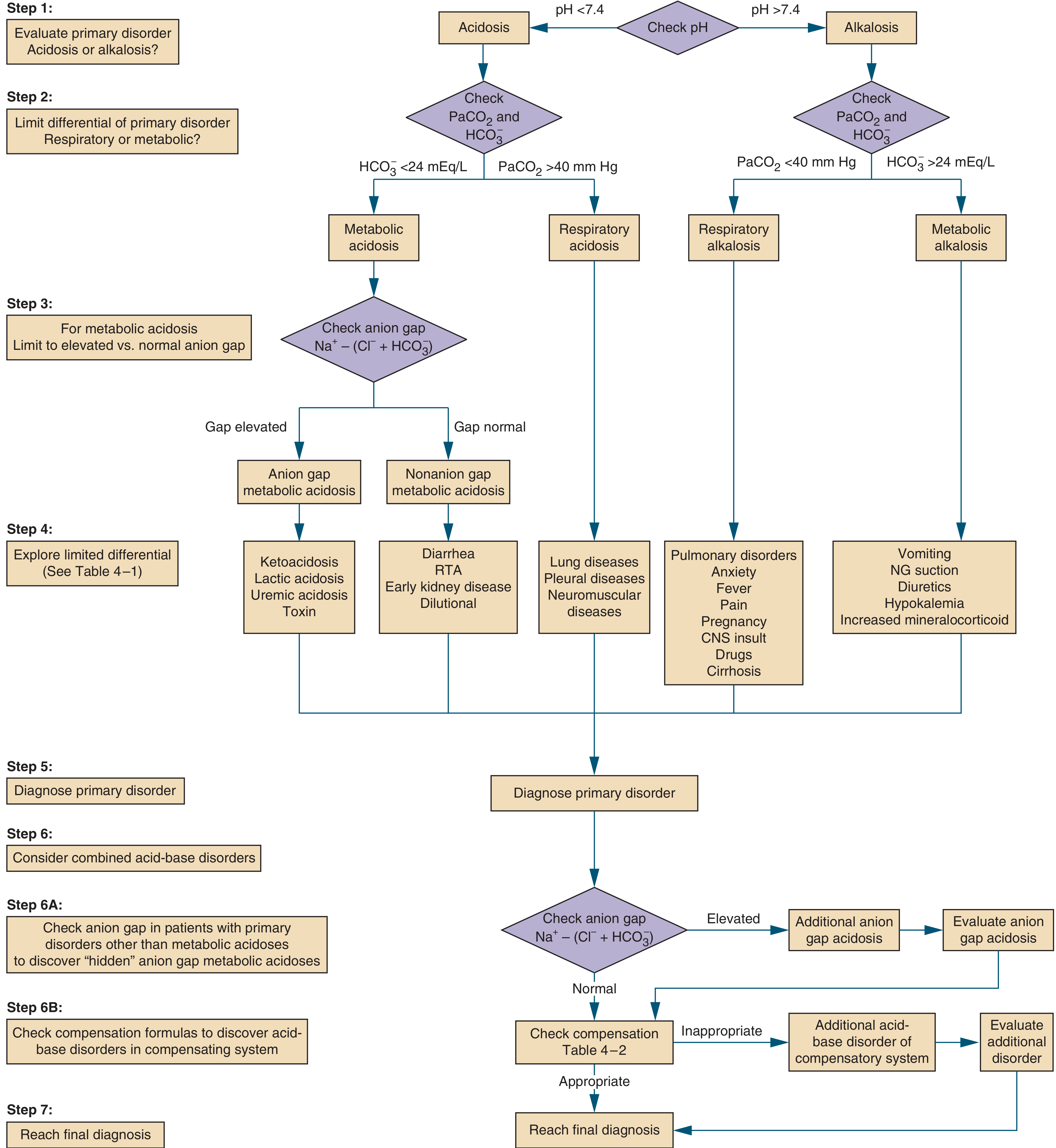
THE 4 RESULTS OF ABG - ONE BY ONE
RESULT 1: RESPIRATORY ACIDOSIS
Definition
pH is low (acidosis) caused by CO2 retention - the lungs are not removing enough CO2.
ABG Values:
| Parameter | Value |
|---|---|
| pH | < 7.35 (LOW) |
| PaCO2 | > 45 mmHg (HIGH) |
| HCO3- | Normal (acute) / High >26 (chronic - compensated) |
Simple Rule:
pH ↓ + PaCO2 ↑ = Respiratory Acidosis
Mechanism:
- Hypoventilation → CO2 builds up in blood
- CO2 + H2O → H2CO3 (Carbonic acid) → pH falls
- Lungs are the problem
Causes (Emergency Medicine):
| Cause | Examples |
|---|---|
| Airway obstruction | Choking, foreign body, laryngospasm |
| CNS depression | Opioid overdose, sedative overdose, head injury |
| Neuromuscular disease | Myasthenia gravis, Guillain-Barre syndrome |
| Chest wall injury | Flail chest, multiple rib fractures |
| Lung disease | COPD exacerbation, severe asthma, pneumothorax |
| Drowning / near-drowning | |
| Cardiac arrest |
Compensation:
- Kidneys compensate (slow - takes 3-5 days)
- Kidneys retain HCO3- → HCO3 rises to correct pH
- Acute: HCO3 rises 1 mEq/L per 10 mmHg rise in PaCO2
- Chronic: HCO3 rises 3.5 mEq/L per 10 mmHg rise in PaCO2
Clinical Features:
- Dyspnoea, confusion
- CO2 narcosis - drowsiness, headache
- Flushed, warm skin (vasodilation due to CO2)
- Cyanosis (in severe cases)
- Papilloedema (raised ICP from CO2-induced cerebral vasodilation)
Emergency Management:
- Open airway - head tilt, chin lift, jaw thrust
- Bag-Valve-Mask (BVM) ventilation
- Intubation if not breathing
- Treat cause (naloxone for opioid OD, bronchodilators for asthma)
- Non-invasive ventilation (NIV/BiPAP) for COPD
- Do NOT give NaHCO3 (controversial, not effective in respiratory acidosis)
RESULT 2: RESPIRATORY ALKALOSIS
Definition
pH is high (alkalosis) due to excessive CO2 blown off - the lungs are over-ventilating.
ABG Values:
| Parameter | Value |
|---|---|
| pH | > 7.45 (HIGH) |
| PaCO2 | < 35 mmHg (LOW) |
| HCO3- | Normal (acute) / Low <22 (chronic - compensated) |
Simple Rule:
pH ↑ + PaCO2 ↓ = Respiratory Alkalosis
Mechanism:
- Hyperventilation → too much CO2 blown out
- Less carbonic acid → pH rises
- Lungs are over-working
Causes (Emergency Medicine):
| Cause | Examples |
|---|---|
| Anxiety / Panic attack | Hyperventilation syndrome |
| Pain | Any severe pain causing tachypnoea |
| Fever / Sepsis | Increased respiratory drive |
| Hypoxia | Any condition causing low O2 (pneumonia, PE, altitude) |
| Pregnancy | Progesterone stimulates breathing |
| Pulmonary embolism (PE) | Very common emergency cause |
| CNS causes | Meningitis, stroke, head injury |
| Drugs | Salicylate (aspirin) toxicity (early), mechanical over-ventilation |
| Liver failure / Cirrhosis |
Compensation:
- Kidneys compensate (slow - 2-3 days)
- Kidneys excrete HCO3- → HCO3 falls
- Acute: HCO3 falls 2 mEq/L per 10 mmHg fall in PaCO2
- Chronic: HCO3 falls 5 mEq/L per 10 mmHg fall in PaCO2
Clinical Features:
- Tingling / numbness of hands, feet, lips (perioral)
- Carpopedal spasm (Chvostek's / Trousseau's sign in severe cases)
- Light-headedness, dizziness
- Palpitations
- Chest tightness
- Syncope (fainting)
Emergency Management:
- Reassure the patient (if anxiety-related)
- Rebreathing into a paper bag (for hyperventilation syndrome - controversial now)
- Treat the underlying cause (PE: anticoagulation; fever: antipyretics)
- For mechanically ventilated patients: reduce respiratory rate or tidal volume
- Pain management if pain is the cause
RESULT 3: METABOLIC ACIDOSIS
Definition
pH is low (acidosis) caused by loss of HCO3- or gain of acid - kidneys/metabolism is the problem.
ABG Values:
| Parameter | Value |
|---|---|
| pH | < 7.35 (LOW) |
| HCO3- | < 22 mEq/L (LOW) |
| PaCO2 | Normal (uncompensated) / Low <35 (compensated - Kussmaul breathing) |
Simple Rule:
pH ↓ + HCO3 ↓ = Metabolic Acidosis
Mechanism:
- Acid accumulates in body OR bicarbonate is lost
- pH falls
- Kidneys or metabolism is the problem
- Lungs try to compensate by blowing off CO2 (deep fast breathing)
ANION GAP - Important Classification:
Anion Gap = Na+ - (Cl- + HCO3-) → Normal = 8-12 mEq/L
High Anion Gap Metabolic Acidosis (HAGMA):
Mnemonic: MUDPILES or KULT
| Mnemonic | Cause |
|---|---|
| M | Methanol poisoning |
| U | Uraemia (renal failure) |
| D | Diabetic Ketoacidosis (DKA) |
| P | Paracetamol / Propylene glycol |
| I | Isoniazid / Iron poisoning |
| L | Lactic acidosis (sepsis, cardiac arrest, shock) |
| E | Ethylene glycol (antifreeze) |
| S | Salicylate (aspirin) toxicity |
Normal Anion Gap Metabolic Acidosis (NAGMA):
Mnemonic: HARDUPS or FUSED
- Diarrhoea (loss of HCO3)
- Renal Tubular Acidosis (RTA)
- Fistulas (pancreatic, small bowel)
- Addison's disease
- Dilutional (excess IV normal saline)
Compensation:
- Lungs compensate (fast - minutes to hours)
- Hyperventilation to blow off CO2
- Winter's Formula: Expected PaCO2 = (1.5 × HCO3) + 8 ± 2
- Classic: Kussmaul breathing - deep, rapid, laboured breathing (seen in DKA)
Clinical Features:
- Deep rapid breathing (Kussmaul)
- Fruity breath (DKA)
- Confusion, drowsiness
- Hypotension, shock
- Weakness, fatigue
- Nausea, vomiting
- Cardiac arrhythmias (severe)
Emergency Management:
- Treat the underlying cause
- DKA: Insulin + IV fluids + K+ replacement
- Sepsis/Lactic acidosis: Antibiotics + fluid resuscitation
- Renal failure: Dialysis
- Poisoning: Specific antidote
- NaHCO3 (Sodium Bicarbonate):
- Give ONLY when pH < 7.1 or severe symptomatic acidosis
- Dose: 1-2 mEq/kg IV slow infusion
- NOT routinely given for DKA
- IV fluids (correct hypovolaemia)
- Oxygen therapy
- Monitor ECG for arrhythmias
RESULT 4: METABOLIC ALKALOSIS
Definition
pH is high (alkalosis) due to gain of HCO3- or loss of acid - kidneys/metabolism is the problem.
ABG Values:
| Parameter | Value |
|---|---|
| pH | > 7.45 (HIGH) |
| HCO3- | > 26 mEq/L (HIGH) |
| PaCO2 | Normal (uncompensated) / High >45 (compensated - hypoventilation) |
Simple Rule:
pH ↑ + HCO3 ↑ = Metabolic Alkalosis
Mechanism:
- Acid is lost from the body OR bicarbonate accumulates
- pH rises
- Kidneys/GI tract is the problem
- Lungs try to compensate by retaining CO2 (slow, shallow breathing)
Causes (Emergency Medicine):
| Cause | Examples |
|---|---|
| Vomiting | Loss of HCl from stomach - most common |
| NG tube suction | Removes gastric acid |
| Diuretics | Frusemide, thiazides (lose H+ and Cl-) |
| Hypokalemia | Low K+ → kidneys retain H+ and lose HCO3 less |
| Excess alkali intake | NaHCO3 overdose, antacid abuse (Milk-alkali syndrome) |
| Corticosteroids | Increase aldosterone-like effect |
| Hyperaldosteronism | Cushing's syndrome, Conn's syndrome |
| Blood transfusion | Citrate in stored blood converts to HCO3 |
Compensation:
- Lungs compensate (limited)
- Hypoventilation to retain CO2
- Expected PaCO2 = 0.7 × HCO3 + 21 ± 2
- Limited compensation - body won't let PaO2 drop too much
Classification by Urine Chloride (Cl-):
| Type | Urine Cl- | Causes | Treatment |
|---|---|---|---|
| Saline-Responsive | < 20 mEq/L | Vomiting, NG suction, diuretics | IV Normal Saline + KCl |
| Saline-Resistant | > 20 mEq/L | Hyperaldosteronism, Cushing's | Treat cause; Spironolactone |
Clinical Features:
- Nausea, vomiting
- Muscle weakness, cramps
- Tetany (in severe cases - low ionised calcium due to high pH)
- Confusion, seizures
- Cardiac arrhythmias (especially with hypokalemia)
- Slow, shallow breathing (hypoventilation compensation)
Emergency Management:
- IV Normal Saline (0.9% NaCl) - first line if volume depleted
- Potassium replacement (KCl IV) - essential, correct hypokalaemia
- Stop the cause - stop diuretics, stop NG suction if possible
- Acetazolamide - used in critically ill, makes kidneys excrete HCO3-
- Spironolactone - for hyperaldosteronism
- HCl via central line - for severe refractory cases
- Dialysis - if in renal failure
QUICK COMPARISON TABLE - ALL 4 ABG RESULTS
| Resp. Acidosis | Resp. Alkalosis | Met. Acidosis | Met. Alkalosis | |
|---|---|---|---|---|
| pH | ↓ Low | ↑ High | ↓ Low | ↑ High |
| PaCO2 | ↑ High | ↓ Low | Normal/↓ | Normal/↑ |
| HCO3- | Normal/↑ | Normal/↓ | ↓ Low | ↑ High |
| Breathing | Slow/shallow | Fast/deep | Deep/fast (Kussmaul) | Slow/shallow |
| Problem | Lungs ↓ | Lungs ↑ | Kidneys/Metabolism | Kidneys/GI |
| Key Example | COPD, overdose | Anxiety, PE | DKA, sepsis, shock | Vomiting, diuretics |
| Compensation | Kidneys ↑ HCO3 | Kidneys ↓ HCO3 | Lungs ↓ CO2 | Lungs ↑ CO2 |
SAMPLE ABG QUESTIONS (Exam Practice)
Q1: pH 7.28, PaCO2 55, HCO3 24
- pH low → Acidosis
- PaCO2 high → Respiratory Acidosis (uncompensated)
Q2: pH 7.50, PaCO2 28, HCO3 22
- pH high → Alkalosis
- PaCO2 low → Respiratory Alkalosis
Q3: pH 7.20, PaCO2 18, HCO3 7
- pH low → Acidosis
- HCO3 low → Metabolic Acidosis (with respiratory compensation)
Q4: pH 7.50, PaCO2 48, HCO3 36
- pH high → Alkalosis
- HCO3 high → Metabolic Alkalosis (with respiratory compensation)
Sources: Barash's Clinical Anesthesia 9e; Symptom to Diagnosis 4e (Evidence-Based Guide)
This covers all 4 ABG results completely - definition, values, causes, compensation, clinical features, and emergency management. Copy each result as a separate section in your log book! Let me know if you want sample patient cases with ABG interpretation or compensation formulas as a separate table.
Emtc hand over and referrals sheet
EMTC (Emergency Medicine and Trauma Care) handover and referral sheets are standardized communication tools used when transferring patient care between staff or departments. The standard format uses SBAR or MIST communication protocols in emergency settings.
Searching sources...
Finding Sources
Searching the Web
EMTC emergency handover referral sheet SBAR MIST format India hospital
Finding Sources
Finding Sources
Here is the complete EMTC Handover and Referral Sheet - fully detailed and ready to use in your log book. This follows standard protocols used in Indian emergency departments and as per MGR University AECT curriculum.
EMTC HANDOVER AND REFERRAL SHEET
Emergency Medicine and Trauma Care
PART A: WHAT IS A HANDOVER?
Handover = The process of transferring responsibility and information about a patient from one healthcare provider to another.
Why is Handover Important?
- Prevents errors in patient care
- Ensures continuity of treatment
- Reduces risk of missed information
- Legally required documentation
- Protects both patient and staff
When is Handover Done?
- Shift change (duty handover)
- Department to department (ED to ICU, ED to OT)
- Inter-hospital transfer (referral)
- Pre-hospital to ED (ambulance to emergency team)
- ED to ward (admission handover)
PART B: HANDOVER PROTOCOLS
PROTOCOL 1: SBAR (Most commonly used in India)
| Letter | Stands For | What to Say |
|---|---|---|
| S | Situation | Who is the patient? What is happening right now? |
| B | Background | Past history, current medications, allergies |
| A | Assessment | Vitals, diagnosis, clinical condition, investigations done |
| R | Recommendation | What needs to be done next, pending tasks, watch points |
PROTOCOL 2: MIST (Used in Trauma / Pre-hospital handover)
| Letter | Stands For | Details |
|---|---|---|
| M | Mechanism of Injury | How did the injury happen? (RTA, fall, assault, stab) |
| I | Injuries found/suspected | Head injury, fractures, internal bleeding |
| S | Signs and Symptoms | Vitals, GCS, pain score, conscious level |
| T | Treatment given | What was done at scene / in ambulance (IV access, O2, immobilisation) |
PROTOCOL 3: IMIST-AMBO (Paramedic to ED Handover)
| Letter | Stands For |
|---|---|
| I | Identification of patient |
| M | Mechanism / Medical complaint |
| I | Injuries / Information relevant |
| S | Signs and vitals |
| T | Treatment given |
| A | Allergies |
| M | Medications |
| B | Background history |
| O | Other information |
PART C: EMTC DUTY HANDOVER SHEET
(Shift change - used at every duty shift handover)
╔══════════════════════════════════════════════════════════╗
║         EMTC DUTY HANDOVER SHEET                         ║
║         Emergency Medicine & Trauma Care                  ║
╚══════════════════════════════════════════════════════════╝

HOSPITAL: ___________________________  DATE: ____________

DEPARTMENT: Emergency / Casualty / Trauma Bay / ICU

HANDOVER FROM: _______________________ DESIGNATION: ________
               (Outgoing Staff)

HANDOVER TO:   _______________________ DESIGNATION: ________
               (Incoming Staff)

SHIFT TIMING:  ___:___ to ___:___ hrs
SECTION 1: ACTIVE PATIENT LIST
| Bed No. | Patient Name | Age/Sex | IP No. | Diagnosis | Current Status | Pending Tasks | Watch Points |
|---|---|---|---|---|---|---|---|
SECTION 2: CRITICAL / HIGH PRIORITY PATIENTS
Patient 1:
Name: _________________ Age/Sex: _____ Bed: ____
Diagnosis: ___________________________________________
Current Vitals: BP: ___ HR: ___ RR: ___ SpO2: ___ Temp: ___
GCS: ___/15
IV Access: _______ Drips running: ______________________
Investigations pending: _______________________________
Action required: _____________________________________

Patient 2:
Name: _________________ Age/Sex: _____ Bed: ____
Diagnosis: ___________________________________________
Current Vitals: BP: ___ HR: ___ RR: ___ SpO2: ___ Temp: ___
GCS: ___/15
Investigations pending: _______________________________
Action required: _____________________________________
SECTION 3: MEDICATIONS / DRIPS TO WATCH
| Patient | Medication | Dose | Rate | Time to Review |
|---|---|---|---|---|
SECTION 4: PENDING INVESTIGATIONS
| Patient | Investigation | Time Sent | Expected Report | Action if Abnormal |
|---|---|---|---|---|
SECTION 5: EVENTS DURING SHIFT
□ Any cardiac arrest / resuscitation: YES / NO
  Details: ___________________________________________

□ Any deaths during shift: YES / NO
  Details: ___________________________________________

□ Any referrals made: YES / NO
  Details: ___________________________________________

□ Any complaints / incidents: YES / NO
  Details: ___________________________________________

□ Equipment issues: YES / NO
  Details: ___________________________________________
SECTION 6: EQUIPMENT STATUS CHECK
| Equipment | Working | Not Working | Remarks |
|---|---|---|---|
| Defibrillator | |||
| BVM (Adult/Paeds) | |||
| Laryngoscope | |||
| Pulse Oximeter | |||
| ECG Machine | |||
| Suction Machine | |||
| Glucometer | |||
| Crash Cart (sealed) | |||
| Oxygen cylinders |
SECTION 7: SIGNATURES
Outgoing Staff Signature: ____________  Time: ________
Name: ________________________________

Incoming Staff Signature: ____________  Time: ________
Name: ________________________________

Supervisor / Sr. Nurse / MO Signature: ______________
PART D: EMTC REFERRAL SHEET
(Used when referring a patient to another department or hospital)
╔══════════════════════════════════════════════════════════╗
║              EMTC PATIENT REFERRAL SHEET                 ║
║         Emergency Medicine & Trauma Care                  ║
╚══════════════════════════════════════════════════════════╝

HOSPITAL NAME: _________________________________
DATE: _______________    TIME: _________________
REFERRAL NO.: __________
SECTION A: PATIENT DETAILS
Patient Name:    ________________________________________
Age:  _____ years    Sex: M / F / Others
IP / OP Number:  _______________
Address:         ________________________________________
Mobile No.:      _______________
Attender Name:   _______________  Relation: ____________
SECTION B: REFERRAL INFORMATION
Referred FROM: Department: ______________
               Hospital:   ______________
               Doctor Name: _____________  Designation: _________
               Contact No.: _____________

Referred TO:   Department: ______________
               Hospital:   ______________
               Doctor Name: _____________  Designation: _________

Reason for Referral:
□ Higher level of care required
□ Specialist consultation needed - Specialty: _______________
□ Facility/equipment not available
□ Patient request
□ ICU bed not available
□ Surgical intervention required
□ Other: _____________________________
SECTION C: CLINICAL SUMMARY (SBAR FORMAT)
━━━━━━━━━━━━━━━━━━━━━━━━━━━━━━━━━━━━━━
S - SITUATION
━━━━━━━━━━━━━━━━━━━━━━━━━━━━━━━━━━━━━━
Chief Complaint: ___________________________________
Duration of Illness: _______________________________
Mechanism (if trauma): ______________________________

━━━━━━━━━━━━━━━━━━━━━━━━━━━━━━━━━━━━━━
B - BACKGROUND
━━━━━━━━━━━━━━━━━━━━━━━━━━━━━━━━━━━━━━
Past Medical History:
□ Diabetes  □ Hypertension  □ CAD  □ Asthma  □ CKD  □ Nil
Others: ____________________________________________

Past Surgical History: _____________________________
Current Medications: _______________________________
Allergies: __________________ (Drug / Food / Others)
Immunisation Status: _______________________________

━━━━━━━━━━━━━━━━━━━━━━━━━━━━━━━━━━━━━━
A - ASSESSMENT (Vitals & Clinical Findings)
━━━━━━━━━━━━━━━━━━━━━━━━━━━━━━━━━━━━━━
Triage Category:  □ Red (Immediate)  □ Yellow (Urgent)
                  □ Green (Minor)    □ Black (Expectant)

VITAL SIGNS AT TIME OF REFERRAL:
Blood Pressure:     ______ / ______ mmHg
Heart Rate:         ______ /min    Rhythm: Regular / Irregular
Respiratory Rate:   ______ /min
SpO2:              ______ %  (on Room Air / O2 ___ L/min)
Temperature:        ______ °C
Blood Sugar (RBS):  ______ mg/dL
GCS:               E___ V___ M___  Total: ___ /15
Pain Score (VAS):  ______ /10
Weight (if known): ______ kg

PUPILS:  Right: ____mm  Reactive: Y/N
         Left:  ____mm  Reactive: Y/N

Airway Status:
□ Patent and self-maintained
□ Maintained with airway adjunct (OPA/NPA)
□ Intubated  ETT size: _____  Depth: _____ cm
□ Surgical airway (Cricothyrotomy / Tracheostomy)

Breathing: □ Spontaneous  □ Assisted  □ Ventilated
           Mode: _________  FiO2: _____  PEEP: _____

Circulation:
IV Access: □ Peripheral  □ Central  □ Intraosseous
           Site: _____________  Size: _____G

Skin: □ Warm  □ Cold  □ Clammy  □ Mottled
Capillary refill: ______ seconds

Working Diagnosis:
___________________________________________________
___________________________________________________

━━━━━━━━━━━━━━━━━━━━━━━━━━━━━━━━━━━━━━
R - RECOMMENDATION / TREATMENT GIVEN
━━━━━━━━━━━━━━━━━━━━━━━━━━━━━━━━━━━━━━
Treatment Given Before Referral:
□ O2 therapy - Type: _________ Flow rate: _____ L/min
□ IV Fluids - Type: _________  Volume: _____ mL  Rate: _____
□ IV/IM Medications Given:
  1. _____________________ Dose: _____ Route: _____ Time: _____
  2. _____________________ Dose: _____ Route: _____ Time: _____
  3. _____________________ Dose: _____ Route: _____ Time: _____

□ CPR performed: YES / NO  Duration: _______
□ Defibrillation: YES / NO  Joules: _______
□ Wound dressing: YES / NO
□ Splinting/immobilisation: YES / NO  Part: _________
□ NG Tube inserted: YES / NO  Size: _____
□ Urinary Catheter: YES / NO  Size: _____
□ Blood transfusion started: YES / NO
  Type: _______  Units: _____ Group: _____

Reason for Continuing Management at Receiving Centre:
___________________________________________________
SECTION D: INVESTIGATIONS DONE
Lab Results Sent with Patient: YES / NO

CBC: Hb: ___ WBC: ___ Platelets: ___ PCV: ___
RFT: Urea: ___ Creatinine: ___ Na: ___ K: ___
LFT: Total Bili: ___ ALT: ___ AST: ___
Coagulation: PT: ___ INR: ___ aPTT: ___
Blood Sugar: _____ mg/dL
ABG: pH: ___ PaCO2: ___ PaO2: ___ HCO3: ___ SpO2: ___
Cardiac Markers: Trop I/T: ___  CK-MB: ___  BNP: ___
Lactate: ___ mmol/L

Imaging:
□ X-ray: _________________ Findings: ___________________
□ ECG: __________________ Findings: ___________________
□ USG/FAST: _____________ Findings: ___________________
□ CT Scan: ______________ Findings: ___________________

Other investigations: ________________________________
SECTION E: TRANSPORT DETAILS
Mode of Transport:
□ Ambulance (BLS)    □ Ambulance (ALS/ACLS)
□ Patient's own vehicle  □ Air ambulance

Oxygen during transport:  YES / NO
Monitoring during transport:
□ Pulse Oximetry   □ ECG   □ BP   □ ETCO2   □ Ventilator

Staff accompanying:
Name: ________________________ Designation: _____________
Contact during transport: _____________________________

Documents sent with patient:
□ This Referral Sheet
□ Discharge Summary / Case Sheet copy
□ All Lab Reports
□ Imaging (X-ray, CD)
□ ECG copy
□ Consent form for transfer
□ Medico-legal documents (if applicable)
SECTION F: CONSENT AND SIGNATURES
Patient / Attender Consent for Transfer: YES / NO

Attender Name: _________________ Signature: ___________
Relationship: __________________

Referring Doctor:
Name: __________________________ Sign: ________________
Designation: ____________________
Registration No.: ________________
Date & Time: ____________________

Receiving Hospital Contact:
Hospital Name: __________________
Doctor Informed: ________________ Time: _______________
Bed confirmed: YES / NO   Ward/ICU: __________________

Senior Doctor Approval for Transfer:
Name: __________________________ Sign: ________________
PART E: INTER-HOSPITAL REFERRAL - KEY POINTS
WHEN TO REFER (Indications):
- Facility or specialist not available at current centre
- Patient requires higher level of care (ICU, Neurosurgery, Cardiac Cath Lab)
- Bed not available in current hospital
- Patient or family request transfer
- MLC (Medico-Legal Cases) - as directed by police/court
BEFORE TRANSFERRING - CHECKLIST:
- Patient stabilised as much as possible
- Receiving hospital informed and confirmed bed
- All documents prepared and handed over
- Patient/attender consent obtained
- Appropriate transport arranged
- Trained staff accompanying patient
- Equipment and drugs loaded in ambulance
- IV access secured, fluids running
- Airway secured if needed before transfer
- Time-sensitive conditions (stroke, STEMI) - notify receiving team early
GOLDEN RULE:
"Stabilise before you mobilise" Never transfer an unstable patient without securing airway, breathing, and circulation first.
PART F: COMMON REFERRAL ACRONYM - ACCEPT
| Letter | Meaning |
|---|---|
| A | Acknowledgement - Receiving hospital acknowledges the referral |
| C | Clinical condition - Patient's condition confirmed |
| C | Critically ill? - Is this a time-critical transfer? |
| E | Equipment needed - What monitoring and equipment during transport |
| P | Personnel - Who will accompany? |
| T | Time - How soon can transfer happen? |
PART G: SAMPLE FILLED REFERRAL (Practice)
Patient: Mr. Rajan, 55yrs Male, IP No. 1234
Date: 20/07/2026  Time: 01:30 hrs

SITUATION:
Patient brought to ED after Road Traffic Accident.
H/O unrestrained driver, frontal collision at high speed.
GCS on arrival: 10/15 (E3V3M4)

BACKGROUND:
Known hypertensive on Amlodipine 5mg OD.
No known drug allergy.

ASSESSMENT:
BP: 90/60  HR: 124  RR: 28  SpO2: 91% on 10L NRM
GCS: 10/15  Pupils: R - 4mm sluggish, L - 2mm reactive
Suspected traumatic brain injury + haemothorax

RECOMMENDATION:
Patient requires emergency CT brain + chest,
Neurosurgical and Cardiothoracic consult.
Facility not available at this centre.
Referring to Government General Hospital Trauma Centre.
This is a complete, exam-quality EMTC Handover and Referral sheet for your log book. You can:
- Copy PART C as your duty handover template
- Copy PART D as your referral sheet template
- Write one sample filled case using PART G format
Let me know if you want a specific case written in SBAR format or a pre-hospital MIST handover example!
🦂 sting Next dog bite with rig
Sting One page Dog bite and rig one 0age
Scorpion sting causes local pain and systemic autonomic effects via venom neurotoxins. Dog bite carries risk of rabies, tetanus, and bacterial infection. Both are common emergency presentations in India.
Searching sources...
Finding Sources
Finding Sources
Finding Sources
Finding Sources
Reading File
Finding Sources
Reading File
Finding Sources
Reading File
Here are both topics - one page each - for your log book.
PAGE 1: SCORPION STING 🦂
DEFINITION
A scorpion sting is envenomation caused by the injection of venom through the stinger on the scorpion's tail. Medically dangerous species include Mesobuthus tamulus (Red Scorpion) - most deadly species found in India, especially Tamil Nadu, Maharashtra, and Andhra Pradesh.
VENOM MECHANISM
- Scorpion venom contains neurotoxins that keep sodium channels open
- This causes massive release of catecholamines (adrenaline, noradrenaline)
- Results in autonomic storm - both sympathetic and parasympathetic overactivation
GRADES OF SCORPION STING (Clinical Grading)
| Grade | Features |
|---|---|
| Grade I | Local pain and swelling only at sting site |
| Grade II | Systemic symptoms - sweating, vomiting, hypertension OR hypotension, tachycardia |
| Grade III | Severe - Pulmonary oedema, cardiogenic shock, multi-organ failure |
CLINICAL FEATURES
Local:
- Severe burning pain at sting site
- Swelling, redness, ecchymosis
- Paresthesia (tingling/numbness)
- Tap test positive - tapping on sting site increases pain
Systemic (Autonomic Storm):
| Sympathetic Overactivation | Parasympathetic Overactivation |
|---|---|
| Hypertension | Hypotension (late) |
| Tachycardia | Bradycardia |
| Sweating (profuse) | Excessive salivation |
| Restlessness, anxiety | Lacrimation (watering eyes) |
| Cold clammy extremities | Vomiting, diarrhoea |
Neuromuscular:
- Muscle twitching, jerking (can look like seizure)
- Blurred vision, abnormal eye movements
- Slurred speech
- Difficulty swallowing secretions
Cardiovascular (Severe):
- Acute pulmonary oedema (most common cause of death)
- Myocarditis
- Cardiogenic shock
Other:
- Hyperthermia
- Rhabdomyolysis
- Acidosis
- Acute kidney injury (Hemiscorpius species - cytotoxic)
INVESTIGATIONS
| Test | Finding |
|---|---|
| ECG | Tachycardia, arrhythmias, ST changes |
| Chest X-ray | Pulmonary oedema |
| Blood Sugar | Hyperglycaemia |
| Serum electrolytes | Hyponatraemia, hypokalaemia |
| ABG | Metabolic acidosis |
| Cardiac enzymes | Elevated (myocarditis) |
| CBC, RFT | Baseline |
EMERGENCY MANAGEMENT
Step 1 - Stabilise (ABC)
- Airway: Maintain; intubate if GCS falls
- Breathing: O2 therapy; mechanical ventilation if pulmonary oedema
- Circulation: IV access, ECG monitoring, fluid management
Step 2 - Local Care
- Do NOT incise or suck the sting site
- Apply cold pack / ice pack to slow venom absorption
- Pressure dressing around site
- Keep patient calm and still
Step 3 - Drug Treatment
| Drug | Dose | Indication |
|---|---|---|
| Prazosin (alpha blocker) | 250-500 mcg oral every 3-4 hrs | Drug of choice in India for Grade II/III - controls hypertension and reverses pulmonary oedema |
| Midazolam IV infusion | 0.1-0.2 mg/kg | Agitation, muscle twitching |
| Atropine IV | 0.01-0.02 mg/kg | Bradycardia, excessive secretions |
| Nifedipine | 5-10 mg sublingual | Hypertension + pulmonary oedema |
| Frusemide IV | 1-2 mg/kg | Pulmonary oedema |
| Dobutamine | Infusion | Cardiogenic shock |
| Analgesics | Paracetamol / Tramadol | Pain relief |
| Antihistamines | Promethazine IV | Allergic component |
Step 4 - Antivenom
- Scorpion Antivenom (Polyvalent, India - Bharat Serums)
- Given IV diluted in 100 mL NS over 30 min
- Effective against Mesobuthus tamulus (Indian Red Scorpion)
- Indicated for Grade II and Grade III envenomation
- Keep adrenaline ready for anaphylaxis
Step 5 - Tetanus Prophylaxis
- TT injection if not vaccinated in last 5 years
DISPOSITION
- Grade I: Observe 4-6 hours, discharge with advice
- Grade II/III: ICU admission, monitor ECG, BP, SpO2 continuously
- Symptoms peak at ~5 hours; resolve in 24-48 hours
- Pain and paresthesia may last weeks
KEY POINT FOR INDIA 🇮🇳
Prazosin is the specific treatment for Indian Red Scorpion (Mesobuthus tamulus) sting. It is NOT available in some countries but is the drug of choice in India. Give early before pulmonary oedema develops.
Source: Harrison's Principles of Internal Medicine 22E
PAGE 2: DOG BITE WITH RABIES (RIG/Vaccine)
DEFINITION
A dog bite is a wound caused by a dog's teeth. In emergency medicine, the main concern is:
- Wound management (infection, tetanus)
- Rabies Post-Exposure Prophylaxis (PEP) - the most critical step
RABIES - KEY FACTS
- Caused by Rabies Lyssavirus - a Rhabdovirus
- Transmitted via saliva of infected animal through bite, scratch, or mucous membrane contact
- 100% fatal once clinical symptoms develop
- Incubation period: 1-3 months (range: 1 week to 1 year)
- Virus travels via peripheral nerves to the brain (retrograde axonal transport)
- India accounts for 36% of global rabies deaths (approximately 20,000/year)
WHO CLASSIFICATION OF BITE (Category System)
| Category | Type of Contact | Action |
|---|---|---|
| Category I | Touching, feeding the animal; licking on intact skin | Wash hands - No PEP needed |
| Category II | Minor scratches/abrasions without bleeding; nibbling of uncovered skin | Wound wash + Vaccine only |
| Category III | Single/multiple transdermal bites or scratches, licks on broken skin; mucous membrane contact; bat bite | Wound wash + Vaccine + RIG |
WOUND MANAGEMENT (First and Most Important Step)
"Immediate wound washing is the single most important first aid measure"
Steps:
- Wash wound immediately with soap and water for minimum 15 minutes
- Apply 70% alcohol / povidone iodine - viricidal
- Do NOT suture the wound immediately (allows virus to escape)
- If suturing needed (cosmetic/large wound), give RIG first, then suture after 2-4 hours
- Do NOT cover tightly - leave slightly open
- Debride devitalised tissue if present
- Assess wound depth and nerve/tendon involvement
RABIES POST-EXPOSURE PROPHYLAXIS (PEP)
TWO COMPONENTS:
1. RIG - Rabies Immunoglobulin (Passive Immunity - Immediate Protection)
| Type | Dose | Route |
|---|---|---|
| Human Rabies Immunoglobulin (HRIG) | 20 IU/kg body weight | Infiltrated into wound + remainder IM at distant site |
| Equine Rabies Immunoglobulin (ERIG) | 40 IU/kg body weight | Same as above (test dose required first) |
Key Rules for RIG:
- Give on Day 0 only (same day as first vaccine)
- Maximum dose of infiltration INTO wound first; remaining IM
- If anatomically impossible to inject all into wound, give remainder IM at a site away from vaccine
- Do NOT give RIG and vaccine in the same syringe or same site
- RIG gives immediate antibodies for 7-10 days until vaccine kicks in
- If RIG not given on Day 0, give up to Day 7 (after that, not needed)
2. RABIES VACCINE (Active Immunity - Long-term Protection)
Schedule - ESSEN Protocol (5 Dose - IM):
| Day | Dose | Site |
|---|---|---|
| Day 0 | 1 vial (1 mL) IM | Deltoid (right) |
| Day 3 | 1 vial (1 mL) IM | Deltoid (left) |
| Day 7 | 1 vial (1 mL) IM | Deltoid |
| Day 14 | 1 vial (1 mL) IM | Deltoid |
| Day 28 | 1 vial (1 mL) IM | Deltoid |
Alternative - Zagreb Protocol (4 Dose - 2-1-1):
| Day | Dose |
|---|---|
| Day 0 | 2 doses (both deltoids simultaneously) |
| Day 7 | 1 dose |
| Day 21 | 1 dose |
Note: In India, WHO recommends the Essen 5-dose IM protocol or the Updated 4-dose protocol (Days 0,3,7,14)
Vaccine Types Available in India:
- PCECV - Purified Chick Embryo Cell Vaccine (Rabipur)
- PVRV - Purified Vero Cell Vaccine (Verorab, Abhayrab)
- Give in deltoid muscle (adults) or anterolateral thigh (children)
- Never give in gluteal region (poor immune response)
TETANUS PROPHYLAXIS (Mandatory for all Dog Bites)
| Vaccination History | Wound Type | Action |
|---|---|---|
| Fully vaccinated (<5 years) | Any | Nothing needed |
| Fully vaccinated (5-10 years) | Clean minor | Nothing needed |
| Fully vaccinated (5-10 years) | Contaminated | TT booster |
| Unknown / Incomplete | Any | TT + TIG (Tetanus Immunoglobulin) |
- TT dose: 0.5 mL IM (deltoid)
- TIG dose: 250 IU IM (if unvaccinated)
ANTIBIOTIC PROPHYLAXIS
| Drug | Dose | Duration |
|---|---|---|
| Amoxicillin-Clavulanate (first choice) | 625 mg TDS (adult) oral | 5-7 days |
| Doxycycline (if penicillin allergy) | 100 mg BD | 5-7 days |
| IV Antibiotics | Piperacillin-Tazobactam | Severe/deep wounds |
- Bacteria in dog mouth: Pasteurella multocida, Staphylococcus, Capnocytophaga canimorsus
ANIMAL OBSERVATION RULE
| Situation | Action |
|---|---|
| Dog is alive and healthy | Observe for 10 days |
| If dog stays healthy for 10 days | Animal is NOT rabid - can stop PEP |
| If dog dies or develops signs in 10 days | Continue full PEP course |
| Dog not available / unknown | Complete full PEP course |
COMPLETE EMERGENCY MANAGEMENT SUMMARY
Dog Bite Patient Arrives
         ↓
1. WOUND WASH - Soap + water 15 min + Povidone iodine
         ↓
2. ASSESS - WHO Category (I / II / III)?
         ↓
Category III → RIG Day 0 + Vaccine Day 0,3,7,14,28
Category II  → Vaccine Day 0,3,7,14,28 (No RIG)
         ↓
3. Tetanus Prophylaxis (TT ± TIG)
         ↓
4. Antibiotics (Amoxicillin-Clavulanate 5-7 days)
         ↓
5. Analgesics (Paracetamol / Ibuprofen)
         ↓
6. Document MLC if required (unprovoked bite)
         ↓
7. Advise patient on vaccine return dates
         ↓
8. Animal observation / report to health authorities
WHEN IS PEP NOT NEEDED?
- Category I exposure only
- Bite by rodent (rat, squirrel, rabbit) - almost never rabid
- Previously fully vaccinated individuals with documented titre (may need 2 booster doses only, no RIG)
QUICK MEMORY TABLE
| Scorpion Sting | Dog Bite | |
|---|---|---|
| Immediate action | Cold pack + Prazosin | Wound wash 15 min |
| Antidote | Scorpion Antivenom | RIG (20 IU/kg HRIG) |
| Drug of choice (India) | Prazosin | Amoxicillin-Clavulanate |
| Vaccine needed? | No | Yes - 5 doses |
| Tetanus | TT injection | TT ± TIG |
| ICU needed? | Grade II/III | Only if anaphylaxis/severe |
| Fatal if untreated? | Grade III - yes | Rabies - 100% fatal |
Sources: Harrison's Principles of Internal Medicine 22E; Red Book 2021 (AAP); WHO Rabies Guidelines
Both pages are complete and log book ready! Want me to also add snake bite or bee/wasp sting as the next page?
This is a shared conversation. Sign in to Orris to start your own chat.