
GDM - Screening methods, Glucose metabolism and Management 30 marks question in PG obs and gynce
Reading File
Now let me search the medical library in parallel across all three topics.
Finding Sources
Finding Sources
Finding Sources
Excellent - found key sources. Let me now read the most relevant textbook sections in parallel.
Reading File
Reading File
Finding Sources
Now let me read the critical GDM sections from Creasy & Resnik and also grab more from Harrison's:
Reading File
Reading File
Now let me get the detailed Creasy & Resnik GDM chapter content plus the insulin types table, and also search for the glucose metabolism section:
Reading File
Finding Sources
Excellent - found the glucose metabolism section. Let me read it now in parallel with more GDM management content:
Reading File
Reading File
I now have rich material. Let me get the screening section and GDM diagnosis criteria from Creasy & Resnik, plus search for images:
Finding Sources
Searching Images
gestational diabetes mellitus screening glucose tolerance test GDM diagnosis
Reading File
Loading Image
I now have comprehensive material to write a thorough 30-mark PG answer. Let me compile the complete answer.
Gestational Diabetes Mellitus (GDM): Screening, Glucose Metabolism & Management
A Complete 30-Mark PG Answer
DEFINITION
Gestational diabetes mellitus (GDM) is defined as carbohydrate intolerance of variable severity with onset or first recognition during pregnancy. It occurs in approximately 8% of all pregnancies and is one of the most common metabolic complications in obstetric practice. (Harrison's Principles of Internal Medicine, 22E)
PART I: GLUCOSE METABOLISM IN PREGNANCY
Understanding GDM requires appreciating the normal physiological changes in glucose metabolism during pregnancy.
A. Normal Glucose Metabolism in Pregnancy
Pregnancy induces a biphasic metabolic pattern:
Early Pregnancy (1st Trimester):
- Increased insulin sensitivity transiently at 12-14 weeks
- Enhanced peripheral glucose uptake
- Decreased fasting glucose and increased hepatic glycogen synthesis
- Rising estrogen and progesterone promote beta-cell hyperplasia
- Net result: mild hypoglycemia in fasting state - "accelerated starvation"
Late Pregnancy (2nd & 3rd Trimester):
- Progressive insulin resistance - glucose infusion rate falls significantly (see Fig. 59.4 - Creasy & Resnik)
- Insulin secretory response increases approximately 3-fold compared to pregravid state (2nd and 3rd phase insulin response)
- Despite increased secretion, insulin sensitivity falls markedly by 34-36 weeks
- The net effect is maintenance of normoglycemia in healthy women through compensatory hyperinsulinemia
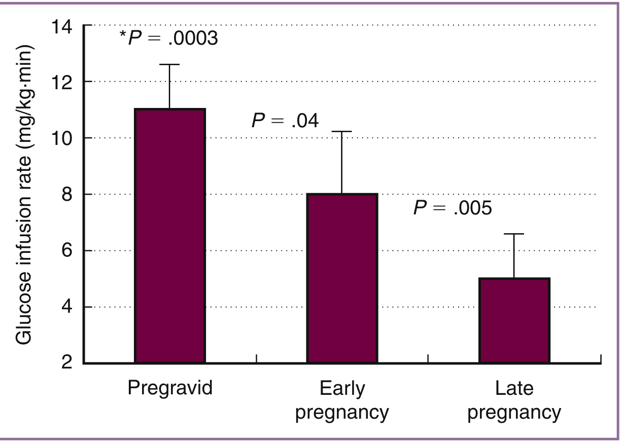
Figure: Longitudinal changes in insulin sensitivity (glucose infusion rate) from pregravid state through early (12-14 weeks) and late (34-36 weeks) pregnancy - Creasy & Resnik
B. The Fetoplacental Unit and Glucose Handling
- The fetoplacental unit shunts glucose and amino acids to the fetus while the mother preferentially uses ketones and triglycerides
- Fetal glucose uptake causes maternal accelerated ketosis during fasting - lower glucose and higher hydroxybutyrate
- Placental hormones responsible for insulin resistance include:
- Human placental lactogen (hPL)
- Placental growth hormone variant
- Progesterone and cortisol
- TNF-alpha and leptin
- hPL peaks progressively and is the dominant anti-insulin factor
C. Mechanisms of Gestational Insulin Resistance (GDM-Specific)
(Creasy & Resnik's MFM, p.1429)
In women who develop GDM, specific metabolic defects are present:
- Increased basal endogenous glucose production - similar to type 2 DM
- Decreased insulin-mediated glucose uptake - insulin infusion assays show insulin's ability to suppress endogenous glucose production is reduced (~80% vs 95% in normal women)
- Impaired first-phase insulin secretion - failure of beta-cell compensation is the key defect
- Decreased insulin sensitivity - persists antepartum and postpartum; women with GDM have significantly lower insulin sensitivity than matched controls even after delivery
- Across the body weight spectrum, fasting insulin concentrations (especially in late gestation) are greater than in normal glucose-tolerant women - reflecting compensatory hyperinsulinemia
Key concept: GDM represents a failure of the beta-cell to fully compensate for the physiological insulin resistance of late pregnancy. Women with GDM carry a pre-existing beta-cell dysfunction or pre-diabetes that is unmasked by pregnancy.
PART II: SCREENING FOR GDM
A. When to Screen
- Universal screening at 24-28 weeks of gestation (recommended by ACOG, ADA, USPSTF - Grade B recommendation)
- High-risk screening at the first prenatal visit for women with risk factors:
- BMI ≥25 kg/m² (or ≥23 kg/m² in Asian populations)
- Prior history of GDM
- Prior macrosomic infant (>4 kg)
- First-degree relative with type 2 DM
- Polycystic ovarian syndrome
- Previous unexplained stillbirth
- Glycosuria on two occasions
- Ethnicity: South Asian, Hispanic, African American, Pacific Islander
B. Screening Strategies
There are two internationally recognized strategies:
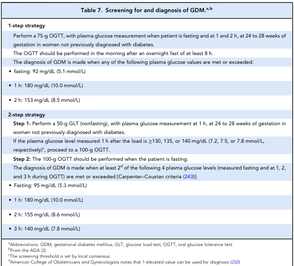
ADA Screening Table: 1-step vs 2-step GDM diagnosis criteria
Strategy 1: One-Step (IADPSG/WHO 2013)
Adopted by IADPSG (International Association of Diabetes and Pregnancy Study Groups) and WHO. Used widely in India (DIPSI is a variant).
Test: 75-g OGTT (fasting), at 24-28 weeks
Diagnosis if ANY ONE value is met or exceeded:
| Timepoint | Threshold |
|---|---|
| Fasting | ≥92 mg/dL (5.1 mmol/L) |
| 1 hour | ≥180 mg/dL (10.0 mmol/L) |
| 2 hours | ≥153 mg/dL (8.5 mmol/L) |
- Based on data from the HAPO study (Hyperglycemia and Adverse Pregnancy Outcomes) - a landmark study of >25,000 pregnant women showing continuous linear relationship between maternal glucose and adverse outcomes
- Increased sensitivity, detects more GDM, but increases treatment burden and resource use
Strategy 2: Two-Step (ACOG / USA standard)
Step 1 - Screening Test (non-fasting):
- 50-g Glucose Challenge Test (GCT) at 24-28 weeks
- Single plasma glucose at 1 hour
- Positive screen: plasma glucose ≥130 mg/dL (or ≥135 or ≥140 mg/dL per local protocol)
- If positive → proceed to Step 2
Step 2 - Diagnostic Test (fasting):
- 100-g OGTT, with plasma glucose fasting and at 1h, 2h, 3h
Carpenter-Coustan Criteria (ACOG recommended):
| Timepoint | Threshold |
|---|---|
| Fasting | ≥95 mg/dL (5.3 mmol/L) |
| 1 hour | ≥180 mg/dL (10.0 mmol/L) |
| 2 hours | ≥155 mg/dL (8.6 mmol/L) |
| 3 hours | ≥140 mg/dL (7.8 mmol/L) |
Diagnosis requires 2 or more elevated values (ACOG allows 1 elevated value in some guidelines)
NDDG Criteria (older, less used):
- Fasting: 105 mg/dL | 1h: 190 | 2h: 165 | 3h: 145 mg/dL
C. DIPSI (Diabetes in Pregnancy Study Group India) Criteria
A practical, single-step, non-fasting test used widely in India - suitable for resource-limited settings:
- 75-g oral glucose regardless of fasting state
- Single 2-hour plasma glucose
- GDM: ≥140 mg/dL
- Impaired GDM: 120-139 mg/dL
D. Other Screening Methods
| Test | Notes |
|---|---|
| Fasting plasma glucose | ≥92 mg/dL = GDM per IADPSG; cheap, simple |
| HbA1c | Not reliable in pregnancy - red cell turnover is increased causing falsely low values; used at booking to detect pre-existing DM (≥6.5% = overt DM) |
| Random blood glucose | ≥200 mg/dL + symptoms = overt DM |
| Urine glucose (glycosuria) | Poor sensitivity and specificity; renal threshold lowers in pregnancy; not recommended as sole screening tool |
| Glycated proteins (fructosamine) | Research use |
E. Key Clinical Points for Screening
- GCT can be done non-fasting (50-g)
- OGTT must be done fasting (overnight fast ≥8 hours)
- Patient must have unrestricted carbohydrate diet for 3 days prior to OGTT
- Patient should remain seated during OGTT; smoking prohibited
- Venous plasma glucose is the standard (fingerstick glucometers are NOT acceptable for diagnosis)
PART III: MANAGEMENT OF GDM
Management follows a stepwise approach: Dietary modification → Physical activity → Medical therapy (insulin/oral agents) → Obstetric surveillance.
(Creasy & Resnik's MFM; Harrison's 22E)
A. Target Glucose Values in GDM
| Timepoint | Target |
|---|---|
| Fasting / pre-meal | 65-95 mg/dL (ADA: <95 mg/dL) |
| 1-hour postprandial | <140 mg/dL |
| 2-hour postprandial | <120 mg/dL |
| Mean blood glucose | <100 mg/dL |
HbA1c monitoring is of minimal utility in pregnancy due to increased red cell turnover causing falsely low values. Blood glucose self-monitoring (SMBG) is the standard.
B. Non-Pharmacological Management
1. Medical Nutrition Therapy (MNT)
- Cornerstone of initial GDM management
- Caloric intake tailored to pre-pregnancy BMI:
- Normal BMI: 30 kcal/kg/day
- Overweight: 25 kcal/kg/day
- Obese: 15-20 kcal/kg/day (minimum 1800 kcal/day)
- Carbohydrate restriction to 35-45% of total calories, spread over 3 meals + 2-3 snacks
- Complex carbs with low glycaemic index preferred
- Adequate protein (20%) and fat (35-40%)
- Approximately 50% of women achieve adequate glucose control with diet alone (McFarland et al.)
2. Physical Activity
- Moderate aerobic exercise for 30 min, 3-5 times/week
- Postprandial walking (15-30 min) significantly reduces postprandial glucose
- Resistance training also beneficial
- Reduces insulin resistance and aids gestational weight management
- Contraindicated in placenta previa, preterm labor risk, severe hypertension
Important timing point: Allow only 1-2 weeks of dietary management before initiating medical therapy. Waiting longer misses the critical window of 28-34 weeks (period of accelerated fetal fat accretion). By 4 weeks of dietary therapy, only an additional 10% of patients achieve control (McFarland et al.).
C. Pharmacological Management
1. Insulin - PREFERRED First-Line Agent
Insulin is the gold standard for GDM treatment due to:
- Does not cross the placenta (no direct fetal effect)
- Proven safety and efficacy
- Lower treatment failure rates than oral agents
- Recommended by both ACOG and ADA as preferred therapy
Key landmark trials:
- ACHOIS Trial (Crowther et al.) - 1000 women; insulin-treated GDM had significantly lower serious perinatal complications (1% vs 4%) and lower leptin concentration (indicating less fetal fat mass)
- Landon et al. multicenter RCT - Treatment of mild GDM with insulin significantly reduced LGA (7.1% vs 14.5%), birth weight >4000g (5.9% vs 14.3%), shoulder dystocia (1.5% vs 4.0%), CS rates (26.9% vs 33.8%), and preeclampsia
Types of Insulin in Pregnancy (Creasy & Resnik, Table 59.12):
| Insulin Type | Onset | Peak | Duration | Notes |
|---|---|---|---|---|
| Regular insulin | 30-60 min | 2-4h | 6-8h | Safe; standard bolus |
| Lispro (Humalog) | 5-15 min | 30-90 min | 3-5h | Rapid-acting; preferred bolus |
| Aspart (Novolog) | 5-15 min | 30-90 min | 3-5h | Rapid-acting; safe in pregnancy |
| NPH | 1-2h | 4-8h | 12-16h | Traditional basal; cheap |
| Detemir (Levemir) | 1-2h | Flat | 18-24h | Long-acting; safe in pregnancy |
| Glargine (Lantus) | 1-2h | Flat | 20-24h | Data supports safety; some concerns |
| Degludec (Tresiba) | Slow | Flat | >42h | Limited pregnancy data |
Insulin Regimens:
- Typical starting dose for GDM: 0.7-0.8 units/kg/day (1st trimester) → 0.8-1.0 units/kg (2nd trimester) → 0.9-1.2 units/kg (3rd trimester)
- Combination regimen: Basal insulin (NPH or detemir) + rapid-acting bolus at meals
- Insulin pump (CSII) may be used in selected patients with pregestational DM
- Doses require weekly or more frequent adjustment as pregnancy progresses
Fasting hyperglycemia → treat with bedtime NPH or long-acting insulin
Postprandial hyperglycemia → treat with rapid-acting insulin before meals
2. Metformin
- Not recommended as first-line by ACOG or ADA for GDM
- Mechanism: Reduces hepatic glucose output, improves insulin sensitivity
- Key concern: Significant transplacental passage - metformin crosses to fetal compartment; inadequate long-term offspring safety data
- Studies show higher adiposity measurements in metformin-exposed children in utero
- MiTy Trial (Feig et al.) - metformin added to insulin in T2DM pregnancy led to lower HbA1c, lower insulin requirements, less weight gain, fewer CS deliveries, fewer LGA infants - but more SGA infants and lower birth weight
- Equivalent to insulin in short-term efficacy for GDM when insulin is not feasible
- Acceptable alternative when patient declines or cannot reliably administer insulin
- Dose: 500 mg once daily, titrate to max 2500 mg/day in divided doses
3. Glyburide (Glibenclamide)
- Currently NOT recommended by ACOG or ADA due to inferior outcomes compared to insulin and metformin
- Crosses placenta; associated with neonatal hypoglycemia
- Higher rates of macrosomia and NICU admissions compared to insulin
- If used: 2.5-5 mg twice daily (given 2 hours before meals), maximum 20 mg/day
D. Antepartum Fetal Surveillance
| Assessment | Frequency |
|---|---|
| Ultrasound for growth | Every 4 weeks from 28 weeks |
| Fetal weight estimation | 32-36 weeks to detect macrosomia |
| Biophysical profile / NST | Weekly from 36 weeks (or 32 weeks if poorly controlled) |
| Amniotic fluid index | For polyhydramnios (sign of poor glycemic control) |
| Umbilical artery Doppler | If IUGR or suspected |
Ultrasound identification of a large for gestational age (LGA) fetus or polyhydramnios are indicators of suboptimal glycemic control.
E. Continuous Glucose Monitoring (CGM)
- CONCEPTT Trial (T1DM in pregnancy) - CGM showed mild improvement in HbA1c, reduced LGA births, shorter NICU stay, and reduced neonatal hypoglycemia
- Target ranges (TIR) for T1DM pregnancy:
- Target range 63-140 mg/dL: goal >70% time in range
- Time below range (<63 mg/dL): goal <4%
- Time above range (>140 mg/dL): goal <25%
- Role in GDM is growing but evidence base developing
F. Obstetric Management - Timing and Mode of Delivery
| Scenario | Management |
|---|---|
| Diet-controlled GDM, no complications | Allow labor up to 40-41 weeks; ACOG suggests delivery by 40+6 |
| Insulin-treated GDM, well controlled | Induce at 39-40 weeks |
| Poorly controlled GDM | Deliver at 37-38 weeks |
| Estimated fetal weight ≥4500 g | Elective CS to prevent shoulder dystocia |
| Macrosomia (EFW ≥4000 g with suboptimal control) | Consider early induction |
Intrapartum management:
- Tight glucose control during labor (target 80-110 mg/dL)
- IV insulin infusion if needed during active labor
- Monitor fetal heart rate continuously
G. Complications of GDM
Maternal:
- Preeclampsia (2-4x increased risk)
- Operative delivery
- Risk of type 2 DM (7-10x increased risk; 10% develop T2DM within 5 years of delivery)
- Recurrence in subsequent pregnancies (30-50%)
Fetal/Neonatal:
- Macrosomia and LGA
- Shoulder dystocia and birth trauma (brachial plexus injury)
- Neonatal hypoglycemia (fetal hyperinsulinemia)
- Polycythemia
- Respiratory distress syndrome
- Hyperbilirubinemia
- Long-term childhood obesity and metabolic syndrome
H. Postpartum Management
- All GDM patients should have a 75-g 2-hour OGTT at 4-12 weeks postpartum to screen for persistent diabetes or impaired glucose tolerance
- If normal, retest every 1-3 years lifelong
- Metformin reduces the risk of T2DM in high-risk GDM patients postpartum
- Breastfeeding encouraged - reduces maternal insulin resistance
- Exercise and weight loss reduce long-term diabetes risk
- Lifestyle modification: most effective intervention to prevent progression to T2DM
SUMMARY TABLE
| Aspect | Key Points |
|---|---|
| Prevalence | ~8% of pregnancies |
| Pathophysiology | Failure of beta-cell compensation for physiological insulin resistance; placental anti-insulin hormones (hPL, cortisol) |
| Screening timing | 24-28 weeks universal; first visit for high-risk |
| One-step (IADPSG) | 75g OGTT: F ≥92, 1h ≥180, 2h ≥153 mg/dL (any one) |
| Two-step (ACOG) | 50g GCT → if ≥130: 100g OGTT (C-C criteria: F 95, 1h 180, 2h 155, 3h 140) |
| DIPSI (India) | 75g non-fasting, 2h value ≥140 mg/dL |
| Glucose targets | Fasting <95, 1h postprandial <140, 2h <120 mg/dL |
| First-line Rx | Diet + exercise → Insulin (if targets not met) |
| Insulin dose | 0.7-1.2 units/kg/day (increases each trimester) |
| Metformin | Acceptable alternative, not first-line; crosses placenta |
| Glyburide | Not recommended (ACOG, ADA 2024) |
| Delivery | Well-controlled: 39-40 weeks; CS if EFW ≥4500g |
| Postpartum | 75g OGTT at 4-12 weeks; lifelong annual screening |
Sources: Creasy & Resnik's Maternal-Fetal Medicine: Principles and Practice (9780323828499); Harrison's Principles of Internal Medicine 22E (9781265977061); ADA GDM Screening Table (ADA Standards of Care)
Tumor markers in gynecology 10 marks question in PG obs and gynec exam
Finding Sources
Finding Sources
Searching Images
tumor markers gynecological cancers ovarian cervical endometrial
Reading File
Finding Sources
Finding Sources
Reading File
Finding Sources
Finding Sources
Reading File
Finding Sources
I have the full gynecological tumor marker table. Let me now get Berek & Novak's detailed content on ovarian cancer markers and read the full gynecological tumor marker section:
Reading File
Finding Sources
I now have comprehensive data from multiple authoritative sources. Let me compile a complete, exam-ready 10-mark PG answer.
Tumor Markers in Gynecology
A Complete 10-Mark PG Answer
DEFINITION
A tumor marker is a substance - protein, enzyme, hormone, or antigen - produced by a tumor cell or by the host in response to a tumor, that can be detected in serum, urine, or tissue, and used for diagnosis, monitoring, and prognosis of malignancy.
Ideal properties of a tumor marker:
- High sensitivity (detect early disease)
- High specificity (positive only in that cancer)
- Correlate with tumor burden
- Short half-life (rapid response to treatment)
- Easy to measure; cost-effective
In practice, NO tumor marker is 100% sensitive or specific. They are best used for monitoring known disease and detecting recurrence, not primary screening.
CLASSIFICATION OF GYNECOLOGICAL TUMOR MARKERS
I. EPITHELIAL OVARIAN CANCER MARKERS
1. CA-125 (Cancer Antigen 125) - Most Important Gynecological Marker
-
Nature: Large membrane glycoprotein of the mucin family; encoded by MUC16 gene
-
Normal value: <35 U/mL
-
Sensitivity:
- Stage I disease: ~50% (poor early detection)
- Advanced disease (Stage III/IV): 80-90% for serous carcinoma
-
Clinical uses:
- Monitoring response to chemotherapy in known ovarian cancer
- Detecting recurrence post-treatment (rising CA-125 precedes clinical recurrence by months)
- Pre-operative assessment of pelvic mass (Risk of Malignancy Index uses CA-125)
- NOT recommended for population screening (USPSTF recommends against routine ovarian cancer screening with CA-125 - no mortality benefit, moderate harms from false positives)
-
Causes of false positive elevation (non-malignant):
- Endometriosis
- Pelvic inflammatory disease
- Uterine fibroids
- Ascites (any cause - hepatic, cardiac, peritoneal TB)
- Menstruation (transient elevation)
- Pregnancy (first trimester)
- Liver cirrhosis, pancreatitis
- Pleural effusion
-
Other malignancies: Endometrial, cervical, pancreatic, breast, lung cancers
(Berek & Novak's Gynecology; Henry's Clinical Diagnosis and Management)
2. HE4 (Human Epididymis Protein 4)
- Nature: Whey acidic protein family; expressed in normal fallopian tube epithelium
- Normal value: <150 pmol/L (varies by menopausal status)
- More specific than CA-125 for epithelial ovarian cancer
- Less affected by endometriosis or benign gynecological conditions
- ROMA Score (Risk of Ovarian Malignancy Algorithm): Combines HE4 + CA-125 + menopausal status
- Pre-menopausal: High risk if ROMA ≥13.1%
- Post-menopausal: High risk if ROMA ≥27.7%
- Superior to CA-125 alone in distinguishing malignant from benign pelvic masses
II. GERM CELL TUMOR MARKERS
These are the most important markers in young women with ovarian masses.
3. Alpha-Fetoprotein (AFP)
- Nature: Major fetal serum glycoprotein synthesized by yolk sac and fetal hepatocytes
- Normal value: <10 ng/mL (adult)
- Tumors: Yolk sac tumor (Endodermal Sinus Tumor) - markedly elevated; also embryonal carcinoma, mixed germ cell tumor
- Important: AFP is also elevated in:
- Hepatocellular carcinoma
- Hepatoblastoma
- Normal pregnancy (used in maternal serum screening)
- Benign liver disease (transient)
- Use: Pre-operative baseline; monitor response to chemotherapy; detect recurrence
4. Beta-hCG (Human Chorionic Gonadotropin)
- Nature: Glycoprotein hormone; beta subunit is tumor-specific
- Tumors:
- Choriocarcinoma - markedly elevated (main marker)
- Gestational Trophoblastic Disease (GTD): Hydatidiform mole, invasive mole, choriocarcinoma, PSTT
- Dysgerminoma (mildly elevated)
- Embryonal carcinoma
- Mixed germ cell tumors
- Unique feature: β-hCG is the most clinically useful marker in gynecology because:
- Quantitative hCG tracks disease accurately
- Used for diagnosis, treatment monitoring, and cure criteria
- In GTD, normalization of hCG = remission; rise = recurrence
- Screening recommended in all women post-molar pregnancy
- Causes of false elevation: Phantom hCG (heterophilic antibody interference), physiological pregnancy
5. LDH (Lactate Dehydrogenase)
- Tumor: Dysgerminoma (pure) - the most common malignant germ cell tumor
- Also elevated in: Other germ cell tumors, non-Hodgkin's lymphoma
- Less specific but useful in combination
6. Placental Alkaline Phosphatase (PLAP)
- Tumor: Dysgerminoma; also seminoma
- Used as additional marker alongside LDH for dysgerminoma
III. SEX CORD-STROMAL TUMOR MARKERS
7. Inhibin A and B
- Nature: Dimeric proteins produced by granulosa cells
- Tumor: Granulosa cell tumor (most specific marker)
- Also Sertoli-Leydig cell tumors (mildly)
- Mucinous cystadenoma/carcinoma (minor elevation)
- Inhibin B is more sensitive than Inhibin A for granulosa cell tumor
- Post-menopausal women: Inhibin should be undetectable; any elevation is suspicious
- Used for monitoring recurrence of granulosa cell tumors (which have a tendency for late recurrence - 5-30 years)
8. AMH (Anti-Müllerian Hormone)
- Produced by granulosa cells of small growing follicles
- Elevated in Adult-type granulosa cell tumors
- More stable than Inhibin; useful for long-term monitoring
- Normal in post-menopausal women → elevated AMH = suspicious for granulosa cell tumor
9. Estradiol / Testosterone
- Granulosa cell tumors: Estradiol elevated (causes isosexual precocious puberty in children; endometrial hyperplasia/carcinoma in adults)
- Sertoli-Leydig cell tumors: Testosterone and DHEAS elevated (causes virilization, amenorrhea, acne, clitoromegaly)
IV. CERVICAL CANCER MARKERS
10. SCC Antigen (Squamous Cell Carcinoma Antigen)
- Normal value: <1.5 ng/mL
- Tumor: Squamous cell carcinoma of the cervix (most important marker for this cancer)
- Correlates with stage, tumor volume, depth of invasion, and lymph node status
- Rising levels predict recurrence
- Other markers for cervical cancer: CEA (cervical adenocarcinoma), CA-125, CYFRA 21-1, TPA
(Henry's Clinical Diagnosis and Management - Gynecological Tumors Table)
V. ENDOMETRIAL CANCER MARKERS
11. CA-125
- Elevated in advanced endometrial cancer (Stage III/IV)
- Predicts extrauterine disease and lymph node involvement pre-operatively
- Not useful for early-stage endometrial cancer
12. HE4
- Superior to CA-125 for endometrial cancer
- Elevated in early-stage disease; correlates with myometrial invasion
- Useful for preoperative risk stratification
VI. GESTATIONAL TROPHOBLASTIC DISEASE (GTD) - Special Mention
| Type | hCG level |
|---|---|
| Hydatidiform mole | Markedly elevated (>100,000 mIU/mL) |
| Invasive mole | Elevated |
| Choriocarcinoma | Very high (can be millions) |
| PSTT (Placental Site Trophoblastic Tumor) | Low hCG (mainly hPL elevated) |
Surveillance protocol post-molar pregnancy:
- Weekly hCG until 3 consecutive normal values
- Then monthly for 6-12 months
- Diagnosis of persistent GTN: hCG plateau for 3 weeks or rise for 2 consecutive weeks
SUMMARY TABLE: GYNECOLOGICAL TUMOR MARKERS
| Tumor Marker | Cancer Type | Normal Value | Clinical Use |
|---|---|---|---|
| CA-125 | Epithelial ovarian, endometrial | <35 U/mL | Monitoring, recurrence |
| HE4 | Epithelial ovarian, endometrial | <150 pmol/L | Pre-op pelvic mass assessment, ROMA |
| AFP | Yolk sac tumor, embryonal ca | <10 ng/mL | Diagnosis + monitoring |
| β-hCG | Choriocarcinoma, GTD, dysgerminoma | <5 mIU/mL | Diagnosis, treatment, cure |
| LDH | Dysgerminoma | Lab-specific | Monitoring |
| Inhibin B | Granulosa cell tumor | Undetectable (post-menopause) | Diagnosis, recurrence |
| AMH | Granulosa cell tumor | Undetectable (post-menopause) | Recurrence monitoring |
| Estradiol | Granulosa cell tumor | - | Clinical effect marker |
| Testosterone | Sertoli-Leydig tumor | - | Virilization assessment |
| SCC antigen | Squamous ca cervix | <1.5 ng/mL | Staging, recurrence |
| CEA | Cervical adenocarcinoma, mucinous ovarian | - | Monitoring |
LIMITATIONS OF TUMOR MARKERS (Important for PG Exam)
- No marker is 100% sensitive or specific
- False positives occur in benign conditions (CA-125 in endometriosis, hCG in normal pregnancy)
- Serial measurements from the same laboratory using the same assay are essential for monitoring
- Half-life must be considered when assessing post-treatment response:
- Wait at least 2 weeks (preferably 1 month) after surgery before re-checking
- Heterogeneous tumors may require multiple markers simultaneously
- Heterophilic antibody interference can cause falsely high values (particularly for hCG - "phantom hCG")
- Markers for screening vs. monitoring are different roles - CA-125 fails as a screening tool but succeeds as a monitoring tool
RISK OF MALIGNANCY INDEX (RMI) - Using CA-125 in Practice
RMI = U × M × CA-125
Where:
- U (ultrasound score): 0 = no features, 1 = one feature, 3 = two or more features
- M (menopausal status): 1 = pre-menopausal, 3 = post-menopausal
- Ultrasound features: Multilocular cyst, solid areas, bilateral lesions, ascites, intra-abdominal metastasis
Interpretation:
- RMI <25: Low risk (benign)
- RMI 25-250: Moderate risk
- RMI >250: High risk - refer to gynae-oncology centre
(RMI >200 has sensitivity ~85%, specificity ~97% for ovarian malignancy)
Sources: Berek & Novak's Gynecology (9781496380357); Henry's Clinical Diagnosis and Management by Laboratory Methods (9780323673204); Robbins & Cotran Pathologic Basis of Disease (9780443264528)
This is a shared conversation. Sign in to Orris to start your own chat.