
Why itching in dialysis patient pathophysiology
uremic pruritus pathophysiology dialysis
PMID: 41598433
PMID: 41251635
uremic pruritus pathophysiology dialysis itching mechanism diagram
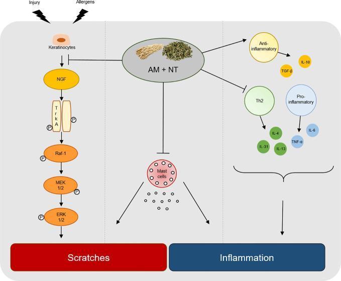
A pathophysiology diagram illustrating the therapeutic mechanism of an herbal mixture (AM + NT) on Allergic Contact Dermatitis (ACD). The diagram is divided into three functional pathways: pruritus (scratches), cellular infiltration, and cytokine regulation. The pruritus pathway shows injury and allergens activating keratinocytes, which release Nerve Growth Factor (NGF). This triggers the TrkA/Raf-1/MEK 1/2/ERK 1/2 phosphorylation cascade, leading to scratching behavior. The central section depicts the herbal mixture inhibiting mast cell activation and subsequent degranulation. The right section illustrates the regulation of the inflammatory response: promoting anti-inflammatory mediators (IL-10, TGF-β) while inhibiting Th2-specific cytokines (IL-4, IL-13, IL-31) and pro-inflammatory markers (IL-6, TNF-α). The overall summary demonstrates how the treatment modulates these molecular pathways to ameliorate skin inflammation and pruritus in dermatological conditions.
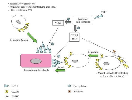
This pathophysiology diagram illustrates the signaling pathways and cellular mechanisms involved in the repair of injured mesothelial cells during continuous ambulatory peritoneal dialysis (CAPD). The schematic shows the interplay between peritoneal adipose tissue and mesothelial regeneration. Peritoneal adipose tissue, influenced by CAPD and up-regulated by VEGF, secretes TGF-̢ and HGF. These factors stimulate the up-regulation of the SDF-1 (stromal-derived factor-1) chemokine on injured mesothelial cells. SDF-1 (blue triangles) acts as a ligand for CXCR4 receptors (yellow U-shapes) on migrating cells, including bone marrow precursors, omental lymphoid progenitor cells, and CD34+ cells from the stromal vascular fraction (SVF). A regulatory mechanism is depicted where DPPIV (represented by scissors) cleaves the CXCR4 receptor to inhibit migration. The diagram highlights the SDF-1/CXCR4 axis as a critical driver for cell trafficking, recruitment, and subsequent repair and re-epithelialization of the peritoneal membrane after injury.
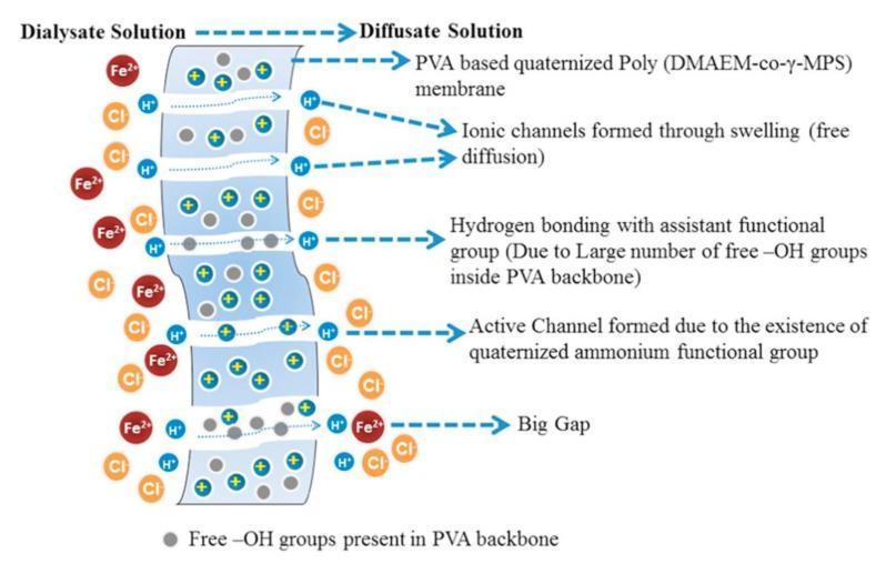
This schematic diagram illustrates the mechanism of diffusion dialysis using a PVA-based quaternized Poly (DMAEM-co-γ-MPS) membrane, a process used in medical and chemical purification for ion separation. The diagram shows two compartments: the Dialysate Solution (left) and the Diffusate Solution (right), separated by a semi-permeable membrane. The dialysate contains Fe2+, H+, and Cl- ions. The membrane structure is detailed with four transport mechanisms: 1) Ionic channels formed through swelling that facilitate free diffusion; 2) Hydrogen bonding sites facilitated by free -OH groups (represented as grey circles) within the PVA backbone; 3) Active channels formed by quaternized ammonium functional groups (indicated by blue circles with positive signs); and 4) Large physical gaps. Selective transport is demonstrated as smaller H+ and Cl- ions successfully transition from the dialysate to the diffusate side via these channels, while larger Fe2+ cations are primarily retained or move through larger gaps. This visual serves as an educational tool for understanding anion exchange membrane (AEM) properties, selective permeability, and the pathophysiology of ion filtration systems.
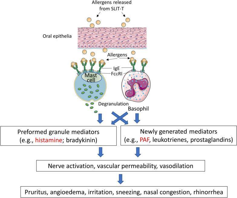
This pathophysiology diagram illustrates the biological mechanism of local allergic reactions induced by sublingual immunotherapy tablets (SLIT-T). The process begins with the release of allergens from SLIT-T into the oral epithelia. These allergens cross the epithelial barrier and bind to IgE antibodies complexed with FcεRI receptors on the surface of mast cells and basophils. This cross-linking triggers mast cell degranulation, releasing preformed granule mediators such as histamine and bradykinin. Simultaneously, it stimulates basophils to produce newly generated mediators, including platelet-activating factor (PAF), leukotrienes, and prostaglandins. The flowchart then maps these inflammatory mediators to their downstream physiological effects: nerve activation, increased vascular permeability, and vasodilation. Finally, the diagram lists the resulting clinical symptoms, such as pruritus, angioedema, irritation, sneezing, nasal congestion, and rhinorrhea. This educational resource summarizes the Type I hypersensitivity response specific to the sublingual route of allergen administration.
Uremic Pruritus (UP) in Dialysis Patients - Pathophysiology
1. Uremic Toxin Accumulation
- Retained toxins directly activate cutaneous sensory nerve endings (C-fibers)
- Middle molecules that are poorly cleared by standard hemodialysis membranes are implicated
- This explains why patients on peritoneal dialysis have less severe pruritus than those on hemodialysis, and why renal transplantation completely eliminates it
2. Xerosis (Skin Dryness) - 60-90% of Dialysis Patients
- Atrophy of sweat glands with impaired sweat secretion
- Sebaceous gland atrophy
- Disturbed stratum corneum hydration
- Abnormal arborization of free cutaneous nerve fiber endings
3. Mineral & Parathyroid Hormone (PTH) Abnormalities
- Elevated PTH, calcium, phosphate, and magnesium are all implicated
- An elevated calcium-phosphate product in the skin triggers mast cell degranulation
- However, serum levels of these minerals do not consistently correlate with UP severity, indicating these are contributing (not sole) factors
4. Mast Cell Activation and Histamine
- The number of dermal mast cells is increased in uremic skin
- Higher tryptase and histamine plasma concentrations are found in severe cases
- Histamine release is triggered by substance P - a key neurotransmitter in itch signaling
- The role of elevated serotonin (5-HT3) levels is debated; clinical trials with 5-HT3 inhibitors (ondansetron) have yielded conflicting results
5. The Opioid Hypothesis (Central Neuromodulation)
- Overexpression of μ-opioid receptors (pro-itch) in dermal cells and lymphocytes
- Decreased κ-opioid receptor expression (anti-itch)
- This imbalance shifts signaling toward itch facilitation
- Supporting evidence: activation of the κ-opioid system using a κ-receptor agonist reduces UP in animal models
- This is the basis for difelikefalin (selective peripheral κ-receptor agonist, FDA-approved for dialysis-associated pruritus) and nalfurafine (used in Japan)
6. The Immune / Inflammatory Hypothesis
- Th1 lymphocyte predominance: numbers of CXCR3-expressing, interferon-γ-secreting CD4+ T cells (Th1 markers) are significantly increased in circulation of dialysis patients with UP vs. those without
- Elevated inflammatory cytokines: high-sensitivity CRP, IL-2, IL-6, and IL-31 are all higher in UP patients (IL-31 is a key itch-promoting cytokine)
- UVB phototherapy benefit is partly explained through this pathway: UVB attenuates Th1 development in favor of Th2 helper cells
7. Neuropathic Component
- Uremia causes peripheral neuropathy, which alters the sensitivity and firing pattern of cutaneous sensory C-fibers (the itch-transmitting neurons)
- Abnormal arborization of nerve endings in the dermis is seen histologically
- This explains why gabapentin and pregabalin (which reduce neuropathic firing) are effective treatments
8. Dialysis-Specific Triggers (Allergic/Reactive)
- Heparin
- Ethylene oxide (ETO) used for membrane sterilization
- Formaldehyde
- Acetate in dialysate
- The dialysis membrane material itself
9. Inadequate Dialysis
- Inadequate clearance of uremic toxins is a direct contributor
- Increasing dialysis dose and optimizing membrane biocompatibility can reduce UP severity
Summary Diagram of Mechanisms
| Mechanism | Key Mediators |
|---|---|
| Uremic toxin retention | Middle molecules, retained solutes |
| Skin xerosis | Sweat/sebaceous gland atrophy, dry stratum corneum |
| Mineral abnormalities | PTH, Ca-P product, Mg |
| Mast cell/histamine | Tryptase, histamine, substance P |
| Opioid dysregulation | ↑μ-receptors, ↓κ-receptors |
| Immune dysregulation | Th1 predominance, IL-31, IL-6, IFN-γ |
| Neuropathic changes | Abnormal C-fiber arborization |
| Dialysis-related allergy | ETO, heparin, acetate, membrane |
Complications of Chronic Scratching
-
Excoriations and superimposed skin infections
-
Prurigo nodularis - treatment-resistant lichenified/excoriated papulonodular eruption
-
Acquired perforating dermatosis - a hallmark CKD skin complication where dermal material is extruded through the epidermis due to repeated trauma
-
Andrews' Diseases of the Skin, CKD section; Comprehensive Clinical Nephrology 7th Ed.
Treatment for that
uremic pruritus treatment dialysis
PMID: 39712488
Treatment of Uremic Pruritus (UP) in Dialysis Patients
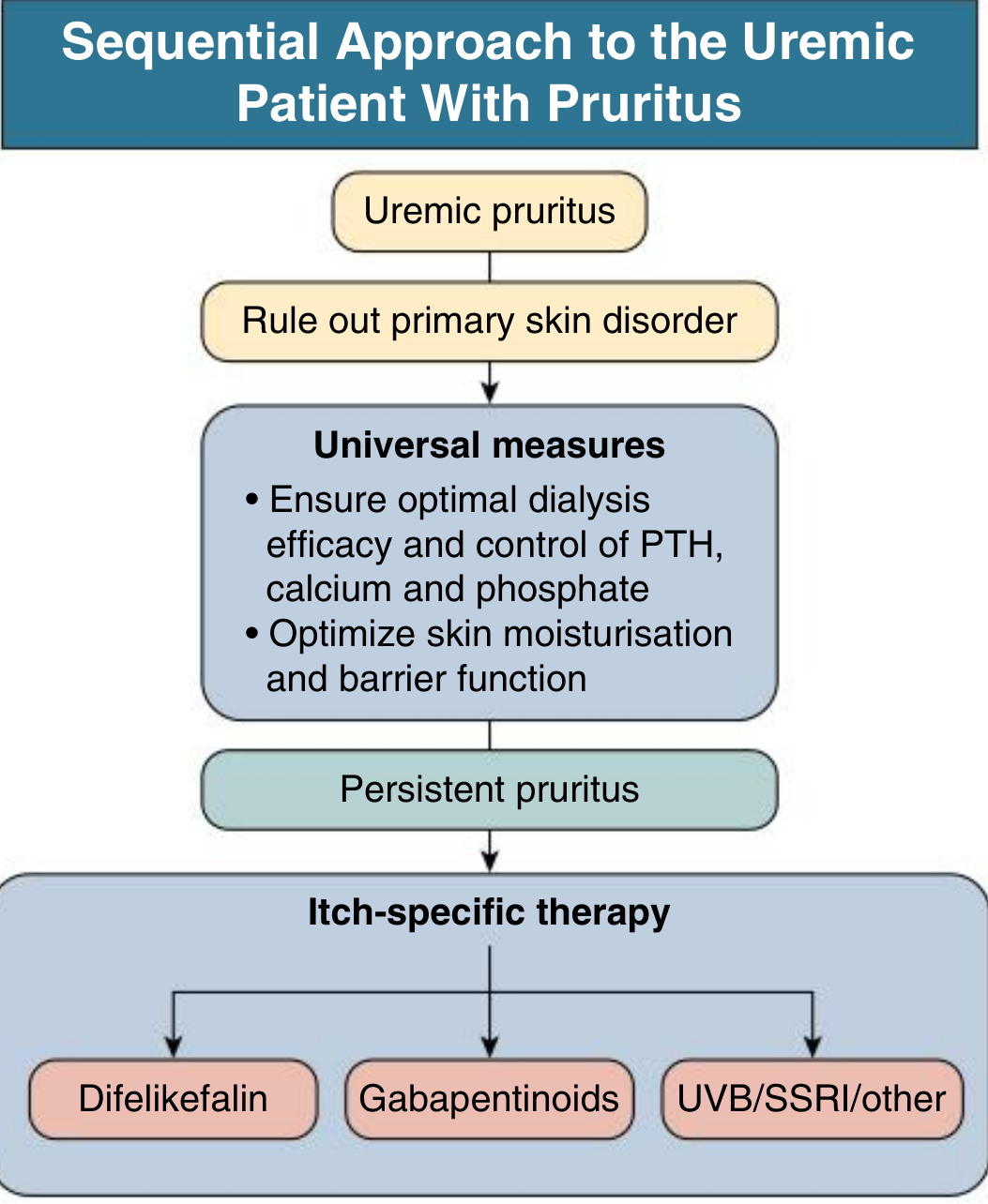
STEP 1 - Universal/Foundation Measures (Start in ALL patients)
A. Optimize Dialysis
- Improve dialysis adequacy (target Kt/V) - inadequate clearance directly worsens pruritus
- Use medium cut-off (MCO) dialyzers for better middle molecule clearance
- Switch to biocompatible membranes (synthetic, gamma-sterilized) rather than cellulose/ETO-sterilized
- Use bicarbonate dialysate instead of acetate
- Use low-calcium / low-magnesium dialysate (short-term; prolonged use risks worsening renal osteodystrophy)
- Discontinue formaldehyde use for dialyzer reprocessing
B. Control Mineral Metabolism
- Control PTH, calcium, phosphate (elevated Ca-P product drives mast cell activation and itching)
- Note: parathyroidectomy is NOT routinely advocated for UP relief - consistent benefit has not been demonstrated
C. Skin Moisturization - Treat Xerosis
- Emollients are the primary first-line topical treatment
- Use simple, fragrance-free/additive-free moisturizers applied consistently
- Soaking and smearing technique (soak in water, immediately apply emollient)
- Gamma-linolenic acid (GLA) cream twice daily - shown effective in trials
- Bath oil with polidocanol (monoether of lauryl alcohol + macrogol) - helps some patients
- Sericin cream (silkworm biopolymer, suppresses pro-inflammatory cytokines) - reduced UP significantly in a 6-week RCT in 50 dialysis patients
STEP 2 - Itch-Specific Pharmacotherapy (for Persistent Pruritus)
1. Kappa-Opioid Receptor Agonists (Most Evidence / Preferred Systemic Agents)
| Drug | Dose | Notes |
|---|---|---|
| Difelikefalin | IV thrice-weekly post-HD | FDA-approved (2021) for moderate-severe CKD-aP on HD; 51.9% vs. 30.9% placebo responders; peripherally restricted - low CNS side effects. ADRs: diarrhea, dizziness, vomiting |
| Nalfurafine | Oral 2.5-5 mg/day or IV post-HD | Approved in Japan; sustained benefit shown at 1 year; ADRs: insomnia (19%), constipation (7%), elevated prolactin |
2. Gabapentinoids (Strong Neuropathic Evidence)
- Gabapentin 100-300 mg given after each dialysis session (thrice-weekly dosing)
- Effective in 2 controlled studies; reduces UP score significantly at 4 weeks
- CAUTION: renally excreted - accumulates in ESKD; start at 100 mg post-dialysis and titrate slowly
- Side effects: dizziness, somnolence, fatigue, nausea
- Pregabalin - similar effects shown in smaller studies
- Andrews' Diseases of the Skin recommends: "low initial dose of 100 mg after each session with slow upward titration"
3. Phototherapy (UVB)
- Broadband UVB (280-315 nm) is effective and well tolerated
- Thrice-weekly total-body UVB sessions (total 8-10 sessions)
- Antipruritic effect may last several months
- Contraindicated for prolonged use (>12 weeks) in skin phototypes I and II (fair skin) due to carcinogenic risk
- NB-UVB: an RCT failed to confirm its efficacy; broadband UVB appears superior for CKD patients
- Mechanism: shifts Th1 → Th2 immune balance
4. Topical Capsaicin (0.025% cream)
- Depletes substance P from cutaneous type-C sensory nerve endings
- Two clinical studies showed significant relief of UP
- Well tolerated; no systemic side effects
- Limitation: burning sensation on application; difficult to use over large areas
STEP 3 - Second-Line / Adjunct Agents
Antihistamines
- Classic antihistamines have similar efficacy to emollients only - limited benefit
- Desloratadine (2nd generation) may have some added effect
- Ketotifen 2-4 mg/day (mast cell stabilizer) showed benefit in one small study
- Generally insufficient as monotherapy for moderate-severe UP
Topical Calcineurin Inhibitors
- Tacrolimus ointment 0.1% - one prospective study showed significant reduction in UP after 6 weeks; well tolerated, no detectable systemic absorption
- However, a subsequent smaller vehicle-controlled trial showed equal benefit with vehicle alone
- Prolonged use is not recommended until more safety data are available
Essential Fatty Acids
- Gamma-linolenic acid (GLA) - primrose oil orally - significant improvement in UP; augments anti-inflammatory eicosanoid synthesis
- Fish oil, olive oil, safflower oil - similar effects reported
Oral Activated Charcoal
- 6 g/day for 8 weeks - UP completely resolved or significantly reduced in two clinical studies
- Inexpensive and well tolerated; considered a valuable alternative
5-HT3 Antagonists (Ondansetron)
- Effective in one small peritoneal dialysis study
- Failed to show benefit over placebo in a larger HD RCT
- Not recommended as routine therapy
STEP 4 - Refractory/Severe UP Only
Thalidomide
- A 7-day course reduced UP intensity by up to 80% in a placebo-controlled crossover study (n=29)
- Reserve for therapy-resistant severe UP in non-reproductive-age patients only
- Contraindications: teratogenic (absolute in women of childbearing age), peripheral neuropathy risk, cardiovascular side effects
Immunomodulatory Approaches
- SSRIs (sertraline) - promising in earlier reports but not consistently effective in controlled trials
- Anti-IL-31 biologics (dupilumab) - preliminary but promising emerging data (2026)
Emerging Therapies (2025-2026)
- AST-120 (spherical carbon adsorbent) - reduces uremic toxin load
- Omega-3 fatty acids - promising preliminary results
- Dupilumab (anti-IL-4/IL-13 biologic) - early trials showing benefit; targets the Th2 cytokine axis
Definitive Treatment
- Renal transplantation eliminates uremic pruritus completely
Summary Table
| Category | Agent | Evidence Level |
|---|---|---|
| Dialysis optimization | MCO membrane, bicarbonate, biocompatible | Clinical practice |
| Topical | Emollients, GLA cream, capsaicin 0.025% | RCT/case series |
| κ-opioid agonist | Difelikefalin (IV), nalfurafine | RCT / FDA-approved |
| Neuropathic | Gabapentin 100-300 mg post-HD | RCT |
| Phototherapy | Broadband UVB x8-10 sessions | RCT |
| Antihistamine | Desloratadine, ketotifen | Limited RCT |
| Charcoal | Activated charcoal 6 g/day | Clinical studies |
| Immunomodulator | Tacrolimus 0.1% topical | Conflicting RCT |
| Refractory only | Thalidomide | RCT (n=29) |
| Definitive | Renal transplantation | Observational |
- Comprehensive Clinical Nephrology 7th Ed., Fig. 91.3; Andrews' Diseases of the Skin