
Explain pancreatitis
Reading File
Now I'll search the medical library and PubMed in parallel for comprehensive pancreatitis coverage.
Finding Sources
Searching PubMed
"pancreatitis"[MeSH Terms] AND "management"[MeSH Terms]
The best sources are Sleisenger & Fordtran's GI textbook and Rosen's Emergency Medicine. Let me read those in parallel.
Reading File
Reading File
Finding Sources
Excellent content so far. Let me read more from Rosen's EM and Sleisenger's dedicated pancreatitis sections in parallel.
Reading File
Reading File
Good coverage. Let me now read the Rosen's EM section for the diagnosis, severity scoring, and management content.
Reading File
Finding Sources
Reading File
Reading File
I now have very rich content. Let me read the management/treatment section from Rosen's EM and fetch the pancreas anatomy image.
Reading File
Loading Image
I now have comprehensive content from multiple authoritative textbooks. Let me compile the full answer.
Pancreatitis
Pancreatitis is inflammation of the pancreas, ranging from mild self-limited disease to life-threatening multiorgan failure. It is the most common pancreatic disease worldwide and a leading GI cause of hospitalization in the United States.
Anatomy of the Pancreas
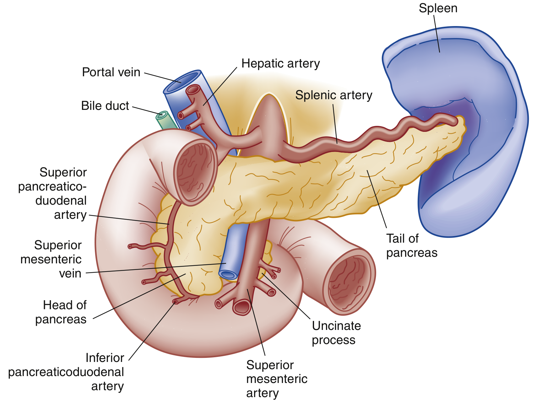
Pancreatic anatomy, anterior view - Rosen's Emergency Medicine
The pancreas is a retroperitoneal organ with three segments:
- Head - sits within the duodenal C-loop; the main pancreatic duct (duct of Wirsung) joins the common bile duct here to form the ampulla of Vater, draining via the sphincter of Oddi
- Body - traverses posterior to the stomach
- Tail - abuts the splenic hilum
Exocrine function: secretes digestive enzymes (e.g., trypsinogen, lipase, amylase) into the duodenum
Endocrine function: secretes insulin, glucagon, and somatostatin
Pathophysiology
Injury begins with an inciting event (gallstone obstruction, alcohol, drugs, etc.) that disrupts normal membrane trafficking in acinar cells. This triggers:
- Inappropriate activation of trypsinogen → trypsin within the gland
- Trypsin activates other digestive enzymes, causing autodigestion of pancreatic tissue
- Macrophage and neutrophil recruitment amplify destruction
- Cytokine release increases vascular permeability → edema, hemorrhage, and necrosis
- A heightened systemic inflammatory response can cause SIRS → sepsis → shock
- Bacteremia may occur via translocation of intestinal flora through a compromised gut barrier
- Extrapancreatic complications include pleural effusions, ARDS, and renal failure
- Rosen's Emergency Medicine, p. 1265
Causes
| Category | Examples |
|---|---|
| Toxic-Metabolic (most common) | Alcohol (25-35%), drugs, hypertriglyceridemia, hypercalcemia, uremia |
| Mechanical-Obstructive (most common) | Gallstones (40-70%), post-ERCP, pancreas divisum, ampullary tumors, trauma |
| Infectious | Viral (mumps, coxsackie, CMV, EBV, HIV); bacterial (TB, Mycoplasma, Salmonella); parasitic (Ascaris) |
| Vascular | Vasculitis, embolism, ischemia, hypercoagulability |
| Other | Idiopathic, hereditary/genetic (SPINK1, CFTR mutations), autoimmune, DKA |
Together, gallstones and alcohol account for ~65-80% of all adult cases.
- Rosen's Emergency Medicine, Box 77.1, p. 1266
Types
1. Acute Pancreatitis (AP)
Sudden inflammatory disease with enzymatic autodigestion. Most episodes are interstitial edematous (resolves within 1 week). Approximately 5-10% develop necrotizing pancreatitis, where the parenchyma and surrounding tissue undergo necrosis - this can remain sterile, liquefy, or become infected, with infected necrosis carrying significantly higher morbidity.
2. Chronic Pancreatitis (CP)
Progressive, permanent damage to pancreatic tissue, usually visible as calcifications on X-ray and CT. Leads to exocrine insufficiency (malabsorption, steatorrhea), endocrine insufficiency (diabetes), and a chronic pain syndrome. Often related to ongoing alcohol use. Many patients develop pseudocysts, ascites, fistulae, and mesenteric venous thrombosis.
- Textbook of Family Medicine, p. 1139
Clinical Features of Acute Pancreatitis
Symptoms:
- Persistent epigastric or LUQ pain radiating to the back, chest, or flanks
- Pain is moderate to severe; classically relieved by sitting forward or leaning ahead
- Nausea, vomiting, and anorexia (oral intake worsens pain)
Signs:
- Vital signs may be normal in mild disease; tachycardia and fever are common
- Jaundice suggests biliary obstruction (gallstone pancreatitis or tumor)
- Shallow respirations, basilar crackles (pulmonary complications)
- Epigastric tenderness with guarding; absent bowel sounds (ileus)
- Cullen sign - bluish periumbilical discoloration (hemoperitoneum) - rare but poor prognosis
- Grey Turner sign - reddish-brown flank discoloration (retroperitoneal bleeding) - rare but poor prognosis
Systemic complications:
-
Pulmonary: pleural effusions (in up to 50%, more often left-sided), atelectasis, ARDS
-
Cardiovascular: hypotension, shock from fluid shifts
-
Renal: failure from hypoperfusion and inflammatory mediators
-
Metabolic: hyperglycemia (decreased insulin), hypocalcemia (from low albumin/magnesium), coagulopathy/DIC
-
Rosen's Emergency Medicine, pp. 1265-1266
Diagnosis
Diagnosis requires at least 2 of 3 criteria (Atlanta criteria):
- Abdominal pain characteristic of acute pancreatitis
- Serum lipase or amylase ≥ 3× the upper limit of normal
- Characteristic findings on abdominal imaging
Laboratory:
- Lipase - preferred; more sensitive and specific than amylase; rises within hours, remains elevated for 1-2 weeks
- Amylase - less specific (also elevated in renal failure, salivary gland disease, cholecystitis, bowel obstruction); stays elevated for 3-5 days
- CBC (leukocytosis), metabolic panel (renal function, LFTs, calcium, glucose), lipid panel
- Note: normal enzyme levels do not exclude pancreatitis in alcohol-related cases
Imaging:
- CT abdomen with contrast - gold standard for severity assessment; shows peripancreatic fat stranding, fluid collections, and necrosis
- Ultrasound - first-line to detect gallstones as etiology; limited for pancreas visualization
- MRCP/MRI - useful to evaluate pancreatic/biliary ducts without radiation
- Plain films and chest X-ray are supportive (may show ileus, pleural effusions, "colon cut-off sign")
Severity Classification (Revised 2012 Atlanta Classification)
| Grade | Criteria |
|---|---|
| Mild | No organ failure; no local or systemic complications |
| Moderately Severe | Transient organ failure (<48 h) OR local/systemic complications |
| Severe | Persistent organ failure (>48 h); modified Marshall score ≥2 for respiratory, cardiovascular, or renal systems |
Note: severe classification cannot be assigned until 48 hours after presentation.
Scoring systems: Ranson criteria (uses admission + 48-hour labs to predict mortality), APACHE II, BISAP score, and CT Severity Index (CTSI) are all used.
- Rosen's Emergency Medicine, Box 77.4, p. 1268
Local Complications
| Setting | Complication | Timing |
|---|---|---|
| Interstitial edematous AP | Acute peripancreatic fluid collection | <4 weeks |
| Interstitial edematous AP | Pancreatic pseudocyst (well-defined wall) | >4 weeks |
| Necrotizing AP | Acute necrotic collection | <4 weeks |
| Necrotizing AP | Walled-off necrosis (well-defined wall) | >4 weeks |
Other late complications: splenic/portal vein thrombosis, GI bleeding, gastric outlet obstruction, bowel necrosis.
Management
Acute Pancreatitis
Supportive care is the cornerstone:
- Fluid resuscitation - aggressive IV hydration (Lactated Ringer's preferred); corrects hypovolemia, maintains pancreatic perfusion, and limits SIRS progression
- Analgesia - IV opioids for pain control (no evidence that any opioid worsens pancreatitis)
- NPO vs. early enteral nutrition (EN):
- Historical practice of NPO (to "rest the pancreas") is no longer standard
- Mild-moderate AP: oral diet should be offered as tolerated; EN considered only if diet cannot be advanced after 4 days
- Severe AP with SIRS/ICU admission: nasogastric (NG) or nasojejunal (NJ) tube feeding should be initiated within 24-36 hours
- Two meta-analyses show EN vs. parenteral nutrition (PN) provides a ~2-fold reduction in infectious complications and a ~2.5-fold reduction in mortality risk
- The AGA technical review of 12 RCTs confirms EN reduces infected peripancreatic necrosis (OR 0.28), single organ failure (OR 0.25), and multiorgan failure (OR 0.41)
- Antibiotics - NOT routinely indicated; reserved for confirmed infected necrosis or other bacterial complications
- ERCP with sphincterotomy - indicated within 24-48 hours for gallstone pancreatitis with concurrent cholangitis or biliary obstruction
Chronic Pancreatitis
-
Pain management: analgesics, antioxidants (selenium, ascorbic acid, β-carotene, vitamin E - evidence mixed), nerve blocks, ERCP, or surgery for selected patients
-
Exocrine insufficiency: pancreatic enzyme replacement with each meal
-
Endocrine insufficiency: insulin therapy for diabetes
-
Alcohol abstinence - essential; continued use drives recurrent attacks
-
Jejunal feeding can improve weight and reduce pain in malnourished patients
-
Sleisenger and Fordtran's Gastrointestinal and Liver Disease, pp. 2935-2941
Differential Diagnosis
Acute pancreatitis must be distinguished from:
- Abdominal: peptic ulcer disease, acute cholecystitis/cholangitis, bowel obstruction, mesenteric ischemia, perforated viscus, ruptured AAA, ectopic pregnancy
- Cardiopulmonary: myocardial infarction, pneumonia, pericarditis
- Systemic: DKA, sickle cell crisis
Key Points
- Gallstones and alcohol together cause ~65-80% of cases
- Lipase is preferred over amylase (more sensitive and specific)
- Atlanta 2012 classifies severity as mild/moderately severe/severe based on organ failure duration
- Early enteral nutrition in severe AP is strongly preferred over parenteral nutrition
- Antibiotics are NOT routinely given - only for infected necrosis
- Chronic pancreatitis causes irreversible damage, malabsorption, and diabetes
Sources:
- Rosen's Emergency Medicine: Concepts and Clinical Practice, Chapter 77
- Sleisenger and Fordtran's Gastrointestinal and Liver Disease
- Textbook of Family Medicine, 9e
This is a shared conversation. Sign in to Orris to start your own chat.