
Pterygium
pterygium eye conjunctival growth cornea
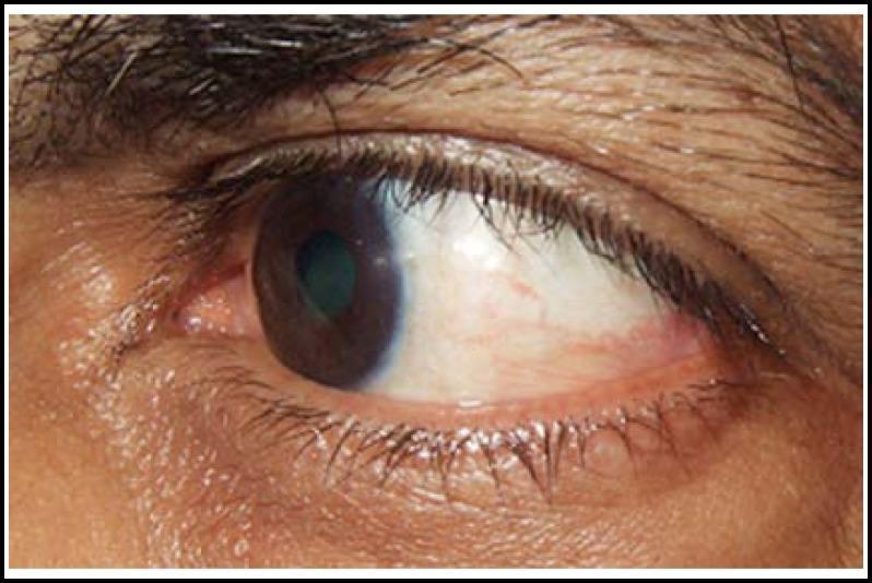
This clinical photograph shows a close-up, lateral view of a human left eye three months after pterygium excision and conjunctival autograft (CAG) surgery. The focus is on the temporal bulbar conjunctiva and the corneal-limbal interface. The cornea is largely clear and transparent, with a smooth, reflective surface. The site of the previous temporal pterygium exhibits a healthy conjunctival autograft that has successfully integrated, showing minimal residual thickening or scarring at the limbus. There is a mild degree of conjunctival injection (vascularization) visible in the temporal region, which is expected during the late postoperative recovery phase. The sclera remains naturally white with some fine episcleral vessels. No significant recurrence of the fibrovascular growth is observed on the corneal surface, demonstrating a stable surgical outcome for temporal pterygium management. The iris and pupil appear normal behind the clear corneal window.
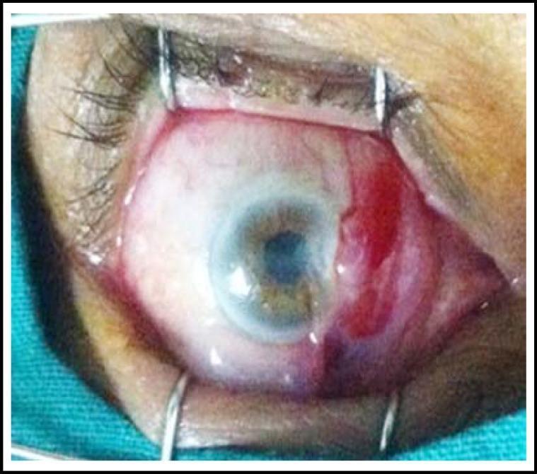
This clinical photograph captures a perioperative view of a human eye during a surgical procedure, likely for pterygium excision. The eye is held open by a wire lid speculum, providing full exposure of the bulbar conjunctiva and cornea. There is prominent focal conjunctival injection and subconjunctival hemorrhage on the nasal side, consistent with the administration of subconjunctival anesthesia (xylocaine 2%). A pterygium is visible as a fleshy, fibrovascular growth extending from the nasal bulbar conjunctiva onto the corneal surface. The cornea itself appears relatively clear except for the site of pterygium encroachment. This image serves as an educational reference for ophthalmology students and residents to understand local anesthesia administration and the preoperative presentation of pterygium within a sterile surgical field. Key landmarks include the limbus, the nasal bulbar conjunctiva showing vascular engorgement, and the eyelid margins retracted by surgical instrumentation.
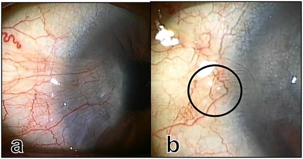
This comparison set of clinical ophthalmology photographs demonstrates the before and after states of a pterygium surgery on the nasal conjunctiva and cornea. Image (a) depicts a preoperative pterygium characterized by a fleshy, triangular fibrovascular tissue growth extending from the nasal conjunctiva over the limbus onto the corneal surface. The vascular pattern is dense and disorganized, with tortuous, engorged vessels concentrated at the pterygium head. Image (b) shows the same eye four months postoperatively following a superficial keratectomy and conjunctival repositioning. The corneal surface is clear, indicating successful removal of the fibrovascular encroachment. A black circle in image (b) highlights a specific vascular loop near the limbus, which is an anatomical hallmark of restored normal limbal vascular architecture. The surrounding conjunctival tissue appears healthy with significantly reduced inflammation and finer, more attenuated blood vessels compared to the preoperative state. These images serve as educational material for assessing surgical outcomes in ocular surface reconstruction and identifying normal versus pathological limbal vascularization.
pterygium treatment management recurrence
Pterygium
Definition
Pathophysiology & Etiology
- Elastotic degeneration of the deep conjunctival stromal collagen, with vascular proliferation and sub-epithelial fibrovascular tissue ingrowth onto the cornea, invading Bowman's layer.
- Unlike a pinguecula, a pterygium actively encroaches onto the cornea.
- Driven by chronic UV exposure (particularly UVB), dry/dusty/windy environments, and chronic ocular surface irritation.
- More common in people living in equatorial regions ("pterygium belt" - latitudes 30° N to 30° S).
- Has a familial tendency.
- Histologically identical to a pinguecula, but distinguished by corneal invasion.
Clinical Features
Anatomy of a Pterygium
| Zone | Description |
|---|---|
| Cap | Avascular, halo-like zone at the advancing edge |
| Head | Apex where the lesion meets the cornea |
| Body | The main vascularized bulk extending to the conjunctiva |
Signs
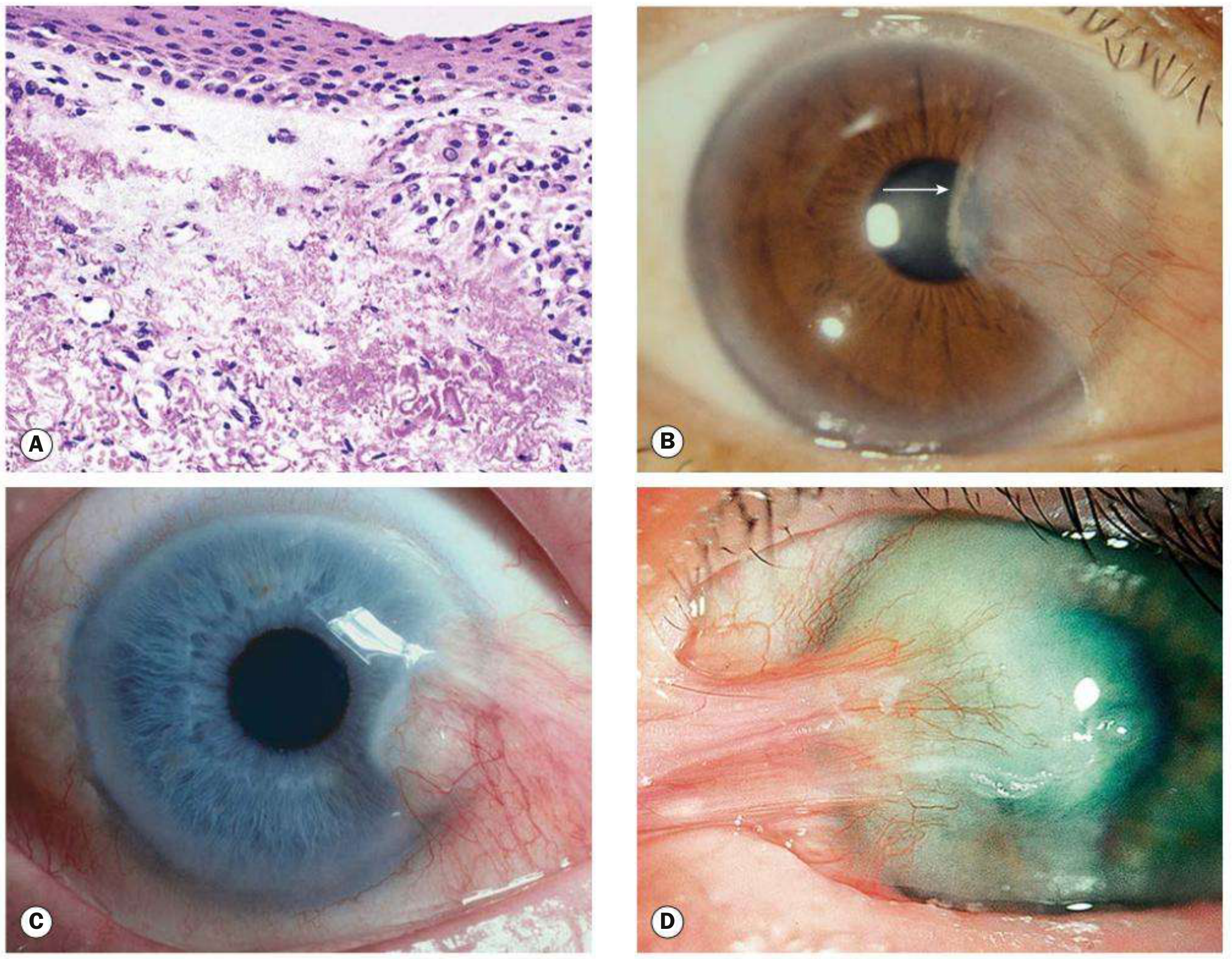
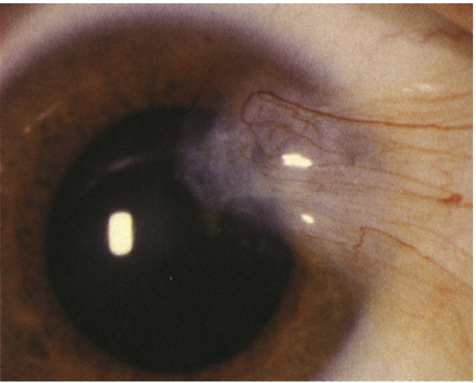
- Wing-shaped fibrovascular fold arising from the interpalpebral conjunctiva, extending onto the cornea (see clinical image below)
- Stocker line: linear iron deposition in the corneal epithelium just anterior to the head of the pterygium - a hallmark sign
- Fuchs' patches: small grey epithelial opacities near the apex in some cases
- May be inflamed (injected and highly vascularized) or quiescent
Symptoms
- Small lesions are often asymptomatic
- Irritation, grittiness, and redness (especially when inflamed)
- Dellen effect: localized drying at the advancing edge due to disruption of the precorneal tear film, particularly if the head is elevated
- Contact lens wearers develop symptoms at an earlier stage (edge lift)
- Visual disturbance: if the lesion encroaches on the visual axis, or induces irregular astigmatism (often with-the-rule)
- Cosmetic concerns
Pseudopterygium vs. True Pterygium
| Feature | True Pterygium | Pseudopterygium |
|---|---|---|
| Cause | UV/chronic irritation - degenerative | Inflammatory - chemical burn, corneal ulcer, trauma, cicatrizing conjunctivitis |
| Corneal thinning | Absent (no underlying thinning) | Often present |
| Probe test | Probe cannot pass under it | Probe can be passed under the tissue |
| History | UV exposure, outdoor work | History of causative acute episode |
Differential Diagnosis
- Pinguecula: yellow-white, flat/slightly raised conjunctival lesion in the interpalpebral fissure - does NOT extend onto the cornea
- Conjunctival intraepithelial neoplasia (CIN): papillomatous, jelly-like, velvety, or leukoplakic unilateral mass - may not be in typical 3/9 o'clock position; atypical pterygia must be biopsied to rule out CIN
- Limbal dermoid: congenital, rounded white lesion usually at the inferotemporal limbus
- Peripheral hypertrophic subepithelial corneal degeneration: bilateral, in Caucasian women, elevated peripheral opacities
- Other conjunctival tumors: papilloma, nevus, melanoma
Workup
- Slit lamp examination: identify the lesion, evaluate corneal integrity, thickness, and limbal anatomy
- Corneal topography: assess the degree of astigmatism induced (often irregular, with-the-rule)
- Biopsy if atypical features are present (to exclude CIN or squamous cell carcinoma)
Treatment
Conservative (for small, asymptomatic lesions)
- UV protection: UV-blocking sunglasses or goggles; minimize dust/wind exposure
- Artificial tears (preservative-free, 4-8x/day) to reduce irritation
- Inflamed pterygium:
- Mild: Artificial tears QID
- Moderate-severe: Mild topical steroid (e.g., fluorometholone 0.1%, loteprednol 0.2-0.5% QID), topical NSAID (ketorolac 0.4-0.5% QID), or topical antihistamine ± mast cell stabilizer (e.g., olopatadine, ketotifen)
- Delle present: artificial tear ointment every 2 hours
Surgical Indications
- Pterygium threatens the visual axis or induces significant astigmatism
- Excessive irritation unresponsive to conservative treatment
- Interfering with contact lens wear
- Causing significant cosmetic concerns
- Prior to cataract or refractive surgery
- Aggressive or atypical growth (excision biopsy)
Surgical Options
| Technique | Notes |
|---|---|
| Bare sclera excision alone | High recurrence rate (up to 80%) - largely abandoned |
| Conjunctival autograft (CAG) | Gold standard - reduces recurrence to ~5-10% |
| Amniotic membrane graft | Alternative when autograft tissue is limited |
| Intraoperative mitomycin C (MMC) | Antimetabolite; reduces recurrence further; more commonly used for recurrent pterygia; risk of corneoscleral thinning/melting if overused |
| Subconjunctival bevacizumab | Emerging adjunct to reduce vascularity; not yet standard |
Key point: Bare sclera excision combined with conjunctival autograft or amniotic membrane graft significantly reduces recurrence. Antimetabolites (MMC) are typically reserved for recurrent cases due to the risk of corneoscleral thinning.
Prognosis & Recurrence
- Recurrence is the main challenge after surgery
- Patients with recent rapid enlargement are more likely to have an aggressive course and early recurrence
- Extensive or recurrent pterygia can develop subconjunctival fibrosis extending to the fornices, potentially causing restricted ocular motility