
Teach me about csom
Reading File
CSOM (Chronic Suppurative Otitis Media) is a chronic ear infection with persistent discharge through a perforated eardrum. It involves the middle ear and mastoid, and is a leading cause of preventable hearing loss worldwide.
Searching sources...
Finding Sources
Searching PubMed
"chronic suppurative otitis media" management
Good results. Now let me read the key textbook sections in parallel.
Reading File
Reading File
Reading File
Let me now pull the core clinical content - diagnosis/treatment sections and the Scott-Brown's overview:
Reading File
Reading File
Finding Sources
Reading File
Finding Sources
Finding Sources
Finding Sources
Finding Sources
Finding Sources
Reading File
Reading File
Loading Image
I now have comprehensive material. Let me compile a thorough, well-structured answer.
Chronic Suppurative Otitis Media (CSOM)
Definition
CSOM is a chronic inflammation of the middle ear and mastoid characterized by persistent or recurrent otorrhea (>6-12 weeks) through a non-intact tympanic membrane (TM). It represents a spectrum of disease ranging from a simple central perforation with mucopurulent discharge to cholesteatoma with bony erosion and life-threatening intracranial complications.
Classification
CSOM is broadly divided into two types:
| Feature | Tubotympanic (Safe) Type | Atticoantral (Unsafe) Type |
|---|---|---|
| Also called | Benign / mucosal | Dangerous / squamous |
| Perforation site | Central (pars tensa) | Marginal / attic / posterior |
| Discharge | Mucopurulent, odorless | Scanty, foul-smelling |
| Cholesteatoma | Absent | Present |
| Bone erosion | Rare | Common |
| Complication risk | Low | High |
| Surgery urgency | Elective | Urgent |
Pathogenesis / Etiology
The underlying driver is Eustachian tube (ET) dysfunction, which leads to a persistent middle ear effusion and chronic mucosal inflammation. The sequence:
- ET dysfunction → negative middle ear pressure → mucosal edema
- Bacterial infection → purulent effusion → inflammatory mediators
- Chronic inflammation → mucosal gland metaplasia (secretory transformation) → perpetuates effusion
- Granulation tissue forms: bacterial toxins rupture the epithelial basement membrane; inflammatory cells and lamina propria extrude into the middle ear lumen
- Angiogenic and epithelial growth factors → fibroblast recruitment, neovascularization, polyp formation
- TM collagen skeleton degraded by enzymes in the granulation tissue → weakening, retraction pockets, perforation
- Deep retraction pockets or frank perforations → cholesteatoma genesis (squamous epithelium migration into the middle ear)
(Shambaugh Surgery of the Ear)
Microbiology
Organisms in CSOM differ from acute otitis media:
- Pseudomonas aeruginosa - the most common gram-negative organism; requires higher-dose or specialized antibiotics
- Staphylococcus aureus (including MRSA)
- Proteus, Klebsiella, anaerobes
- Fungal overgrowth is surprisingly common - Candida species grow in ~10% of ears with purulent otorrhea, and in up to 35% of ears treated with topical ciprofloxacin for 3 weeks
- Biofilm formation (bacterial and fungal) is well-established and contributes to treatment resistance
(Shambaugh Surgery of the Ear, p. 527)
Clinical Features
Symptoms
- Otorrhea - intermittent, sometimes foul-smelling (hallmark symptom)
- Hearing loss - typically conductive; sensorineural component possible
- Otalgia and headache are uncommon - if present, raise suspicion for intracranial complication or malignancy
- Vertigo - suggests labyrinthitis or perilymphatic fistula
Examination
- Tympanic membrane perforation (central or marginal/attic)
- Otorrhea may obscure the TM
- EAC polyps, granulation tissue
- Scutal or ossicular erosion (in advanced/cholesteatoma disease)
Audiometry
- Conductive hearing loss (CHL) is common
- CHL >30 dB suggests ossicular erosion
- Note: hearing can occasionally be preserved despite ossicular erosion if the cholesteatoma itself transmits sound to the oval window
- Sensorineural hearing loss (SNHL) ranging 5-33 dB has been documented and must be recorded preoperatively
Cholesteatoma
Cholesteatoma is an accumulation of desquamating stratified squamous epithelium within the middle ear or mastoid. It is "unsafe" because:
- It is expansile, locally destructive
- Erodes ossicles, tegmen, labyrinth, facial nerve canal
- Creates pathways for intracranial spread
Types:
- Acquired (most common) - from retraction pocket deepening or epithelial migration through a marginal perforation
- Congenital - white pearly mass behind an intact TM, typically anteromedial quadrant
Investigations
- Otomicroscopy - gold standard for TM and middle ear evaluation
- Audiometry (pure tone + tympanometry) - mandatory baseline
- CT temporal bone (HRCT) - extent of disease, ossicular chain, mastoid pneumatization, tegmen integrity, sigmoid sinus position
- MRI - superior for soft tissue; DWI-MRI detects cholesteatoma (restricted diffusion) and is useful for follow-up after canal-wall-down mastoidectomy
- Ear swab - for culture and sensitivity to guide topical/systemic antibiotic choice
Management
Medical (First-line for CSOM without cholesteatoma)
- Aural toilet - meticulous microsuction/dry mopping to clear discharge
- Topical antibiotic ear drops - first-line; fluoroquinolone drops (ciprofloxacin, ofloxacin) are preferred as they are non-ototoxic and have excellent gram-negative coverage including Pseudomonas
- Systemic antibiotics - reserved for acute exacerbations, systemic spread, or when topical therapy fails; guided by culture results
- Antifungal - if fungal overgrowth suspected (clotrimazole ear drops)
- Water precautions - protect ear from water ingress
2025 Cochrane evidence update: Three simultaneous Cochrane systematic reviews (Brennan-Jones et al., Head et al., Chong et al., June 2025 - PMIDs 40484407, 40484400, 40484405) confirm that topical antibiotics outperform topical antiseptics and systemic antibiotics for CSOM, reinforcing current practice of prioritizing ototopical fluoroquinolones.
Surgical
Indications:
- Cholesteatoma (near-absolute indication)
- Medically refractory CSOM (failed multiple medical treatments)
- Complications (vertigo, facial weakness, headache)
- Hearing restoration (ossiculoplasty, tympanoplasty)
The three priorities of CSOM surgery (Shambaugh Surgery of the Ear, p. 528-529):
- Eradication of disease
- Prevention of recurrence
- Preservation or restoration of hearing
Surgical options:
| Procedure | Description | Indication |
|---|---|---|
| Myringoplasty | TM repair alone (no mastoid work) | Central perforation, dry ear |
| Tympanoplasty | TM repair ± ossicular reconstruction | CSOM ± ossicular damage |
| Canal-wall-up (CWU) mastoidectomy | Mastoid exenteration preserving posterior EAC wall | Cholesteatoma, limited disease |
| Canal-wall-down (CWD) mastoidectomy | Posterior EAC wall removed; creates open cavity | Extensive cholesteatoma, recurrent disease |
- CWD offers better disease control and lower recurrence but requires lifelong cavity maintenance and restricts water exposure
- CWU preserves anatomy and shorter recovery but higher recurrence; needs second-look surgery
Complications
Complications arise when infection erodes the bony barriers of the middle ear cleft. Cholesteatoma and granulation tissue are the main culprits.
Extracranial
- Mastoiditis (most common)
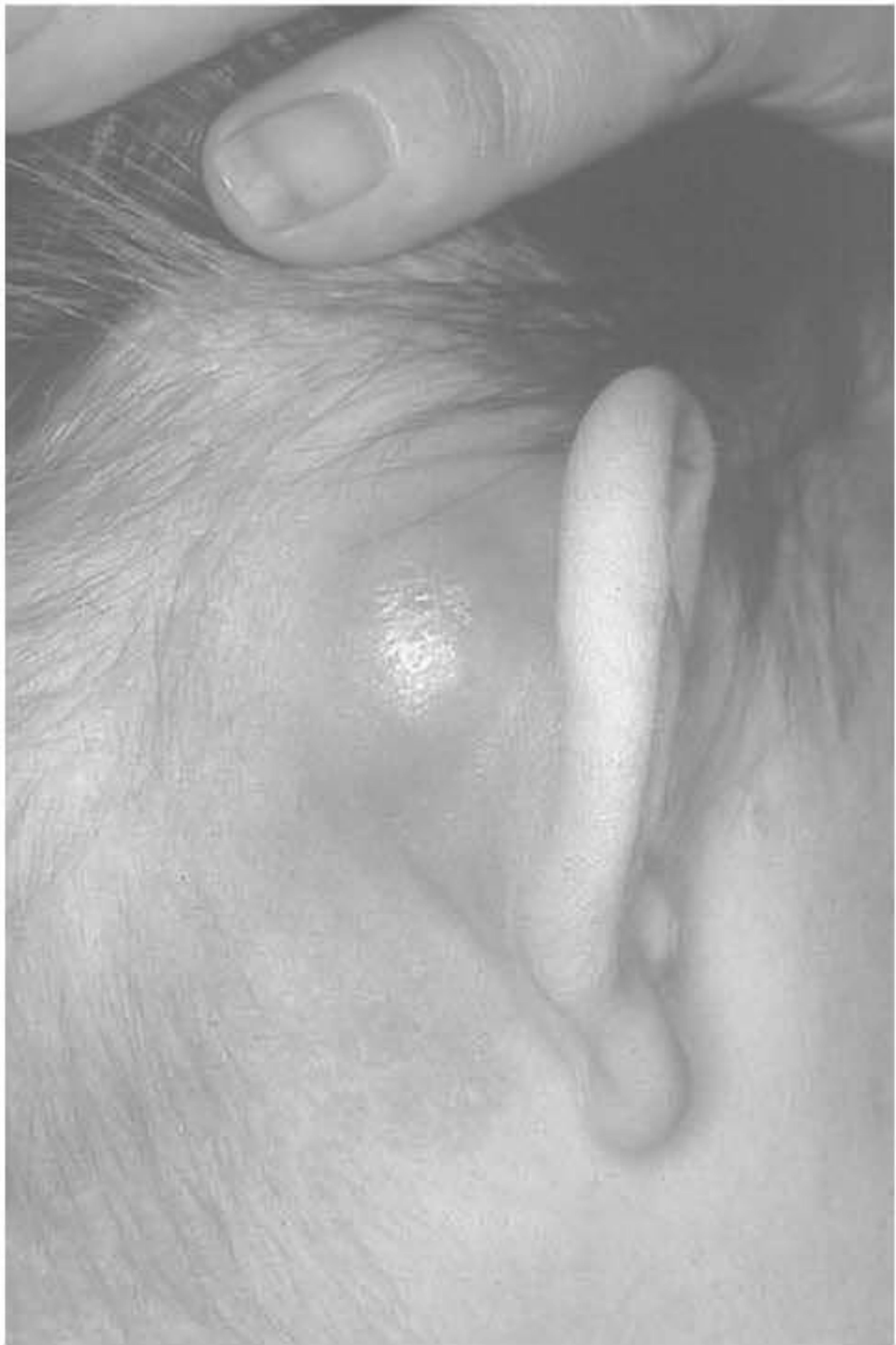
- Subperiosteal abscess (postauricular swelling - see image below)
- Facial nerve palsy (VII nerve canal erosion)
- Labyrinthitis / labyrinthine fistula
- Bezold's abscess (pus tracks along sternocleidomastoid)
- Petrositis (Gradenigo's syndrome: otorrhea + diplopia + facial pain)
Intracranial
- Meningitis (most common intracranial complication)
- Brain abscess
- Epidural abscess
- Subdural empyema
- Lateral (sigmoid) sinus thrombosis
- Otitic hydrocephalus
(Cummings Otolaryngology, Table 141.1 & 141.2)
Here is an example of postauricular swelling from mastoid involvement - a key extracranial sign:
Special Situation: CSOM and Cochlear Implantation
CSOM was historically a contraindication to cochlear implantation (CI). Current practice advocates:
- Dry ear at time of implantation is the key criterion
- Staged approach: (1) mastoidectomy ± ET obliteration to dry the ear; (2) CI 2-6 months later
- Risk of cholesteatoma formation in a closed cavity must be monitored long-term
- Biofilm eradication before electrode insertion is essential to prevent device infection
(Cummings Otolaryngology, p. 3072; Scott-Brown's Vol 2)
Summary Table
| Aspect | Key Points |
|---|---|
| Core defect | ET dysfunction → chronic mucosal inflammation |
| Hallmark | Persistent otorrhea through perforated TM |
| Key organisms | Pseudomonas aeruginosa, S. aureus, anaerobes, Candida |
| Safe type | Central perforation, no cholesteatoma, low risk |
| Unsafe type | Marginal/attic perforation, cholesteatoma, high risk |
| First-line Rx | Aural toilet + topical fluoroquinolone drops |
| Surgery trigger | Cholesteatoma, failed medical Rx, complications |
| Dreaded complication | Brain abscess, meningitis, lateral sinus thrombosis |
Sources: Shambaugh Surgery of the Ear | Cummings Otolaryngology Head and Neck Surgery | Scott-Brown's Otorhinolaryngology | Cochrane Reviews 2025 (PMIDs 40484407, 40484400, 40484405)
This is a shared conversation. Sign in to Orris to start your own chat.