Biochemistry of blood
Reading File
Finding Sources
Finding Sources
Finding Sources
Finding Sources
Finding Sources
Reading File
Reading File
Reading File
Reading File
Reading File
Finding Sources
Reading File
Reading File
Finding Sources
Loading Image
Loading Image
Here is a comprehensive overview of the biochemistry of blood, drawn from Ganong's Review of Medical Physiology and Guyton and Hall Textbook of Medical Physiology.
Biochemistry of Blood
Blood is a specialized connective tissue composed of a liquid matrix (plasma) and formed cellular elements. Its biochemistry spans protein chemistry, gas transport, enzymatic reactions, coagulation cascades, and acid-base regulation.
1. Composition of Blood
Blood consists of:
- Plasma (~55%): an aqueous solution of proteins, electrolytes, nutrients, hormones, and waste products
- Formed elements (~45%): red blood cells (RBCs), white blood cells (WBCs), and platelets (thrombocytes)
The hematocrit (percentage of blood volume occupied by cells) is normally ~47% in males and ~42% in females.
2. Hematopoiesis - Origin of Blood Cells
All formed elements arise from a single pluripotent hematopoietic stem cell in the bone marrow. Differentiation is driven by cytokines including:
- Erythropoietin (EPO) - stimulates red cell production
- GM-CSF, G-CSF, M-CSF - drive granulocyte/monocyte lineages
- Thrombopoietin - drives platelet production
- IL-3, IL-4, IL-5, IL-6 - various supporting roles
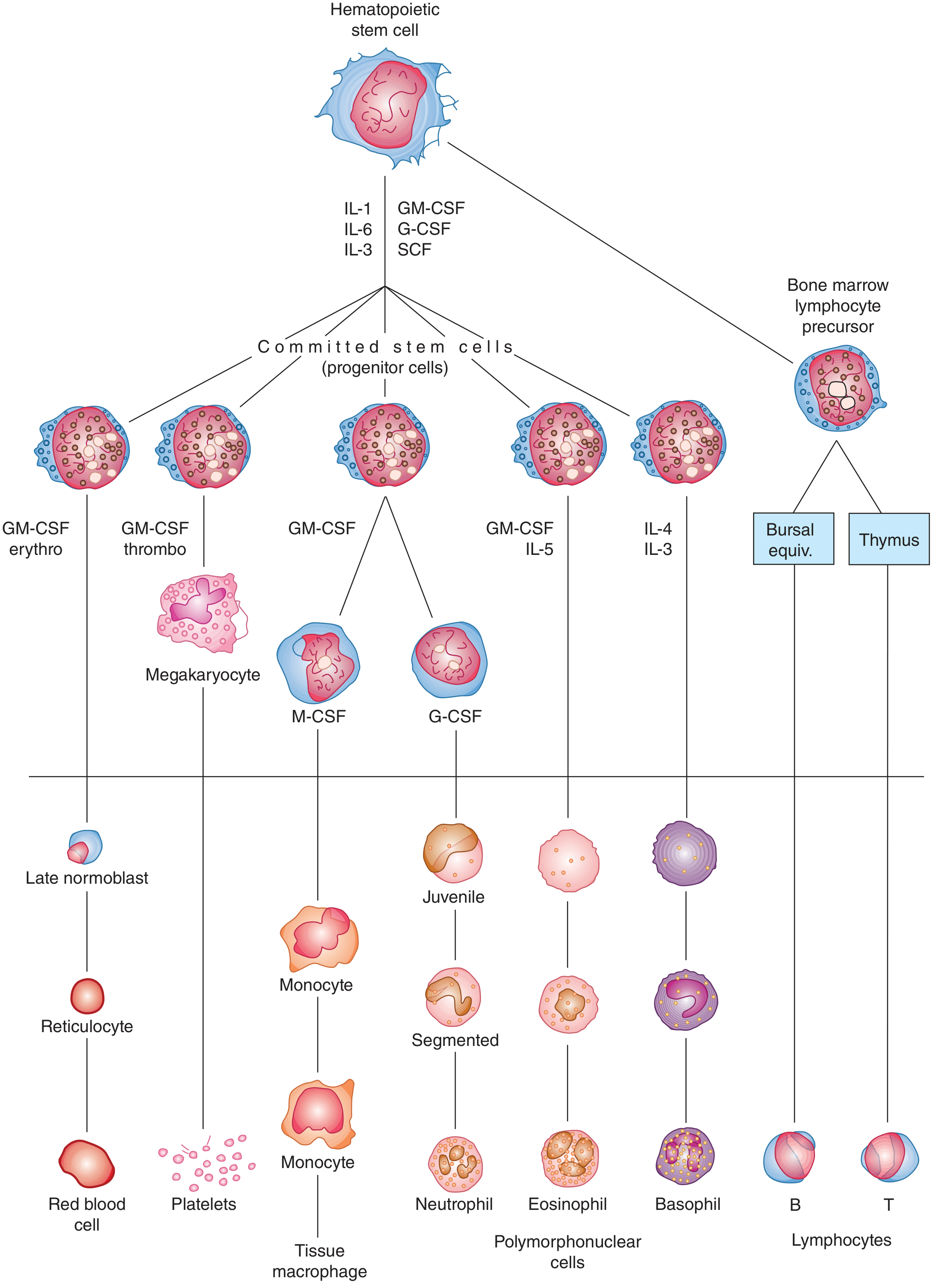
Fig. 31-3: Development of blood cell types from bone marrow. Cells below the line are found in normal peripheral blood. (Ganong's Review of Medical Physiology)
3. Red Blood Cells (Erythrocytes)
Structure and Size
- Biconcave discs, ~7.8 µm diameter, 90-95 µm³ volume
- No nucleus or mitochondria in mature form
- Mean count: 5.2 million/µL (men), 4.7 million/µL (women)
- Lifespan: ~120 days
Key RBC Functions
- Oxygen transport via hemoglobin
- CO₂ transport via carbonic anhydrase: CO₂ + H₂O ⇌ H₂CO₃ ⇌ H⁺ + HCO₃⁻ (this enzyme accelerates the reaction ~1000-fold, enabling bicarbonate-mediated CO₂ transport)
- Acid-base buffering: RBC proteins (especially hemoglobin) account for a significant portion of blood's buffering capacity
Red Cell Indices (Normal Values)
| Parameter | Male | Female |
|---|---|---|
| Hemoglobin (Hb) | 16 g/dL | 14 g/dL |
| Hematocrit (Hct) | 47% | 42% |
| MCV | 87 fL | 87 fL |
| MCH | 29 pg | 29 pg |
| MCHC | 34 g/dL | 34 g/dL |
Source: Ganong's Review of Medical Physiology, Table 31-2
4. Hemoglobin - Structure and Function
Hemoglobin is a globular tetrameric protein:
- 4 subunits, each containing a heme group (iron-containing porphyrin derivative) bound to a globin polypeptide
- Hemoglobin A (HbA, adult): α₂β₂ (2 alpha + 2 beta chains)
- Hemoglobin A₂: α₂δ₂ (~2.5% of adult Hb)
- Each gram of hemoglobin can carry 1.34 mL O₂ when fully saturated
- 100 mL whole blood carries ~20 mL O₂ (in men)
Hemoglobin Reactions
- Oxyhemoglobin: Hb + O₂ → HbO₂ (O₂ binds to Fe²⁺)
- Deoxyhemoglobin: quaternary structure shifts ("T-state" vs. "R-state")
- Carboxyhemoglobin: Hb + CO → HbCO (CO has ~250x greater affinity for Hb than O₂)
- Methemoglobin: Fe²⁺ oxidized to Fe³⁺; cannot carry O₂
Allosteric Regulation of O₂ Affinity
The oxygen-hemoglobin dissociation curve is sigmoidal due to cooperative binding. Affinity is decreased (curve shifts right) by:
- ↑ CO₂ (Bohr effect)
- ↓ pH (acidosis)
- ↑ Temperature
- ↑ 2,3-bisphosphoglycerate (2,3-BPG) - competes with O₂ for binding to deoxygenated Hb
Glycated Hemoglobin (HbA1c)
A glucose molecule attaches to the terminal valine of each β chain. HbA1c reflects average blood glucose over ~3 months and is the standard marker for monitoring diabetes mellitus control.
5. Plasma Proteins
Plasma contains approximately 7 g/dL of protein total. The major fractions are albumin, globulins, and fibrinogen.
Major Functions of Plasma Proteins
- Oncotic pressure (colloid osmotic pressure): ~25 mmHg, maintains fluid within the vasculature
- Buffering: ~15% of blood's total buffering capacity from COOH and NH₂ groups (proteins are mostly anionic at pH 7.4)
- Transport: nonspecific carriers for hormones, fatty acids, drugs, steroids, vitamins
- Specific defense and clotting: antibodies (immunoglobulins), clotting factors
Key Plasma Proteins Synthesized by the Liver
| Protein | Function | Concentration |
|---|---|---|
| Albumin | Osmotic regulation, carrier protein for hormones/fatty acids/drugs | 4500-5000 mg/dL (3.5-5.0 g/dL) |
| Orosomucoid (α1-acid glycoprotein) | Uncertain; acute phase reactant | - |
| Transferrin | Iron transport | - |
| Ceruloplasmin | Copper transport | - |
| Haptoglobin | Binds free Hb, prevents renal loss | - |
| Fibrinogen | Clot formation (converted to fibrin) | - |
| Prothrombin | Clotting factor II | - |
| Complement proteins | Innate immunity | - |
Circulating antibodies (immunoglobulins) are produced by lymphocytes, not hepatocytes.
Albumin Dynamics
- Normal plasma albumin: 3.5-5.0 g/dL
- Total exchangeable pool: 4.0-5.0 g/kg body weight; 38-45% is intravascular
- 6-10% of the pool is degraded daily, replaced by hepatic synthesis of 200-400 mg/kg/day
- Synthesis is decreased in fasting and increased in protein-losing states (e.g., nephrosis)
Hypoproteinemia
Low plasma protein levels occur in:
- Prolonged starvation / malabsorption
- Liver disease (impaired synthesis)
- Nephrotic syndrome (urinary albumin loss)
- Result: decreased oncotic pressure → edema
6. Hemostasis and Blood Coagulation
Hemostasis is the process preventing blood loss after vascular injury. It involves four sequential events:
6.1 Vascular Constriction
Smooth muscle contraction immediately reduces blood flow. Triggered by local myogenic spasm, neural reflexes, and thromboxane A₂ released from activated platelets.
6.2 Platelet Plug Formation
- Platelets are anucleate discs (1-4 µm), formed from megakaryocytes, with a lifespan of 8-12 days
- Normal count: 150,000-450,000/µL
- On vascular injury, platelets adhere to exposed subendothelial collagen (via von Willebrand factor), then aggregate, forming a plug
- Platelet cytoplasm contains: actin/myosin (contractile proteins), calcium stores, ATP/ADP-generating mitochondria, prostaglandin-synthesizing enzymes, fibrin-stabilizing factor, and platelet-derived growth factor (PDGF)
6.3 Coagulation Cascade
The cascade has two converging pathways:
Extrinsic pathway (rapid, triggered by tissue injury):
- Tissue damage releases tissue factor (TF/Factor III) - a lipoprotein complex
- TF + Factor VII + Ca²⁺ → activates Factor X
- Factor Xa + Factor V + phospholipids → prothrombin activator
Intrinsic pathway (slower, triggered by blood trauma or collagen contact):
- Factor XII activated by contact with collagen
- XII → XI → IX → (with Factor VIII) → Factor X
- Factor Xa → same prothrombin activator as above
Final common pathway:
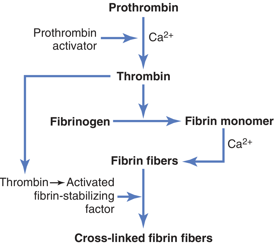
Conversion of prothrombin to thrombin, and fibrinogen to cross-linked fibrin fibers. (Guyton and Hall)
- Prothrombin activator + Ca²⁺ → Prothrombin → Thrombin
- Thrombin cleaves Fibrinogen → Fibrin monomers → Fibrin fibers (Ca²⁺ dependent)
- Fibrin-stabilizing factor (Factor XIII), activated by thrombin, cross-links fibrin fibers → stable clot
6.4 Anticoagulant Mechanisms (Normal Homeostasis)
Under normal conditions, anticoagulants dominate to prevent spontaneous clotting:
- Antithrombin III - inhibits thrombin and other serine proteases
- Protein C/S system - inactivates Factors Va and VIIIa
- Tissue factor pathway inhibitor (TFPI) - limits extrinsic pathway
- Prostacyclin (PGI₂) from endothelium - inhibits platelet aggregation
7. Blood as a Buffer System
Blood maintains pH 7.35-7.45 through three main buffer systems:
- Bicarbonate buffer (HCO₃⁻/CO₂) - the dominant extracellular buffer
- Hemoglobin buffer - deoxyHb is a better buffer than oxyHb; accounts for the majority of RBC buffering
- Plasma protein buffers - ~15% of total blood buffering capacity
The chloride shift (Hamburger shift): In peripheral tissues, CO₂ enters RBCs, is hydrated to HCO₃⁻, which exits into plasma in exchange for Cl⁻, allowing efficient CO₂ transport.
8. Other Key Biochemical Components of Blood
| Component | Normal Range | Function |
|---|---|---|
| Glucose | 70-110 mg/dL (fasting) | Primary energy substrate |
| Sodium (Na⁺) | 135-145 mEq/L | Main extracellular cation; osmolality |
| Potassium (K⁺) | 3.5-5.0 mEq/L | Main intracellular cation; membrane potential |
| Calcium (Ca²⁺) | 8.5-10.5 mg/dL | Coagulation, neuromuscular function |
| Bicarbonate (HCO₃⁻) | 22-28 mEq/L | Acid-base balance |
| Urea (BUN) | 7-20 mg/dL | Nitrogen waste product |
| Creatinine | 0.6-1.2 mg/dL | Muscle metabolism waste |
| Total protein | ~7 g/dL | Multiple functions (see above) |
Summary
The biochemistry of blood integrates structural proteins (hemoglobin, plasma proteins), enzymatic machinery (carbonic anhydrase, coagulation proteases), signaling molecules (cytokines, thromboxane, prostacyclin), and electrolytes into a tightly regulated system. Key themes include:
- Cooperative allosterism in hemoglobin O₂ binding
- Protease cascade amplification in coagulation
- Oncotic-hydrostatic balance governed by albumin
- Acid-base homeostasis via bicarbonate and hemoglobin buffers
Sources: Ganong's Review of Medical Physiology (26th ed.), Guyton and Hall Textbook of Medical Physiology
This is a shared conversation. Sign in to Orris to start your own chat.