
Trace the visual pathway with a neat diagram. Write about the effects of lesions at various levels in visual pathway Physiology Give me ready to write answer ,give flow chart and tables if necessary
Reading File
Finding Sources
Finding Sources
Finding Sources
Reading File
Reading File
Finding Sources
Loading Image
Loading Image
Reading File
Reading File
Reading File
Loading Image
Reading File
Reading File
Loading Image
I now have comprehensive content from the textbooks. Let me compile the full answer.
THE VISUAL PATHWAY
Overview
The visual pathway transmits light signals from the photoreceptors of the retina to the primary visual cortex (area 17, striate cortex) in the occipital lobe. Understanding its anatomy is essential for localizing lesions based on visual field defects.
FLOWCHART: The Visual Pathway
LIGHT → RETINA (rods & cones)
           ↓
    BIPOLAR CELLS (1st neuron synapse)
           ↓
    GANGLION CELLS (axons form optic nerve)
           ↓
    OPTIC NERVE (CN II)
           ↓
    OPTIC CHIASMA
    ┌──────────────────┐
    │ Nasal fibers     │ → DECUSSATE to opposite side
    │ Temporal fibers  │ → Stay IPSILATERAL
    └──────────────────┘
           ↓
    OPTIC TRACT (each carries:
    - Temporal fibers from ipsilateral eye
    - Nasal fibers from contralateral eye)
           ↓
    LATERAL GENICULATE NUCLEUS (LGN) of thalamus
    (2nd order neuron synapse)
           ↓
    OPTIC RADIATIONS (Geniculocalcarine tract)
    ┌─────────────────────────────────────────────┐
    │ Upper fibers   → parietal lobe → upper cortex │
    │ Lower fibers   → Meyer's loop (temporal lobe) │
    │                → lower cortex                 │
    └─────────────────────────────────────────────┘
           ↓
    PRIMARY VISUAL CORTEX
    (Area 17 / Striate cortex, calcarine sulcus,
    Brodmann's Area 17, occipital lobe)
A small collateral from the optic tract goes to the pretectal area and superior colliculus - mediating the pupillary light reflex (does NOT reach visual cortex).
ANATOMY IN DETAIL
1. Retina
- Photoreceptors (rods for dim light, cones for color/acuity) synapse on bipolar cells, which synapse on retinal ganglion cells
- Ganglion cell axons coalesce at the optic disc (blind spot - no photoreceptors)
2. Optic Nerve (CN II)
- Axons acquire myelin from oligodendrocytes (CNS-type)
- Covered by cranial meninges
- Passes through the optic canal in the sphenoid bone
3. Optic Chiasma
- Situated anterior to the infundibular stalk, ~10 mm above the pituitary gland
- In shape, resembles the letter H (not the Greek letter chi)
- 50% of fibers decussate: nasal retinal fibers cross to the opposite optic tract
- Temporal retinal fibers remain ipsilateral
- Note: Nasal retina receives light from the temporal visual field; temporal retina receives from the nasal visual field
4. Optic Tract
- Each optic tract carries:
- Temporal fibers from the ipsilateral eye
- Nasal fibers from the contralateral eye
- Together they represent the contralateral visual hemifield
- Courses around the midbrain to reach the LGN
5. Lateral Geniculate Nucleus (LGN)
- Located in the posterior thalamus
- Has 6 laminae (layers 1,4,6 from contralateral eye; layers 2,3,5 from ipsilateral eye)
- 2nd order neuron synapse occurs here
6. Optic Radiations (Geniculocalcarine Tract)
- Upper fibers: pass through parietal lobe → terminate on upper bank of calcarine fissure (represents lower visual field)
- Lower fibers: loop anteriorly through the temporal lobe as Meyer's loop → terminate on lower bank of calcarine fissure (represents upper visual field)
7. Primary Visual Cortex (Area 17)
- Located in the calcarine sulcus, occipital lobe
- Macula (central vision) is represented at the posterior pole (occipital tip)
- Peripheral vision is represented more anteriorly
- Upper bank of calcarine → receives lower visual field signals
- Lower bank of calcarine → receives upper visual field signals
DIAGRAM: Visual Pathway (from textbook)
(Representation of the visual field through the pathway - from Gray's Anatomy for Students)
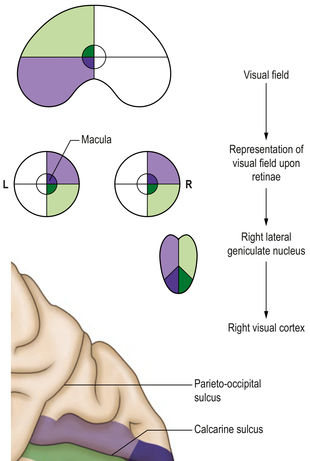
The diagram shows how the left visual hemifield (green and purple) is represented at the retinal level, then carried through the right LGN and projected onto the right visual cortex in the calcarine sulcus.
DIAGRAM: Optic Radiations and Meyer's Loop
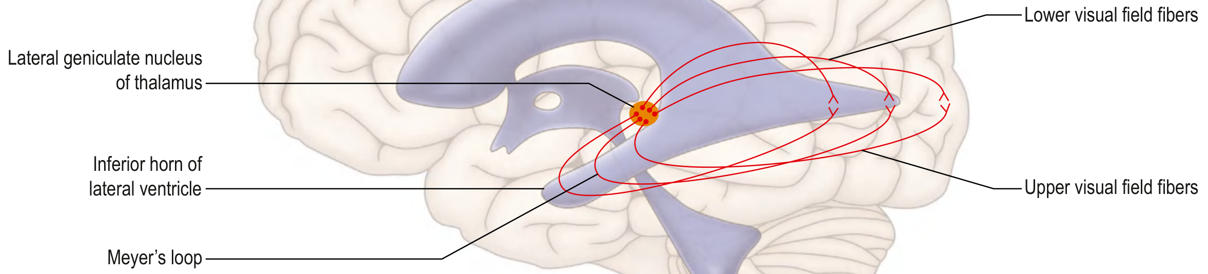
Shows Meyer's loop curving around the inferior horn of the lateral ventricle. Upper visual field fibers loop through the temporal lobe (Meyer's loop); lower visual field fibers pass through the parietal lobe.
EFFECTS OF LESIONS AT VARIOUS LEVELS
Master Diagram: Lesions and Visual Field Defects
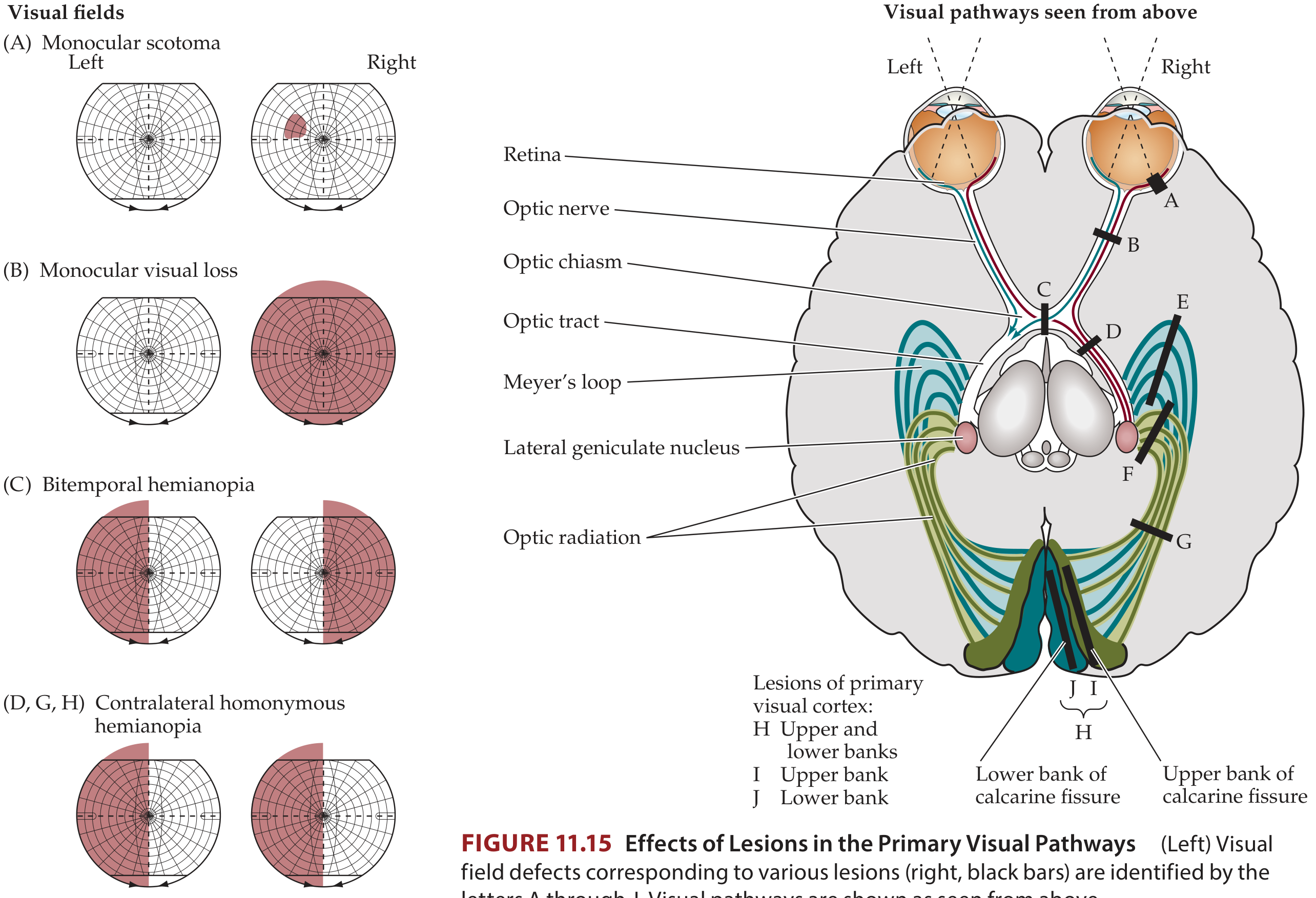
Lesions A-J correspond to visual field defects shown on the left. Visual pathways seen from above.
TABLE: Effects of Lesions at Various Levels in the Visual Pathway
| Site of Lesion | Visual Field Defect | Key Features | Common Causes |
|---|---|---|---|
| 1. Retina (partial) | Monocular scotoma (small patch in one eye) | Shape/location matches retinal lesion; monocular | Retinal infarct, hemorrhage, degeneration, infection |
| 2. Optic nerve (complete) | Complete monocular blindness (one eye) | Ipsilateral total loss; contralateral eye normal; RAPD present | Optic neuritis, glaucoma, AION, trauma, optic glioma, meningioma, elevated ICP |
| 3. Optic chiasma (central) | Bitemporal hemianopia ("tunnel vision") | Loss of both temporal fields; heteronymous; does NOT respect macula as midpoint | Pituitary adenoma (most common), craniopharyngioma, meningioma, hypothalamic glioma |
| 4. Optic tract | Contralateral homonymous hemianopia (incongruous) | Incomplete/incongruous - fibers not yet well-mixed; contralateral RAPD | Tumors, infarct, demyelination (less common) |
| 5. LGN (lateral geniculate nucleus) | Contralateral homonymous hemianopia | Occasionally "keyhole" sectoranopia (wedge-shaped); incongruous | Tumors, infarct, hemorrhage, toxoplasmosis |
| 6. Meyer's loop (temporal lobe fibers) | Contralateral superior homonymous quadrantanopia ("Pie in the Sky") | Only lower optic radiations interrupted | MCA inferior division infarct, temporal lobe tumor, temporal lobectomy |
| 7. Parietal lobe (upper radiations) | Contralateral inferior homonymous quadrantanopia ("Pie on the Floor") | Only upper optic radiations interrupted | MCA superior division infarct, parietal tumor |
| 8. Entire optic radiation | Contralateral complete homonymous hemianopia | Congruous (fibers now well-mixed) | Large MCA infarcts, trauma, hemorrhage |
| 9. Visual cortex (upper bank of calcarine) | Contralateral inferior quadrantanopia | Upper bank → lower visual field | PCA infarct, tumor, trauma |
| 10. Visual cortex (lower bank of calcarine) | Contralateral superior quadrantanopia | Lower bank → upper visual field | PCA infarct, tumor, trauma |
| 11. Entire primary visual cortex | Contralateral complete homonymous hemianopia with macular sparing | Macular sparing due to dual MCA/PCA supply to occipital pole; most CONGRUOUS defect | PCA infarct (most common), tumors, trauma |
| 12. Bilateral occipital poles | Bilateral central scotomas | Both foveal representations destroyed | Bilateral PCA infarcts |
| 13. Bilateral visual cortex (complete) | Cortical blindness (Anton's syndrome) | No light perception; pupils normal (light reflex intact); patient may deny blindness | Bilateral PCA infarcts |
KEY CONCEPTS TABLE
| Concept | Explanation |
|---|---|
| Scotoma | Area of lost/diminished vision surrounded by normal vision |
| Hemianopia | Loss of half the visual field |
| Quadrantanopia | Loss of one quarter of visual field |
| Homonymous | Same side of visual field lost in both eyes (retrochiasmal lesion) |
| Heteronymous | Different sides lost in each eye (at chiasma) |
| Congruous | Defect identical in shape/size in both eyes (more posterior = more congruous) |
| Incongruous | Defect differs between the two eyes (optic tract and LGN lesions) |
| Macular sparing | Central vision preserved despite hemianopia; seen with occipital cortex lesions due to dual blood supply |
| RAPD (Marcus Gunn pupil) | Relative afferent pupillary defect; seen in optic nerve and optic tract lesions (contralateral in tract lesion) |
SPECIAL NOTES FOR EXAM
1. Optic chiasma lesions - why bitemporal?
- Nasal retinal fibers (carrying temporal field information) from both eyes cross at the chiasma
- A central chiasmal lesion (e.g., pituitary tumor pressing from below) destroys these crossing fibers
- Result: both temporal visual fields are lost
2. Meyer's loop - clinical importance
- The lower fibers of the optic radiation loop anteriorly into the temporal lobe
- Temporal lobectomy (e.g., for epilepsy) can cause "pie in the sky" superior quadrantanopia
3. Macular sparing in cortical lesions
- The macular area (central 5-10° of vision) has large cortical representation at the occipital pole
- The occipital pole may receive dual blood supply (MCA + PCA)
- A PCA infarct causing hemianopia typically spares the macula
4. Pupillary light reflex vs. vision
- Fibers leave the optic tract BEFORE reaching LGN to go to the pretectal nucleus
- Therefore: cortical blindness (bilateral occipital lesion) has normal pupils (light reflex intact)
- Lesions at or before the optic tract affect the light reflex
5. Rule of congruity
- More POSTERIOR the lesion → more CONGRUOUS the defect
- Optic tract = most incongruous homonymous defect
- Visual cortex = perfectly congruous (identical) defect
SUMMARY FLOWCHART: Lesion Localization
Monocular vision loss
    → Ipsilateral eye, optic nerve, or retina

Bitemporal hemianopia
    → Optic chiasma (central crossing fibers)

Binasal hemianopia (rare)
    → Lateral compression of chiasma (bilateral)

Contralateral homonymous hemianopia (incongruous)
    → Optic tract or LGN

Contralateral homonymous superior quadrantanopia ("pie in sky")
    → Meyer's loop / temporal lobe

Contralateral homonymous inferior quadrantanopia ("pie on floor")
    → Parietal lobe / upper optic radiation

Contralateral homonymous hemianopia (congruous) WITH macular sparing
    → Primary visual cortex (PCA territory)

Bilateral visual loss, normal pupils
    → Cortical blindness (bilateral occipital cortex)
Sources: Neuroanatomy through Clinical Cases, 3rd Ed. (Fig. 11.15) | Gray's Anatomy for Students (eFig. 9.65-9.68) | Localization in Clinical Neurology, 8th Ed. | Scott-Brown's Otorhinolaryngology Head & Neck Surgery
Refer guyton Trace the visual pathway with a neat diagram. Write about the effects of lesions at various levels in visual pathway Physiology Give me ready to write answer ,give flow chart and tables if necessary
Finding Sources
Finding Sources
Finding Sources
Finding Sources
Finding Sources
Finding Sources
Finding Sources
Finding Sources
Reading File
Loading Image
Reading File
Reading File
Now let me get the specific section on lesions/abnormalities in visual fields from Guyton, and also fetch the key Guyton visual pathway image:
Reading File
Loading Image
Loading Image
Finding Sources
I now have all content directly from Guyton and Hall Textbook of Medical Physiology (ISBN 9780443111013). Let me compose the complete, exam-ready answer.
THE VISUAL PATHWAY
(As per Guyton and Hall Textbook of Medical Physiology)
DIAGRAM: Principal Visual Pathways (Guyton Fig. 52.1)
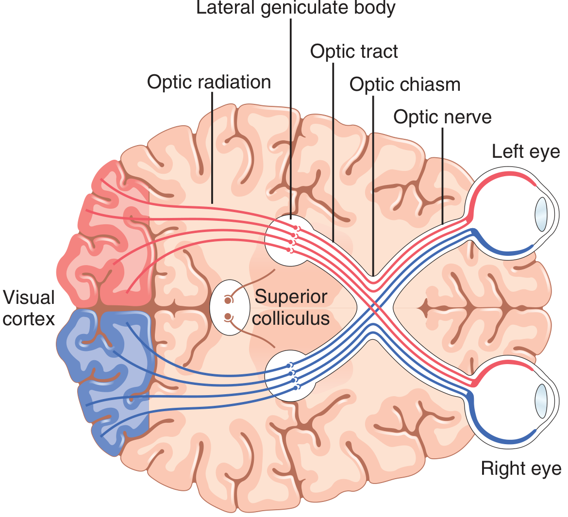
Figure 52.1 from Guyton & Hall: Principal visual pathways from the eyes to the visual cortex. Red = right eye fibers; Blue = left eye fibers. Note crossing at the optic chiasm, relay in the lateral geniculate body, and projection via the optic radiation to the visual cortex.
FLOWCHART: The Visual Pathway (Guyton)
LIGHT
  ↓
RETINA
  ├── Rods (dim light / peripheral vision)
  └── Cones (colour / central acuity)
         ↓ (synapse)
    BIPOLAR CELLS
         ↓ (synapse)
    RETINAL GANGLION CELLS
         ↓
    OPTIC NERVE (CN II)
    [Axons of ganglion cells, myelinated, CNS structure]
         ↓
    OPTIC CHIASMA
    ┌─────────────────────────────────────────────────────┐
    │  Nasal retinal fibers (temporal visual field)       │
    │       → CROSS to opposite optic tract               │
    │  Temporal retinal fibers (nasal visual field)       │
    │       → REMAIN IPSILATERAL in optic tract           │
    └─────────────────────────────────────────────────────┘
         ↓
    OPTIC TRACT
    [Each tract = temporal fibers from same eye
                + nasal fibers from opposite eye
     = represents contralateral visual hemifield]
         ↓
    DORSAL LATERAL GENICULATE NUCLEUS (LGN)
    of the thalamus
    [6 layers; "gates" transmission to cortex]
         ↓
    OPTIC RADIATION
    (Geniculocalcarine tract)
    ┌───────────────────────────────────────────────────┐
    │ Upper fibers → Parietal lobe → Upper calcarine    │
    │ Lower fibers → Meyer's loop (temporal lobe)       │
    │              → Lower calcarine                    │
    └───────────────────────────────────────────────────┘
         ↓
    PRIMARY VISUAL CORTEX (Area 17)
    [Calcarine fissure, medial occipital lobe]
         ↓
    SECONDARY VISUAL AREAS (Areas 18, 19)
    [Higher-order processing: form, color, movement]
Collateral Projections from the Optic Tract (Guyton)
| Destination | Function |
|---|---|
| Suprachiasmatic nucleus of hypothalamus | Controls circadian rhythms (day-night synchrony) |
| Pretectal nuclei (midbrain) | Pupillary light reflex; reflex eye movements |
| Superior colliculus | Rapid directional movements of both eyes |
| Ventral lateral geniculate + basal forebrain | Behavioral visual functions |
THE VISUAL PATHWAY - STEP BY STEP
1. Retina
- Contains rods (120 million; for dim light and peripheral vision) and cones (6 million; for color and fine acuity)
- Signal pathway: Photoreceptors → Bipolar cells → Retinal Ganglion cells
- Ganglion cell axons converge at the optic disc (blind spot - no photoreceptors here, ~15° lateral to central point of vision)
2. Optic Nerve (CN II)
- Formed by axons of retinal ganglion cells
- Covered by cranial meninges; myelin provided by oligodendrocytes
- Physiologically a CNS tract, not a peripheral nerve
3. Optic Chiasma
- Fibers from the nasal half of each retina (receiving light from the temporal visual field) decussate to the contralateral optic tract
- Fibers from the temporal half of each retina remain ipsilateral
- Result: Each optic tract carries information from the contralateral visual hemifield
4. Dorsal Lateral Geniculate Nucleus (LGN)
Guyton describes two key functions:
Function 1 - Relay: Point-to-point transmission with high spatial fidelity from retina to cortex.
Function 2 - Gating: Controls how much signal passes to cortex, via:
- Corticofugal fibers from primary visual cortex (backward)
- Reticular areas of the mesencephalon Both are inhibitory and help highlight important visual information.
Six Laminar Layers of LGN:
| Layers | Cell Type | Input from | System |
|---|---|---|---|
| I, II (ventral) | Magnocellular (large) | M-type ganglion cells | Fast, black-and-white; no color; poor spatial resolution |
| III - VI (dorsal) | Parvocellular (small-medium) | P-type ganglion cells | Slower, COLOR vision; accurate point-to-point spatial detail |
| II, III, V | - | Ipsilateral (temporal) retina | - |
| I, IV, VI | - | Contralateral (nasal) retina | - |
Signals from the two eyes are kept separate in LGN and remain so all the way to the visual cortex.
5. Optic Radiation (Geniculocalcarine Tract)
- Upper fibers: Pass through parietal lobe → terminate on upper bank of calcarine fissure
- Lower fibers: Loop anteriorly through temporal lobe (Meyer's loop) → terminate on lower bank of calcarine fissure
6. Primary Visual Cortex (Area 17 / Striate Cortex)
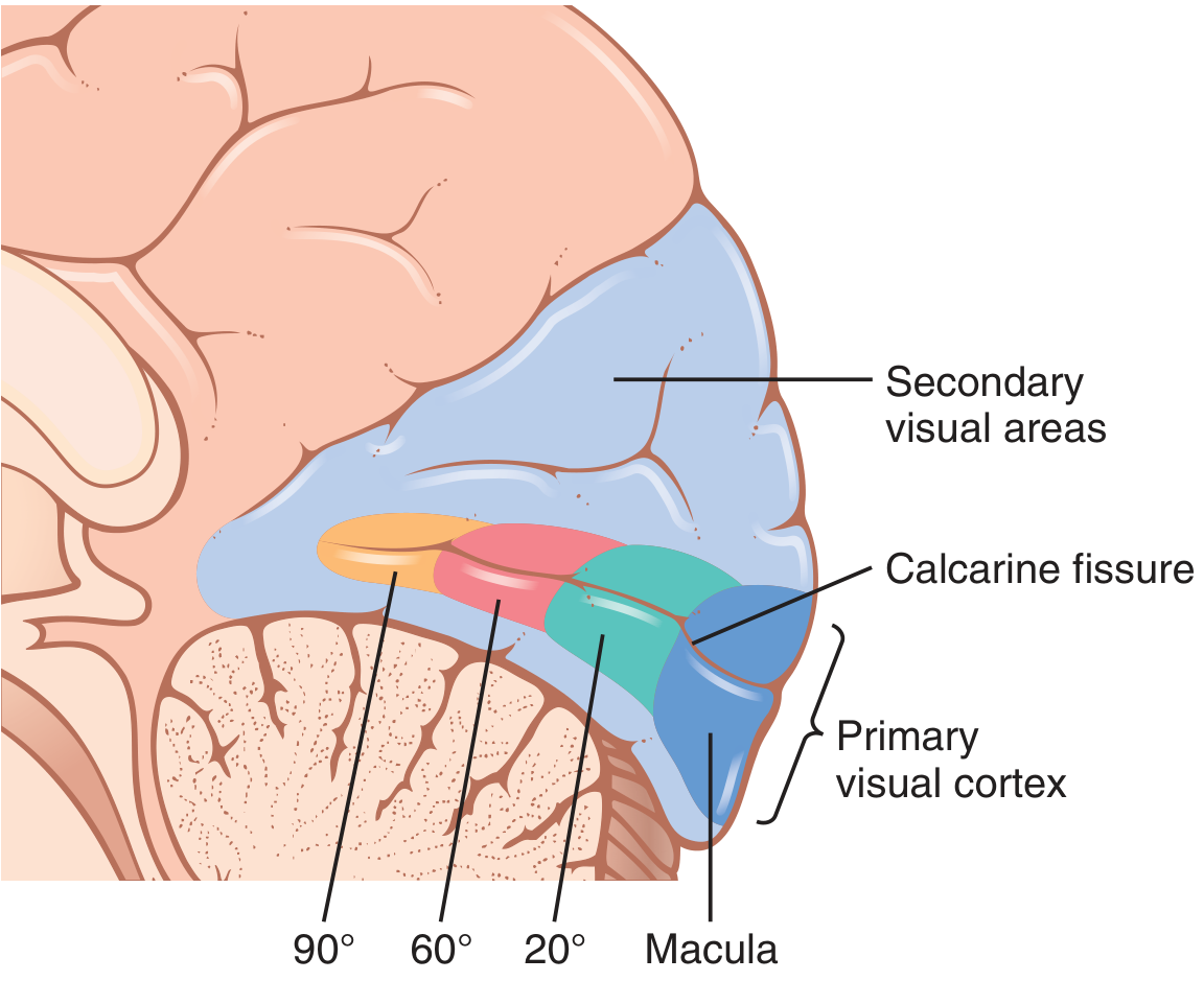
Figure 52.2 from Guyton & Hall: The visual cortex in the calcarine fissure area of the medial occipital cortex. Macula (central vision) is projected most posteriorly; peripheral fields (90°) project more anteriorly.
- Located in the calcarine fissure, medial occipital lobe
- Macula (central 5-10° of vision) has disproportionately large representation at the posterior occipital pole
- Peripheral fields project anteriorly along the calcarine fissure
- Has 6 layers with columns detecting orientation, movement, and color
Effect of removing primary visual cortex (Guyton): Causes loss of conscious vision (blindness). However, such patients can still react subconsciously to changes in light intensity, movement, or gross patterns via the superior colliculi and older visual system - this is "blindsight."
PERIMETRY AND THE VISUAL FIELD
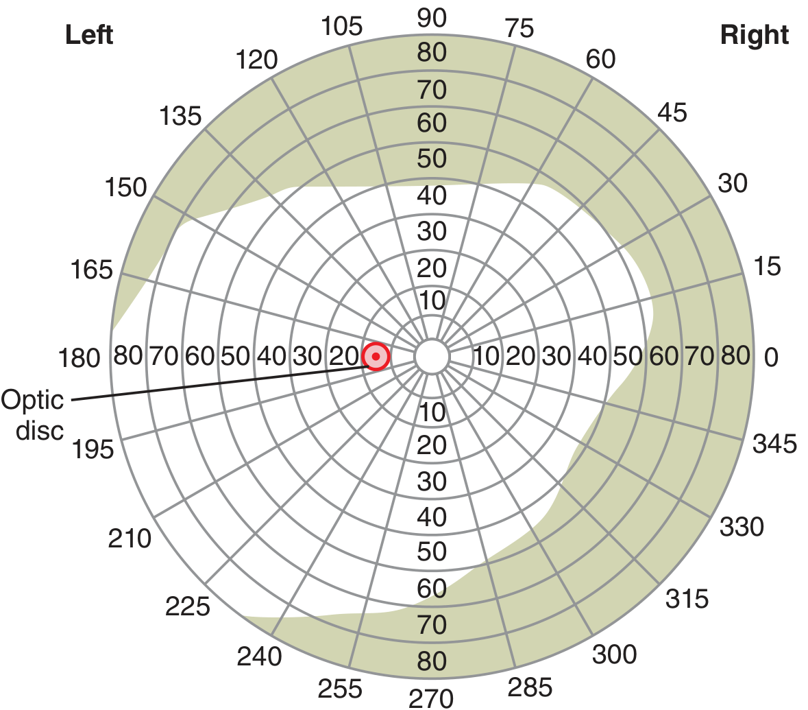
Guyton Fig. 52.6: Perimetry chart for the left eye. The red dot marks the blind spot (optic disc), located ~15° lateral to the central point of fixation.
- Perimetry maps the visual field by moving a small light or object through all areas while the subject fixes on a central point
- The blind spot is ~15° lateral to the central point of vision in each eye
EFFECTS OF LESIONS AT VARIOUS LEVELS IN THE VISUAL PATHWAY
Master Table (Guyton-based)
| Lesion Site | Visual Field Defect | Name of Defect | Key Point |
|---|---|---|---|
| Optic nerve (one side) | Loss in one eye only (monocular) | Monocular blindness / Monocular scotoma | Ipsilateral total loss; other eye unaffected |
| Optic chiasm (central - crossing nasal fibers) | Loss of both temporal fields | Bitemporal hemianopia | Heteronymous; classic for pituitary tumour compressing from below |
| Optic chiasm (lateral - temporal fibers) | Loss of nasal field same side | Nasal hemianopia (monocular) | Rare; e.g., aneurysm pressing laterally |
| Optic tract (one side) | Loss of opposite half field in both eyes (incongruous) | Contralateral homonymous hemianopia | Retrochiasmal; incongruous as fibers not fully intermingled |
| Lateral geniculate nucleus | Contralateral homonymous hemianopia | Homonymous hemianopia | Rare; keyhole sectoranopia occasionally |
| Meyer's loop (lower optic radiation - temporal lobe) | Loss of opposite upper quadrant in both eyes | Contralateral superior homonymous quadrantanopia ("Pie in the Sky") | Temporal lobe lesions (e.g., temporal lobectomy, MCA inferior division) |
| Upper optic radiation (parietal lobe) | Loss of opposite lower quadrant in both eyes | Contralateral inferior homonymous quadrantanopia ("Pie on the Floor") | Parietal lobe lesions |
| Entire optic radiation | Loss of entire opposite half field | Contralateral homonymous hemianopia (congruous) | More congruous than optic tract lesion |
| Primary visual cortex (one side, entire) | Contralateral homonymous hemianopia with macular sparing | Homonymous hemianopia + macular sparing | PCA infarct; macular sparing due to dual blood supply (MCA + PCA) to occipital pole |
| Upper bank of calcarine fissure | Contralateral inferior quadrantanopia | Inferior quadrantanopia | Upper cortex → lower visual field |
| Lower bank of calcarine fissure | Contralateral superior quadrantanopia | Superior quadrantanopia | Lower cortex → upper visual field |
| Bilateral visual cortex | No conscious vision; normal pupils | Cortical blindness | Light reflex intact (pretectal pathway bypasses cortex); may have Anton syndrome (denial of blindness) |
VISUAL FIELD DEFECT DIAGRAM
VISUAL FIELDS          LESION SITE
L Eye    R Eye
[●   ]  [   ●]   A. Optic nerve right → Right monocular blindness
[■   ]  [   ■]   B. Optic chiasm (central) → Bitemporal hemianopia
[   ■]  [   ■]   C. Right optic tract → Left homonymous hemianopia
[  ▲ ]  [  ▲ ]   D. Right Meyer's loop → Left superior quadrantanopia
[  ▼ ]  [  ▼ ]   E. Right parietal optic rad → Left inferior quadrantanopia
[ ■○ ]  [ ■○ ]   F. Right visual cortex → Left homonymous hemianopia + macular sparing (○)

(■ = field lost; ○ = macula spared; ▲ = upper quadrant lost; ▼ = lower quadrant lost)
KEY CONCEPTS TABLE
| Term | Definition (Guyton) |
|---|---|
| Scotoma | Localized area of blindness surrounded by normal vision; caused by damage to optic nerve (e.g., glaucoma, tobacco toxicity) |
| Hemianopia | Loss of half the visual field |
| Bitemporal hemianopia | Loss of both temporal fields; caused by central chiasmal lesion (e.g., pituitary tumor pressing from below) |
| Homonymous | Same half field lost in both eyes; all retrochiasmal lesions produce this |
| Macular sparing | Central (macular) vision preserved in cortical hemianopia; due to dual MCA/PCA blood supply to occipital pole |
| Congruous defect | Identical defect in both eyes; more posterior the lesion = more congruous |
| Incongruous defect | Slightly different defect in each eye; seen with optic tract/LGN lesions |
| Cortical blindness | Bilateral occipital cortex destruction; no conscious vision; pupils react normally |
| Retinitis pigmentosa | Progressive peripheral visual field loss; melanin deposited in degenerated areas |
| Perimetry | Method of charting visual fields; blind spot is 15° lateral to fixation point |
SUMMARY FLOWCHART: Lesion Localization
MONOCULAR visual loss (one eye only)
    → Retina / Optic nerve (prechiasmal)

BITEMPORAL hemianopia (both temporal fields lost)
    → Optic chiasma (central - crossing nasal fibers)
    → Classic cause: Pituitary adenoma

BINASAL hemianopia (both nasal fields lost)
    → Lateral pressure on both sides of chiasm (rare)

CONTRALATERAL HOMONYMOUS HEMIANOPIA
    → Optic tract / LGN / Optic radiation / Visual cortex

    + INCONGRUOUS → Optic tract or LGN (anterior)
    + "PIE IN SKY" (superior quadrant) → Meyer's loop (temporal lobe)
    + "PIE ON FLOOR" (inferior quadrant) → Parietal lobe (upper radiation)
    + MACULAR SPARING → Primary visual cortex (PCA territory)
    + CONGRUOUS, no macular sparing → Optic radiation (entire)

BILATERAL LOSS + NORMAL PUPILS
    → Cortical blindness (bilateral occipital cortex)
IMPORTANT PHYSIOLOGICAL POINTS (Guyton)
-
The "New" vs. "Old" visual system: Guyton distinguishes a new system (LGN → visual cortex, for conscious perception of form/color) and an older system (to superior colliculus, pretectal nuclei, hypothalamus) for reflex responses and circadian rhythms.
-
LGN gating function: The LGN does not merely relay - it gates signals via corticofugal feedback and mesencephalic reticular inputs. This "highlights" important visual information.
-
Magno vs. Parvo channels: Magnocellular (M, layers I-II) = fast, color-blind, poor spatial resolution. Parvocellular (P, layers III-VI) = slow, color, fine spatial detail. Both remain segregated through LGN to cortex.
-
Removing primary visual cortex → Loss of conscious vision but NOT loss of pupillary reflex (pupillary light reflex is mediated via pretectal nuclei, which receive fibers from the optic tract before the LGN).
-
Macular representation: The macula (central 5-10°) has a disproportionately large representation at the posterior occipital pole and is supplied by both MCA and PCA - hence macular sparing in PCA territory infarcts.
Source: Guyton and Hall Textbook of Medical Physiology, Chapter 52 - "The Eye: III. Central Neurophysiology of Vision," Figs. 52.1, 52.2, 52.6.
This is a shared conversation. Sign in to Orris to start your own chat.