
Gastrointestinal lymphomas pathology note
MALT lymphoma gastric histology lymphoepithelial lesion
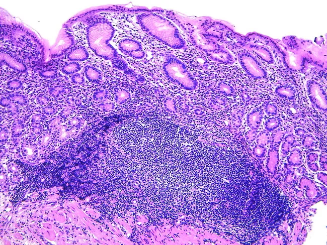
Gastric biopsy histology showing acquired MALT following Helicobacter pylori infection. Hematoxylin and eosin stained sections reveal dense lymphoid infiltration in the lamina propria with formation of lymphoid aggregates and germinal centers, partially effacing normal mucosal architecture. The overlying gastric epithelium remains intact in areas, but lymphoepithelial lesions may be present, indicating invasion of ducts or epithelium by neoplastic B-cells. The stomach normally lacks organized lymphoid tissue; the observed lymphoid proliferation represents mucosa-associated lymphoid tissue expansion, consistent with extranodal marginal zone B-cell lymphoma of mucosa-associated lymphoid tissue (MALT lymphoma). The cellular population is typically small to medium-sized lymphocytes with irregular chromatin, sometimes with plasmacytoid differentiation; scattered immunoblasts may occur. Chronic inflammation, such as Helicobacter pylori gastritis, provides the antigenic stimulus driving MALT development and potential regression after microbial eradication. Clinically, MALT lymphoma of the stomach may respond to antibiotic therapy, with lesion regression upon H. pylori eradication, or require staging and systemic therapy if refractory or advanced. Differential includes reactive lymphoid hyperplasia and transformation to diffuse large B-cell lymphoma. Diagnostic significance lies in recognizing MALT patterns to guide treatment, prognosis, and surveillance; correlate with endoscopic findings and serology for infection status.
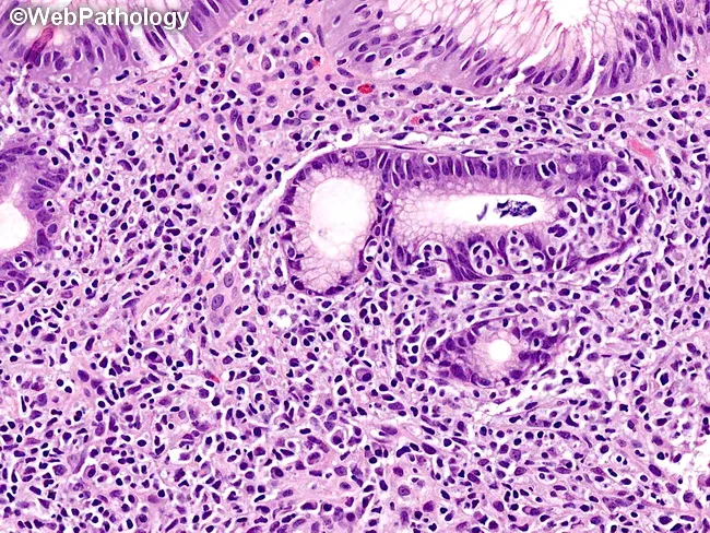
This histopathology image depicts a high-magnification hematoxylin and eosin stained section of gastric mucosa, illustrating a lymphoepithelial lesion characteristic of gastric mucosa-associated lymphoid tissue (MALT) lymphoma. Dense infiltration by small to intermediate-sized lymphocytes disrupts normal gastric glands, with marked invasion of glandular epithelium and glandular destruction. A modest plasmacytic component is present within the infiltrate, giving rise to plasmacytoid cells and occasional plasma cells adjacent to lymphoid aggregates. The surrounding lamina propria is expanded with lymphoid tissue, including rare intracellular plasma cells. MALT lymphoma etiologies include Helicobacter pylori infection; tumor cells are typically marginal zone B-cells that may form lymphoepithelial lesions within gastric glands. In this image, glands are variably spared, but architectural effacement and epithelial damage reflect neoplastic lymphoid infiltration rather than simple chronic gastritis. The cytology shows cohesive lymphoid sheets with relatively uniform nuclei, inconspicuous nucleoli, and scant mitotic activity in well-differentiated areas; occasional reactive lymphoid follicles may be seen elsewhere. Overall, the image exemplifies characteristic histopathologic features used to diagnose gastric MALT lymphoma and to distinguish it from chronic gastritis and other gastric lymphomas; the finding supports targeted clinical management decisions. Correlate with H. pylori status and molecular tests for complete diagnostic characterization and therapeutic planning guidance.
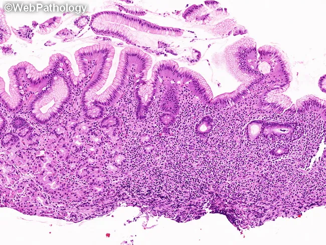
Imaging modality and technique: This image is a histopathology micrograph obtained from a gastric mucosal biopsy and viewed under light microscopy after formalin fixation and Hematoxylin and Eosin (H&E) staining. The specimen shows gastric mucosa with intact surface epithelium and deep lamina propria infiltrated by a dense, monomorphic lymphoid population that distorts and displaces the native gastric glands. The infiltrate is composed of small to medium-sized lymphocytes with relatively uniform nuclear contours and scant cytoplasm, characteristic of marginal zone B-cells seen in MALT lymphoma. Lymphoepithelial lesions are present as lymphoid cells invade and disrupt gastric glands, a hallmark of extranodal marginal zone lymphoma of MALT. There is minimal overt cytologic atypia and preserved overall tissue architecture aside from glandular distortion, a pattern that supports an early or low-grade MALT process rather than high-grade transformation. The image highlights a localized gastric mucosal process; in the clinical setting, such histology often correlates with Helicobacter pylori infection and response to antibiotic eradication therapy in localized disease. Differential considerations include gastritis with reactive lymphoid aggregates, other gastric B-cell lymphomas, and gastric adenocarcinoma with reactive lymphoid infiltrates. Overall, the finding confirms gastric MALT lymphoma pathology and informs staging, surveillance, and therapeutic planning.
diffuse large B-cell lymphoma intestinal small bowel gross pathology
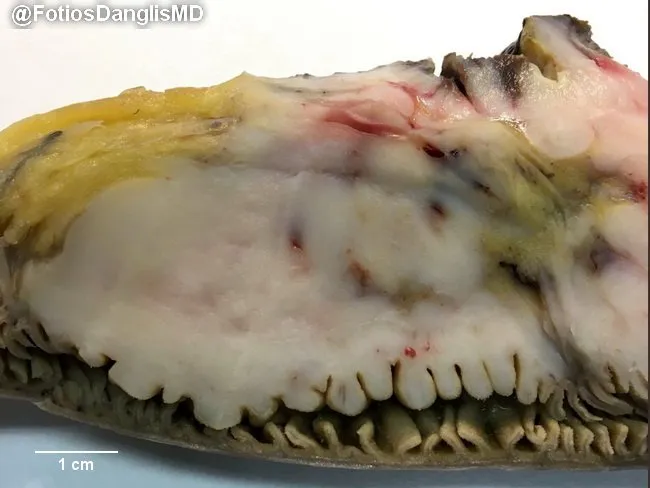
Macroscopic photograph of a surgically resected small bowel segment illustrating transmural infiltration by diffuse large B‑cell lymphoma. The intestinal wall is diffusely thickened; mucosal folds are effaced and markedly expanded by tumoral tissue, producing a plaque‑like, bulky appearance along the luminal surface. The serosa shows no distinct gross ulceration in this view, though focal friability and areas of pale yellow to tan coloration reflect tumor replacement of normal wall layers. The tumor mass extends through the mucosa and submucosa with involvement of the deeper wall, consistent with lymphomatous infiltration. The surrounding mesenteric fat appears unremarkable in this slice, and vascular demand is not assessable in this plane. A centimeter scale bar is provided for size reference, indicating that the thickening spans several centimeters in length and a few millimeters in wall thickness. This gross pattern, combined with the clinical diagnosis of small bowel diffuse large B‑cell lymphoma (DLBCL), correlates with aggressive non-Hodgkin lymphoma of the gastrointestinal tract. Clinically, DLBCL of the small intestine presents with obstructive symptoms, abdominal pain, and weight loss; management typically combines surgical resection with systemic chemotherapy (e.g., R‑CHOP). This image is valuable for education on gross morphology of lymphomatous involvement, differential with other small bowel neoplasms, and radiologic correlation.
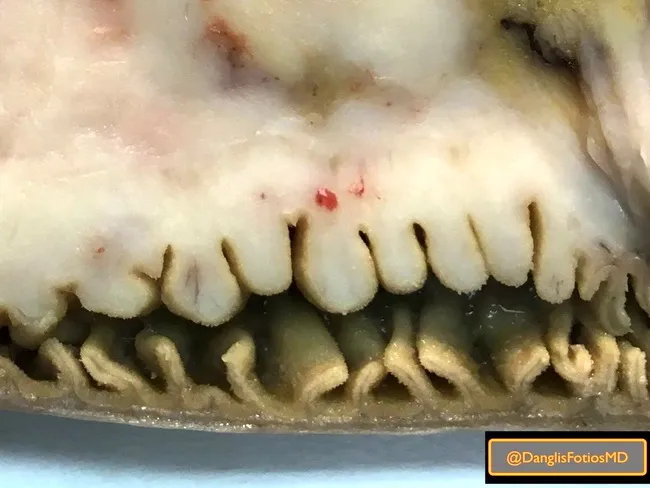
This macroscopic photograph depicts a surgically resected segment of small intestine infiltrated by diffuse large B-cell lymphoma (DLBCL). The gross appearance is characterized by diffuse thickening of the mucosal folds with marked expansion of each mucosal villus-like folding by tumor tissue, leading to a bulky, convoluted luminal surface. The serosa remains intact but areas of irregular, firm nodularity correspond to tumor infiltration within the mucosa and submucosa. The lesion effaces normal mucosal architecture, producing a mottled whitish-yellow to tan coloration with focal hemorrhagic streaks. The image captures a longitudinal or near-longitudinal open view, enabling assessment of transmural involvement and luminal contour changes. In DLBCL of the small bowel, malignant lymphoid cells form sheets that disrupt epithelial integrity and may produce focal ulceration or obstruction; immunohistochemical workup (CD20, CD79a, BCL6, MUM1) is essential for subclassification and guiding therapy. Clinically, such lesions present with abdominal pain, weight loss, GI bleeding, or obstruction; radiologic correlation can show circumferential bowel wall thickening and aneurysmal dilation. This image is valuable for education, differential diagnosis, and research on intestinal lymphomas, helping trainees recognize gross patterns of mucosa-infiltrating lymphoid neoplasms and the anatomic distribution within the small intestine. Reference clinical correlation and biopsy confirmation for definitive diagnosis purposes.
Gastrointestinal Lymphomas - Pathology Note
Overview
- No palpable peripheral lymphadenopathy
- Normal leukocyte count and differential on peripheral blood smear
- No mediastinal nodes on chest X-ray
- Involvement limited to the GI tract and proximal regional nodes
- No hepatic or splenic involvement (unless by direct extension)
Classification
B-Cell Lymphomas (majority)
- Extranodal marginal zone lymphoma (MALT lymphoma)
- Diffuse large B-cell lymphoma (DLBCL)
- Mantle cell lymphoma (Multiple Lymphoid Polyposis)
- Follicular lymphoma
- Immunoproliferative small intestinal disease (IPSID / Mediterranean lymphoma)
- Small noncleaved cell lymphoma (including Burkitt)
T-Cell Lymphomas (minority, mostly small bowel)
- Enteropathy-associated T-cell lymphoma (EATL - Type I)
- Monomorphic epitheliotropic intestinal T-cell lymphoma (MEITL - Type II, formerly EATL Type II)
1. Extranodal Marginal Zone (MALT) Lymphoma
- The stomach normally contains no organized lymphoid tissue. MALT is acquired in response to chronic antigenic stimulation, most commonly H. pylori infection (in gastric MALT)
- H. pylori drives a T-cell dependent, antigen-stimulated B-cell proliferation that is initially polyclonal, then oligoclonal, then monoclonal
- The Cag pathogenicity island-expressing strains are especially implicated
- In IPSID (a MALT subtype), Campylobacter jejuni is the implicated pathogen
- Other MALT sites: salivary glands (Sjogren's), thyroid (Hashimoto's), ocular adnexa
- Infiltrate of small to medium-sized marginal zone B-cells with abundant pale cytoplasm ("centrocyte-like" cells)
- Hallmark: Lymphoepithelial lesions (LELs) - neoplastic B-cells invade and destroy gastric glands/crypts
- Reactive germinal centers may be colonized by neoplastic cells
- Plasmacytic differentiation is common
- Background chronic active gastritis with H. pylori organisms often visible
- CD20+, CD22+, CD79a+, LN-1+
- CD5-, CD10-, CD23-, cyclin D1- (helps distinguish from mantle cell and follicular lymphoma)
- Trisomy 3 is the most common cytogenetic finding
- Recurrent translocations: t(11;18)(q21;q21) - API2-MALT1 most common in gastric MALT; t(14;18); t(1;14)

2. Diffuse Large B-Cell Lymphoma (DLBCL)
- May arise de novo or by high-grade transformation from MALT lymphoma ("MALT-to-DLBCL transformation")
- Associated with HIV/AIDS, post-transplant immunosuppression, EBV
- Large, exophytic or ulcerating masses
- Transmural wall thickening, often with effacement of mucosal folds
- Diffuse sheets of large lymphoid cells with vesicular nuclei, prominent nucleoli, and frequent mitoses
- Often with central necrosis
- "Starry sky" pattern may be seen (tingible body macrophages)
- CD20+, CD79a+ (B-cell markers)
- BCL6, MUM1, BCL2 expression varies by subtype (GCB vs. ABC/non-GCB)
- Immunoglobulin genes rearranged
3. Mantle Cell Lymphoma (Multiple Lymphoid Polyposis / MLP)
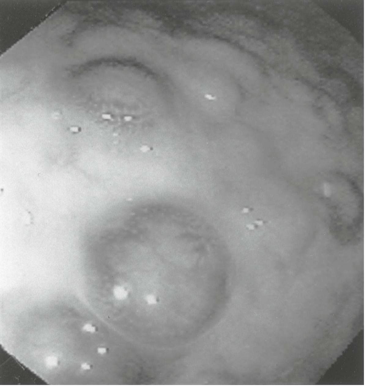
- Characteristic multiple lymphomatous polyposis - mucosa studded with numerous small polypoid nodules throughout the GI tract (see endoscopic image below)
- Monotonous small lymphocytes with irregular, cleaved nuclei
- Arrangement similar to the mantle zone of lymph follicles
- Mucosa and submucosa most frequently involved
- Epithelial invasion is uncommon despite aggressive behavior
- CD5+, CD20+, cyclin D1+, SOX11+
- CD23-, CD10- (helps distinguish from CLL/SLL and follicular lymphoma)
- t(11;14)(q13;q32): IGH-CCND1 in nearly 100% of cases
- Juxtaposition of CCND1 (cyclin D1) gene to IGH enhancer → cyclin D1 overexpression → uncontrolled cell cycle progression
- Detectable by FISH (most sensitive, ~100%) or immunohistochemistry for cyclin D1
- Additional: loss of 1p, 13q, 17p; gain of 3q
4. Enteropathy-Associated T-Cell Lymphoma (EATL)
- Arises from intraepithelial lymphocytes (IELs) as a complication of celiac disease
- Sequence: Celiac disease → Type II refractory celiac disease (RCD II) → ulcerative jejunoileitis → EATL
- Monoclonal T-cell receptor rearrangement is present both in the tumor and in the surrounding "cryptic" mucosa
- "Cryptic lymphoma" refers to clonally expanded T-cells in macroscopically normal surrounding mucosa
- EATL Type I (classic): associated with celiac disease; CD4+/CD8- or CD4-/CD8-
- MEITL (Type II): NOT associated with celiac disease; CD8+, CD56+; more common in Asia
- Often multifocal and diffuse
- Mucosal ulceration, hemorrhage, perforation, and stricture with obstruction are common complications
- Large cells resembling histiocytes (pleomorphic to anaplastic large cells)
- Intense surrounding inflammatory infiltrate (eosinophils, histiocytes)
- Adjacent mucosa shows celiac-type changes: villous atrophy, crypt hyperplasia, abundant IELs
- T-cell markers: CD3+, CD7+, CD103+, HML-1+
- CD4+/CD8- (EATL Type I); CD8+, CD56+ (MEITL/Type II)
- T-cell receptor genes rearranged
- NKp46 overexpression is a hallmark of malignant intestinal T-cells in Type II RCD and EATL
5. Immunoproliferative Small Intestinal Disease (IPSID / Mediterranean Lymphoma)
- Associated with Campylobacter jejuni infection (molecular/immunohistochemical evidence)
- Antigen-driven clonal proliferation of IgA-secreting B-cells in the lamina propria
- Alpha-heavy chains (without light chains) are secreted and detectable in serum in up to 69% of early-stage patients
| Phase | Histology |
|---|---|
| Early | Dense infiltrate of plasma cells and small lymphocytes in mucosa and submucosa throughout affected jejunum |
| Late | Dysplastic changes in small cells; increasing proportion of large cells → transformation to diffuse large cell lymphoma |
6. Follicular Lymphoma (GI)
- Relatively rare primary GI presentation; duodenum most common site
- Gross: Small, pale nodules in mucosa
- Histology: Nodular/follicular growth pattern of small cleaved follicle center cells (centrocytes + centroblasts)
- IHC: CD20+, CD10+, BCL2+, BCL6+
- Genetics: t(14;18)(q32;q21) - IGH-BCL2 (same as nodal follicular lymphoma)
- Generally indolent
Staging of Primary GI Lymphoma
| Stage | Description |
|---|---|
| I | Tumor confined to GI tract (single primary or multiple non-contiguous) |
| II | Tumor extending to abdomen: II-1 = local node involvement; II-2 = distant nodal involvement |
| IIE | Penetration of serosa to involve adjacent organs/tissues |
| IV | Disseminated extranodal involvement or concomitant supradiaphragmatic nodal involvement |
Summary Table: Features of Major GI Lymphomas
| Type | Histology | IHC | Genetics |
|---|---|---|---|
| DLBCL | Diffuse large cells, necrosis | CD20+, B markers | Ig gene rearranged |
| MALT (low-grade B-cell) | Centrocyte-like cells, lymphoepithelial lesions | CD20+, CD22+, LN-1+; CD5-, CD10- | Ig gene rearranged; trisomy 3; t(11;18) |
| Mantle cell (MLP) | Monotonous small cells, irregular nuclei | CD20+, CD5+, cyclin D1+; CD23- | t(11;14) - IGH-CCND1 |
| EATL | Large cells + intense inflammation; adjacent villous atrophy | CD3+, CD103+, HML-1+, CD4+/CD8- | TCR gene rearranged |
| IPSID | Early: plasma cells; Late: large atypical cells | B markers, KB61+, alpha-heavy chain | - |
| Small noncleaved (Burkitt) | Medium cells, "starry sky" | CD20+, CD10+, sIg+ | MYC translocation t(8;14) |
Risk Factors for GI Lymphoma
| Condition | Associated Lymphoma |
|---|---|
| H. pylori infection | Gastric MALT, DLBCL |
| Celiac disease | EATL (7% incidence with long-standing disease) |
| Campylobacter jejuni | IPSID |
| HIV/AIDS | DLBCL (30% risk of lymphoma; 10-25% are intestinal) |
| Post-transplant immunosuppression | PTLD (90% EBV-associated B-cell; highest risk in intestinal transplant recipients ~30%) |
| Crohn's disease (male, small bowel) | DLBCL |
| Nodular lymphoid hyperplasia | B-cell intestinal lymphoma (in primary immunodeficiency) |
Key Pathology Pearls
- MALT lymphoma hallmark = lymphoepithelial lesions (neoplastic B-cells invading glands)
- Mantle cell lymphoma hallmark = t(11;14); cyclin D1 overexpression; multiple polyposis gross pattern
- EATL hallmark = adjacent villous atrophy + intraepithelial lymphocytosis (celiac changes)
- IPSID hallmark = alpha-heavy chain paraprotein; diffuse proximal jejunal disease
- Burkitt lymphoma hallmark = "starry sky" pattern; MYC translocation
- EATL and MLP carry the worst prognosis among primary GI lymphomas
- B-cell lymphomas predominate in the GI tract; T-cell lymphomas are strongly associated with the small bowel and celiac disease