
1. Discuss and classify tumors of testis. Discuss Seminoma in detail. 2. Endometrial biopsy in health and disease 3. Classify ovarian tumours. Discuss functional ovarian tumors. 4. Endometriosis 5. Contrasting features of serous and mucinous tumors or epithelial tumours of ovary. 6. Placental site of trophoblastic tumors
Reading File
Now let me run parallel searches for all 6 topics simultaneously:
Finding Sources
Finding Sources
Finding Sources
Finding Sources
Finding Sources
Finding Sources
Finding Sources
Finding Sources
Reading File
Reading File
Excellent! I have rich pathology content. Let me now read the testis tumor and seminoma sections, endometriosis, and trophoblastic tumor content:
Reading File
Reading File
Reading File
Now let me read the testis tumor section from Robbins Basic Pathology, and the endometrial biopsy and endometriosis sections:
Finding Sources
Reading File
Reading File
Reading File
Reading File
Reading File
Now let me read the key testis tumor / seminoma section and the endometriosis and sex cord sections:
Reading File
Reading File
Now let me get the testis tumor content from Robbins basic pathology and the endometriosis and endometrial biopsy pathology sections:
Finding Sources
Reading File
Now let me read the key seminoma and sex cord sections from Robbins PBD and the endometriosis content:
Reading File
Reading File
I now have comprehensive content. Let me also read the endometriosis pathology section and more of the sex cord tumor/ovarian classification:
Reading File
Reading File
I now have extensive content from the textbooks. Let me read the serous tumor section and sex cord stromal tumors, plus the trophoblastic tumor section from Berek & Novak:
Reading File
Reading File
Reading File
I now have sufficient content to write comprehensive answers for all 6 topics. Let me now compile everything:Here are comprehensive answers to all six topics:
1. Tumors of the Testis - Classification and Seminoma
Classification of Testicular Tumors
Testicular neoplasms are the most common cause of painless testicular enlargement. Germ cells are the source of 95% of testicular tumors; the remainder arise from Sertoli or Leydig cells.
WHO 2022 Pathologic Classification (Robbins, Cotran & Kumar PBD)
A. Germ Cell Tumors (GCTs) - Derived from Germ Cell Neoplasia In Situ (GCNIS)
Noninvasive:
- Germ cell neoplasia in situ (GCNIS) - the precursor lesion
- Gonadoblastoma
Pure (single histologic type):
- Seminoma - most common (~50% of all GCTs), peak 4th decade
Nonseminomatous Germ Cell Tumors (NSGCTs):
- Embryonal carcinoma
- Yolk sac tumor (postpubertal type)
- Choriocarcinoma
- Teratoma (postpubertal type)
- Teratoma with somatic-type malignancy
- Mixed germ cell tumor (more than one histologic type)
B. Germ Cell Tumors Unrelated to GCNIS
- Spermatocytic tumor
- Teratoma (prepubertal type)
- Yolk sac tumor (prepubertal type)
- Mixed teratoma and yolk sac tumor (prepubertal type)
C. Sex Cord-Stromal Tumors
- Leydig cell tumor
- Sertoli cell tumor
Clinical Division (for management purposes)
- Seminoma - confined to testis for a long time, spreads mainly to para-aortic lymph nodes, rare distant spread, better prognosis
- Nonseminomatous tumors - tend to spread earlier via both lymphatics and blood vessels
Seminoma - Detailed Discussion
Incidence and Overview
- Most common type of GCT, making up about 50% of all GCTs
- Peak incidence: fourth decade of life
- An identical tumor in the ovary is called dysgerminoma; in the CNS (pineal gland) it is called germinoma
- Associated with undescended testes (cryptorchidism) and testicular dysgenesis
Gross Morphology
- Produces bulky masses, sometimes 10 times the size of the normal testis
- Homogeneous, gray-white, lobulated cut surface
- Usually devoid of hemorrhage or necrosis
- Tunica albuginea generally not penetrated
- Occasionally extends to epididymis, spermatic cord, or scrotal sac
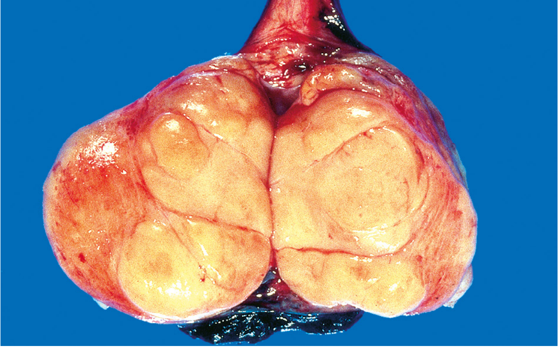
Seminoma of the testis - well-circumscribed, pale, fleshy, homogeneous mass on cut surface
Microscopic Morphology
- Sheets of uniform cells divided into poorly demarcated lobules by delicate fibrous septa
- Septa contain a prominent lymphocytic infiltrate
- In some tumors, ill-defined granulomas are present (host immune response)
- Classic seminoma cell features:
- Round to polyhedral shape
- Distinct cell membrane
- Clear/watery-appearing cytoplasm (contains glycogen)
- Large, central nucleus with one or two prominent nucleoli
Immunohistochemistry
- Positive: KIT, OCT3/4, podoplanin (D2-40)
- Negative: cytokeratin (distinguishes from embryonal carcinoma)
- ~15% of seminomas contain syncytiotrophoblasts → elevated serum hCG (not as high as choriocarcinoma)
- AFP is NOT elevated in pure seminoma
Precursor Lesion
- Arises from Germ Cell Neoplasia In Situ (GCNIS) - intratubular malignant germ cells that do not yet invade
- GCNIS cells are OCT3/4 and KIT positive; they line the basement membrane of seminiferous tubules
Molecular/Genetic Features
- Characteristic cytogenetic change: isochromosome 12p [i(12p)] - gain of short arm of chromosome 12
- Present in virtually all GCTs derived from GCNIS
Staging
- Stage I - confined to testis
- Stage II - retroperitoneal (para-aortic) lymph node involvement
- Stage III - distant metastases (lung, liver, brain)
Clinical Features & Prognosis
- Presents as painless testicular swelling/heaviness
- Serum markers: hCG elevated in ~15% (those with syncytiotrophoblasts); AFP always normal
- LDH may be elevated
- Radiosensitive - responds excellently to radiotherapy
- 5-year survival >95% for stage I; overall excellent prognosis compared to NSGCTs
2. Endometrial Biopsy in Health and Disease
Overview
Endometrial biopsy (EMB) is the sampling of endometrial tissue for histologic examination. It is a key procedure in the evaluation of abnormal uterine bleeding and in the detection of endometrial pathology.
Indications for Endometrial Biopsy
(Bailey & Love's Short Practice of Surgery, 28th ed.)
Endometrial biopsy should be considered in:
- Women with suspected endometrial pathology
- All women >45 years in whom medical treatment has been unsuccessful
- Persistent intermenstrual bleeding
- Endometrial thickness >4 mm in postmenopausal women, or >7 mm in women with PCOS
- Persistently thickened/abnormal endometrial appearance in premenopausal women
- Irregular or unscheduled bleeding while on HRT after the initial 3 months
- Younger women with major risk factors for endometrial hyperplasia/cancer:
- Polycystic ovarian syndrome (PCOS)
- Obesity
- Treatment with tamoxifen
- Irregular bleeding on unopposed estrogen
- Family history of endometrial/colon cancer (hereditary non-polyposis colorectal cancer - HNPCC)
- Lynch syndrome patients - lifetime risk of endometrial cancer up to 60%; annual screening with TVS + EMB from age 35
Normal Endometrial Histology (Cyclic Changes)
Proliferative Phase (Days 5-14)
- Estrogen-driven
- Straight, tubular, elongated glands with pseudostratified columnar epithelium
- Mitotic figures present in glands and stroma
- Stroma: compact with small, oval to spindle-shaped cells
Secretory Phase (Days 15-28)
- Progesterone-driven (after ovulation)
- Glands become tortuous, saw-toothed ("corkscrew" glands)
- Subnuclear vacuoles appear first (day 16-17), then supranuclear secretion
- Predecidual change in stroma (days 23-24)
- Spiral arteries become prominent
Menstrual Phase
- Breakdown of stroma and glands
- Neutrophilic infiltration, fibrin thrombi
- "Blue balls" of stroma (dense stromal breakdown)
Endometrial Pathology Detected on Biopsy
1. Endometrial Hyperplasia
Caused by unopposed estrogen stimulation:
| Type | Features | Risk of Carcinoma |
|---|---|---|
| Simple hyperplasia without atypia | Increased gland:stroma ratio, no architectural complexity | ~1% |
| Complex hyperplasia without atypia | Gland crowding, budding, branching | ~3% |
| Simple hyperplasia with atypia | Cytologic atypia + simple architecture | ~8% |
| Complex hyperplasia with atypia (EIN) | Gland crowding + cytologic atypia | ~25-30% |
Endometrial Intraepithelial Neoplasia (EIN) is the modern term for complex atypical hyperplasia.
2. Endometrial Carcinoma
- Endometrioid adenocarcinoma (Type I) - most common; estrogen-related; mutations in PTEN, PIK3CA, KRAS, ARID1A, mismatch repair genes
- Serous carcinoma (Type II) - non-estrogen dependent; TP53 mutations; aggressive
- Grading: Grade 1 (well-differentiated), Grade 2 (moderately), Grade 3 (poorly)
3. Endometrial Polyp
- Focal overgrowth of endometrial glands and stroma with fibrous cores
- Irregular vascular channels
- Non-cycling glands (lack of secretory change)
4. Endometritis
- Acute: neutrophilic infiltration (postpartum, post-abortion, PID)
- Chronic: plasma cells in stroma (key diagnostic feature) - seen in PID, IUD use, tuberculous endometritis
5. Anovulatory Cycles / Irregular Shedding
- Out-of-phase endometrium
- Mixed proliferative and secretory pattern
- Common in perimenopause and PCOS
3. Classification of Ovarian Tumors and Functional Ovarian Tumors
Classification
WHO Classification of Ovarian Neoplasms
(Robbins, Cotran & Kumar PBD)
I. Surface Epithelial-Stromal Tumors (~65-70% of all ovarian tumors)
- Serous tumors (benign cystadenoma, borderline, malignant)
- Mucinous tumors, intestinal and endocervical type
- Endometrioid tumors
- Clear cell tumors
- Brenner tumors (transitional cell type)
- Epithelial-stromal tumors (Adenosarcoma, Carcinosarcoma)
II. Sex Cord-Stromal Tumors (~8%)
- Granulosa cell tumors
- Fibromas, Fibrothecomas, Thecomas
- Sertoli-Leydig cell tumors
- Steroid (lipid) cell tumors
III. Germ Cell Tumors (~15-20%)
- Teratoma (mature/immature; solid/cystic - dermoid cyst; monodermal - struma ovarii, carcinoid)
- Dysgerminoma
- Yolk sac tumor (endodermal sinus tumor)
- Choriocarcinoma
- Mixed germ cell tumors
IV. Metastatic Tumors (~5%)
- From colorectal, gastric, pancreaticobiliary, breast primaries
- Krukenberg tumor (bilateral mucin-secreting "signet-ring" cells, usually gastric origin)
Non-Neoplastic / Functional Lesions
- Follicular cysts
- Corpus luteum cysts
- Theca-lutein cysts
- Polycystic ovarian syndrome (PCOS)
Functional Ovarian Tumors (Hormonally Active Tumors)
These are sex cord-stromal tumors that secrete hormones and cause clinical syndromes.
A. Granulosa Cell Tumors (Feminizing)
Incidence: ~5% of all ovarian tumors; 95% are adult type
Gross:
- Usually unilateral
- Vary from microscopic foci to large solid-cystic encapsulated masses
- Yellow coloration on cut surface (due to intracellular lipids in hormonally active tumors)
Microscopic:
- Small cuboidal to polygonal cells in anastomosing cords, sheets, or strands
- Call-Exner bodies: small, distinctive gland-like structures filled with acidophilic material, resembling immature follicles - pathognomonic
- Express high levels of inhibin (IHC marker)
Molecular: FOXL2 mutation in 97% of adult granulosa cell tumors
Clinical effects (due to estrogen secretion):
- Prepubertal girls: precocious sexual development (juvenile granulosa cell tumor)
- Reproductive age: menstrual irregularities, endometrial hyperplasia
- Postmenopausal: abnormal uterine bleeding, endometrial hyperplasia
- ~10-15% develop endometrial carcinoma (due to prolonged estrogenic stimulation)
- Occasionally may secrete androgens causing virilization
Behavior: Low-grade malignancy; may recur 10-20 years after primary resection
Marker: Inhibin (positive); also AMH
B. Thecoma (Feminizing)
- Composed of lipid-laden theca cells
- Usually unilateral, solid, yellow-cut surface
- Almost always benign
- Estrogenic symptoms: endometrial hyperplasia/carcinoma risk
- Often associated with fibroma (fibrothecoma)
C. Fibroma
- Most common benign sex cord-stromal tumor
- Composed of fibroblasts
- Usually hormonally inactive
- Associated with Meigs syndrome: ovarian fibroma + ascites + pleural effusion (right-sided)
- Resolves with tumor removal
D. Sertoli-Leydig Cell Tumors (Androblastoma - Virilizing)
- Contain Sertoli cells (form tubular structures) and Leydig cells (with Reinke crystalloids)
- Secrete androgens → virilization in females:
- Defeminization: amenorrhea, breast atrophy
- Masculinization: clitoral enlargement, hirsutism, voice deepening
- Usually low-grade malignant; <5% recur or metastasize
- Most common in women of reproductive age
E. Leydig Cell Tumors (Hilus Cell Tumors - Virilizing)
- Located in ovarian hilus
- Large lipid-laden Leydig cells with Reinke crystalloids (cytoplasmic rod-shaped structures)
- Secrete testosterone → virilization
- Almost always benign
F. Steroid/Lipid Cell Tumors
- Contain lipid-rich cells resembling adrenal cortex, Leydig, or lutein cells
- May be virilizing or feminizing depending on hormone produced
4. Endometriosis
Definition
Endometriosis is the presence of ectopic endometrial tissue (glands and/or stroma) outside the uterus.
Sites of Involvement (in descending frequency)
- Ovaries (most common) - form "chocolate cysts" (endometriomas)
- Uterine ligaments
- Rectovaginal septum
- Cul-de-sac (pouch of Douglas)
- Pelvic peritoneum
- Serosa of large/small bowel and appendix
- Mucosa of cervix, vagina, fallopian tubes
- Laparotomy scars (rare)
Epidemiology
- Affects ~10% of women of reproductive age
- Most common in 3rd and 4th decades
- Major cause of infertility (30-40% of infertile women)
Types
- Superficial peritoneal endometriosis - implants on peritoneal surface
- Ovarian endometriosis - "chocolate cyst" or endometrioma
- Deep infiltrating endometriosis - penetrates >5 mm below peritoneum; rarely undergoes malignant transformation
Pathogenesis (Theories)
1. Regurgitation Theory (Sampson's Theory) - Most Accepted
- Retrograde flow of menstrual endometrium through fallopian tubes implants at ectopic sites in peritoneal cavity
- Explains pelvic endometriosis
- Limitation: retrograde menstruation occurs in 90% of women; only ~10% develop endometriosis
2. Benign Metastasis Theory
- Endometrial tissue "spreads" to distant sites (bone, lung, brain) via blood vessels and lymphatics
- Explains rare extrapelvic locations
3. Metaplastic Theory (Coelomic Metaplasia)
- Endometrium arises from metaplastic transformation of coelomic epithelium (mesothelium)
- Explains endometriosis in amenorrheic women and even in males (rare cases)
4. Extrauterine Stem/Progenitor Cell Theory
- Bone marrow-derived stem cells differentiate into endometrial tissue
Molecular Differences in Endometriotic Implants
Compared to normal endometrium, endometriotic tissue shows:
- Increased aromatase activity (local estrogen production)
- Resistance to progesterone (progesterone receptor B downregulation)
- Increased inflammatory cytokines (IL-1, IL-6, IL-8, TNF-alpha)
- Reduced apoptosis
- ARID1A mutations in endometriosis-associated ovarian cancer
Morphology
Gross
- Superficial: red, blue, or black "powder burn" peritoneal spots
- Ovarian endometriomas ("chocolate cysts"): filled with thick, brown, altered blood; walls contain endometrial glands and stroma
- Fibrosis and adhesions cause organs to become matted together ("frozen pelvis")
- Rectosigmoid involvement: low-signal fibrotic plaques on MRI; contraction of bowel wall
Microscopic
- Endometrial glands + endometrial stroma
- Hemosiderin-laden macrophages (key feature in old lesions)
- Fibrosis in surrounding tissue
- Cyclic changes (proliferative/secretory) may be present
Clinical Features
- Dysmenorrhea (painful menstruation - most common)
- Dyspareunia (deep)
- Pelvic pain (chronic)
- Infertility (tube/ovary adhesions, impaired implantation)
- Abnormal uterine bleeding (menorrhagia, menometrorrhagia)
- Rectal bleeding, bowel symptoms if bowel involved
- Cyclical hematuria if bladder involved
Complications
- Infertility (30-40% of cases)
- Chocolate cyst rupture → acute abdomen
- Endometriosis-associated ovarian cancer (clear cell and endometrioid carcinoma; risk ~0.5%)
- Frozen pelvis from adhesions
Diagnosis
- Clinical history + examination
- Transvaginal ultrasound (homogeneous low-level echo in endometrioma)
- MRI (best for deep infiltrating disease; low signal fibrotic plaques)
- Definitive: Laparoscopy with biopsy
Treatment
- Medical: NSAIDs, combined OCP, progestogens, GnRH agonists, danazol, levonorgestrel IUS
- Surgical: Conservative (laparoscopic excision/ablation), definitive (hysterectomy + bilateral salpingo-oophorectomy for severe cases)
5. Contrasting Features of Serous and Mucinous Tumors (Epithelial Ovarian Tumors)
(Source: Robbins, Cotran & Kumar PBD; Robbins & Kumar Basic Pathology)
| Feature | Serous Tumors | Mucinous Tumors |
|---|---|---|
| Frequency | Most common epithelial ovarian tumor (47% of malignant ovarian tumors) | Less common (~3% of malignant ovarian tumors) |
| Bilateral involvement | Common (benign 25%, borderline 30%, malignant 65%) | Rare (benign 5%, borderline 10%, malignant <5%) |
| Surface involvement | Frequently involves surface of ovary | Surface of ovary rarely involved |
| Size | Moderate | Very large (some >25 kg); multiloculated |
| Cyst content | Clear, watery, serous fluid | Thick, sticky, gelatinous fluid rich in glycoproteins |
| Gross appearance | Smooth or papillary; thin-walled | Multiloculated, filled with mucin |
| Cell type | Resembles tubal/serous epithelium; columnar cells with cilia | Tall columnar cells with apical mucin; no cilia; gastric or intestinal-type differentiation |
| Psammoma bodies | Present (concentric calcified laminations) - characteristic | Absent |
| Papillary projections | Common, both intracystic and external | Less prominent |
| Borderline tumor histology | Increased architectural complexity, epithelial stratification; complex micropapillary growth | Epithelial stratification, tufting, papillary intraglandular growth; resembles intestinal adenoma |
| Carcinoma (malignant) pattern | High-grade: solid sheets, necrosis; Low-grade: micropapillary pattern | Confluent glandular ("expansive") invasion; desmoplastic stromal invasion |
| Key molecular alterations | - High-grade: TP53 mutations (96%), BRCA1/BRCA2 - Low-grade: KRAS, BRAF mutations | KRAS mutations (58% benign, 75-86% borderline, 85% carcinoma) |
| Origin (current understanding) | High-grade serous carcinoma - likely arises from fallopian tube fimbriated end (STIC) | May arise from mucinous glands of teratomas/Brenner tumors, or Müllerian epithelium |
| Bilateral frequency | High (malignant: 65%) | Low (malignant: <5%) |
| Spread outside ovary | Common at presentation | Rare for primary mucinous carcinoma; must rule out metastasis from GI tract |
| Pseudomyxoma peritonei | Does not cause | Can cause (rupture of mucinous tumor → mucin in peritoneal cavity) - usually appendiceal origin |
| Prognosis (stage I) | Low-grade borderline: excellent; High-grade carcinoma: poor | Stage I invasive: ~90% 10-year survival; extraovarian spread: poor |
| IHC markers | WT1 positive, PAX8 positive | CK20 positive, CDX2 positive (if intestinal type); WT1 negative |
| Stromal reaction | Fibrovascular cores with papillary projections | Mucin-filled glands with minimal stroma |
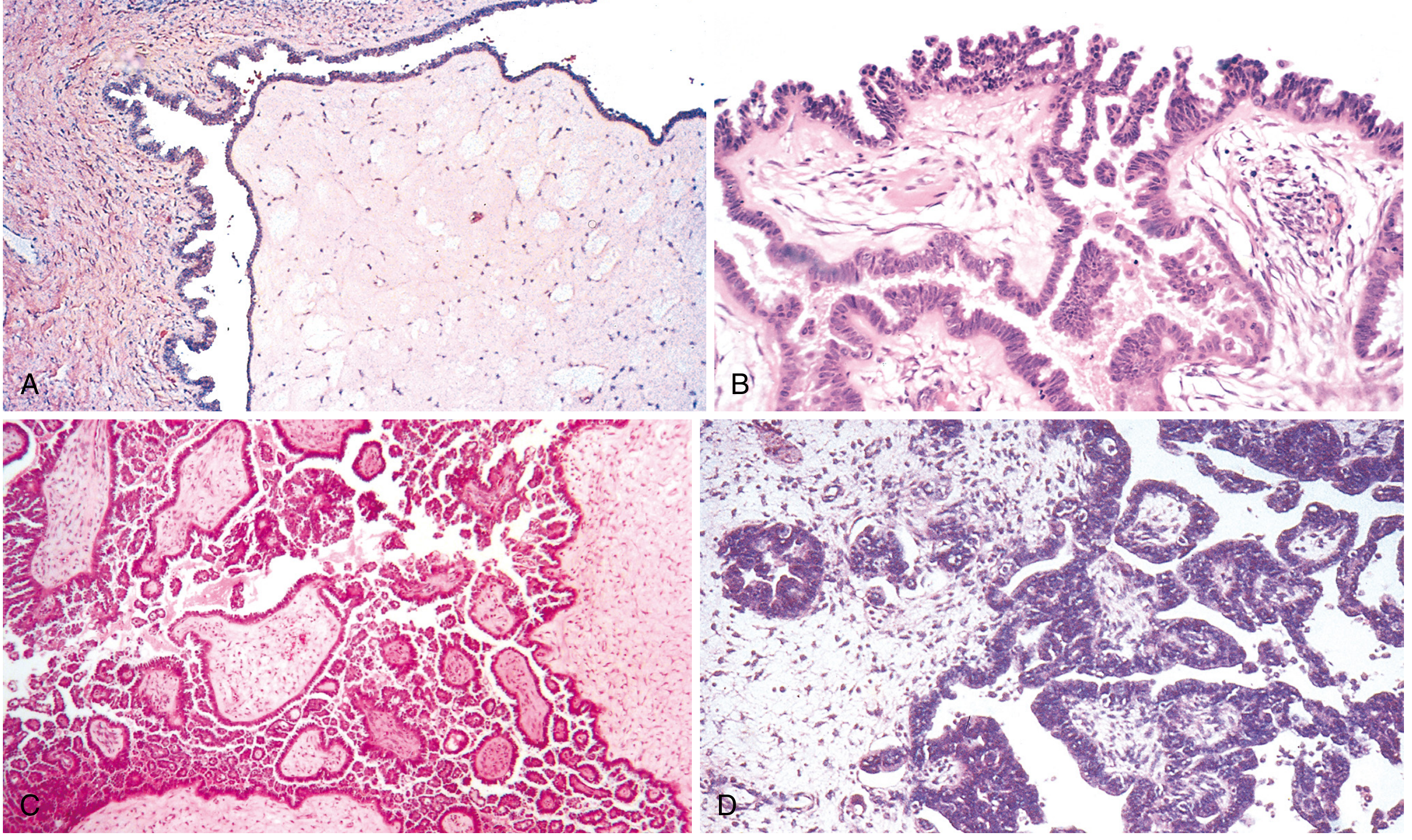
Serous tumors: (A) Serous cystadenoma - stromal papillae with columnar epithelium; (B) Borderline serous tumor - increased complexity and stratification; (C) Low-grade "micropapillary" serous carcinoma; (D) High-grade serous carcinoma with stromal invasion
6. Placental Site Trophoblastic Tumor (PSTT)
Definition and Overview
Placental site trophoblastic tumors (PSTT) are rare gestational trophoblastic neoplasms (GTN) comprising less than 2% of all GTN. They are neoplastic proliferations of extravillous (intermediate) trophoblasts - cells that normally proliferate and migrate from the cytotrophoblast of the placenta to invade the maternal decidua and myometrium.
(Robbins & Kumar Basic Pathology; Berek & Novak's Gynecology)
Cell of Origin
- Derived from intermediate trophoblasts (extravillous trophoblasts)
- These cells have features overlapping with both cytotrophoblasts and syncytiotrophoblasts
- They are the cells that normally invade the myometrium at the placental implantation site
PSTT vs Epithelioid Trophoblastic Tumor (ETT)
Both PSTT and ETT are variants of GTN consisting predominantly of intermediate trophoblast. They share:
- Small amounts of hCG production relative to tumor mass
- Tendency to remain confined to uterus
- Late metastasis
- Relative insensitivity to chemotherapy
Hormone Production
- hCG: LOW levels (in contrast to choriocarcinoma, which has very high hCG)
- Intermediate trophoblasts do not produce hCG in large amounts
- Human Placental Lactogen (hPL): Elevated - key diagnostic marker
- Inhibin A may also be elevated
Pathology
Gross
- Usually infiltrates myometrium as a soft, tan-yellow mass
- May appear as an ill-defined intramural thickening
- No villi present (distinguishes from hydatidiform mole)
Microscopic
- Sheets and cords of intermediate trophoblastic cells infiltrating between smooth muscle fibers of myometrium
- Cells are mononucleate, with abundant eosinophilic cytoplasm
- Characteristic vascular invasion: tumor cells replace vascular smooth muscle (fibrinoid necrosis of vessel walls)
- No chorionic villi
- Focal syncytiotrophoblast cells may be present
IHC
- hPL positive (strongly)
- Mel-CAM (CD146) positive
- hCG: Weakly/focally positive (in contrast to choriocarcinoma which is diffusely positive)
- Ki-67: Low proliferative index in PSTT vs. high in choriocarcinoma
- p63: Negative in PSTT; positive in ETT (key distinction)
Karyotype
- Usually diploid, XX karyotype
- Arises from the trophoblastic component of a previous pregnancy
Clinical Features
- Usually arises months to years after a preceding pregnancy (most commonly a normal term delivery; less often after abortion or mole)
- Presents with:
- Irregular vaginal bleeding
- Amenorrhea
- Uterine enlargement
- Serum hCG mildly elevated (often <1000 mIU/mL)
Natural History
- Generally indolent course
- Usually confined to endometrium and myometrium initially
- Once extrauterine spread occurs, prognosis becomes poor
- NOT sensitive to chemotherapy (unlike choriocarcinoma, which responds dramatically to MTX/actinomycin-D)
Contrast with Choriocarcinoma
| Feature | PSTT | Choriocarcinoma |
|---|---|---|
| Cell of origin | Intermediate trophoblast | Cytotrophoblast + Syncytiotrophoblast |
| hCG | Low | Very high |
| hPL | High | Low |
| Villi | Absent | Absent |
| Chemosensitivity | Poor | Excellent |
| Prognosis (localized) | Favorable | Excellent with chemo |
| Prognosis (metastatic) | Poor | Good (with aggressive chemo) |
| Bilateral involvement | No | No |
Metastatic Pattern of GTN (including PSTT)
When GTN spreads beyond uterus:
- Lungs: 80% (most common) - "snowstorm" pattern on CXR
- Vagina: 30%
- Pelvis: 20%
- Liver: 10%
- Brain: 10%
Treatment
- Hysterectomy is the primary treatment (unlike other GTN where chemotherapy is first-line)
- Chemotherapy (EMA-CO or EP-EMA regimen) for metastatic disease, though less effective than in choriocarcinoma
- Complete surgical excision results in excellent prognosis when confined to uterus
Sources: Robbins, Cotran & Kumar Pathologic Basis of Disease (10th ed.); Robbins & Kumar Basic Pathology; Bailey & Love's Short Practice of Surgery (28th ed.); Berek & Novak's Gynecology; Robbins, Cotran & Kumar PBD Chapter 22 (Female Genital Tract), Chapter 21 (Male Genital Tract).
This is a shared conversation. Sign in to Orris to start your own chat.