
Pathophysiology of high blood pressure
I'll check the medical library skill first as required for this medical question.
Reading File
Now I'll search the medical library for hypertension pathophysiology content in parallel.
Finding Sources
Finding Sources
Searching PubMed
"hypertension"[MeSH Terms] AND "pathophysiology"[MeSH Terms]
Excellent - I found two key sources. Let me read the most relevant ones: Fuster and Hurst's The Heart and the National Kidney Foundation Primer, in parallel.
Reading File
Reading File
Great content. Let me read more from Fuster & Hurst's Heart section on primary hypertension and also the RAAS/sympathetic nervous system sections.
Reading File
Reading File
Excellent content. Let me read more from both sources - the sympathetic nervous system section and primary hypertension section.
Reading File
Reading File
I now have rich, detailed content. Let me also quickly read the SNS and RAAS sections from Kidney Foundation Primer to round out the answer.
Reading File
I now have comprehensive textbook content. Let me compose the full answer.
Pathophysiology of High Blood Pressure (Hypertension)
Hypertension is defined as a sustained elevation in blood pressure (BP) above 130/80 mmHg (ACC/AHA 2017 guidelines) or 140/90 mmHg (older/WHO definition). Over 90% of cases are primary (essential) hypertension, where no single reversible cause is identified. The remaining ~10% are secondary hypertension (renal artery stenosis, primary aldosteronism, pheochromocytoma, etc.).
1. The Fundamental Formula
BP = Cardiac Output (CO) × Total Peripheral Resistance (TPR)
Long-term BP regulation requires more than just this formula. It also depends on:
- Kidney regulation of body fluid and sodium balance
- Local autoregulation of tissue blood flow
- Time-dependent interactions between multiple control systems
In most patients with established primary hypertension, CO is normal and TPR is elevated - not as a primary driver, but as an autoregulatory response to maintain normal tissue perfusion at higher pressures.
- Fuster and Hurst's The Heart, 15th Ed.
2. Renal Mechanisms and Pressure Natriuresis
The kidney is the most important long-term regulator of BP. It does this via pressure natriuresis: a rise in mean arterial pressure (MAP) of as little as 1-3 mmHg causes the kidney to increase sodium and water excretion, reducing blood volume and returning BP toward normal.
How it works:
- Rising renal perfusion pressure increases medullary blood flow (less tightly autoregulated than cortical flow), which washes out medullary interstitial NaCl, reducing the osmotic gradient for tubular water reabsorption
- Elevated pressure reduces proximal tubular reabsorption by altering peritubular capillary Starling forces (lower oncotic pressure, higher hydrostatic pressure)
How it breaks down in hypertension:
-
In essential hypertension, the pressure-natriuresis curve is shifted rightward - the kidney requires a higher BP set-point to excrete a given sodium load
-
This shift can result from: increased renal sympathetic activity, excess angiotensin II, structural nephron loss, reduced nitric oxide generation, or oxidative stress
-
Salt sensitivity (BP rises with sodium intake) is present in ~30% of normotensives and ~60% of hypertensives, and is especially prevalent in Black individuals, the elderly, and those with chronic kidney disease (CKD)
-
National Kidney Foundation Primer on Kidney Diseases, 8e
3. The Renin-Angiotensin-Aldosterone System (RAAS)
The RAAS is a central pressor pathway:
- Decreased renal perfusion or sodium delivery → juxtaglomerular cells secrete renin
- Renin cleaves angiotensinogen → Angiotensin I
- ACE converts Ang I → Angiotensin II (Ang II)
- Ang II acts on:
- Vascular smooth muscle (AT1 receptors): potent vasoconstriction → raises TPR
- Adrenal cortex: stimulates aldosterone secretion → sodium and water retention → raises CO
- Renal tubules: directly promotes sodium reabsorption
- SNS: facilitates norepinephrine release, amplifying sympathetic tone
- CNS: stimulates thirst and ADH release
In primary hypertension, renin levels are variable (low, normal, or high), meaning RAAS overactivation is not universal, but it remains a key amplifier of hypertension, especially when volume expansion is already present.
4. Sympathetic Nervous System (SNS) Activation
Increased sympathetic activity contributes to hypertension via:
- Cardiac: increased heart rate and contractility → raises CO
- Vascular: alpha-1 receptor-mediated vasoconstriction → raises TPR
- Renal: enhanced tubular NaCl reabsorption + increased renin release from juxtaglomerular apparatus → sodium retention
Obesity is a key driver of SNS activation: adipose tissue increases leptin, which directly stimulates central sympathetic outflow. This explains why overweight and obesity account for 65-75% of the risk for primary hypertension.
- Fuster and Hurst's The Heart, 15th Ed.
5. Vascular Mechanisms
Endothelial Dysfunction
- Normal endothelium produces nitric oxide (NO) via nitric oxide synthase (NOS), which causes vasodilation and inhibits tubular NaCl reabsorption
- In hypertension, NOS activity is reduced, causing impaired vasodilation and increased peripheral resistance
- Reactive oxygen species (ROS) - especially superoxide (O₂⁻) - inactivate NO by forming peroxynitrite (ONOO⁻), further reducing vasodilatory capacity and promoting oxidative vascular injury
Vascular Remodeling
- Chronic elevated BP triggers hypertrophic remodeling of vessel walls: smooth muscle hypertrophy, increased wall-to-lumen ratio
- Capillary rarefaction (reduced capillary density) occurs in tissues, raising TPR structurally
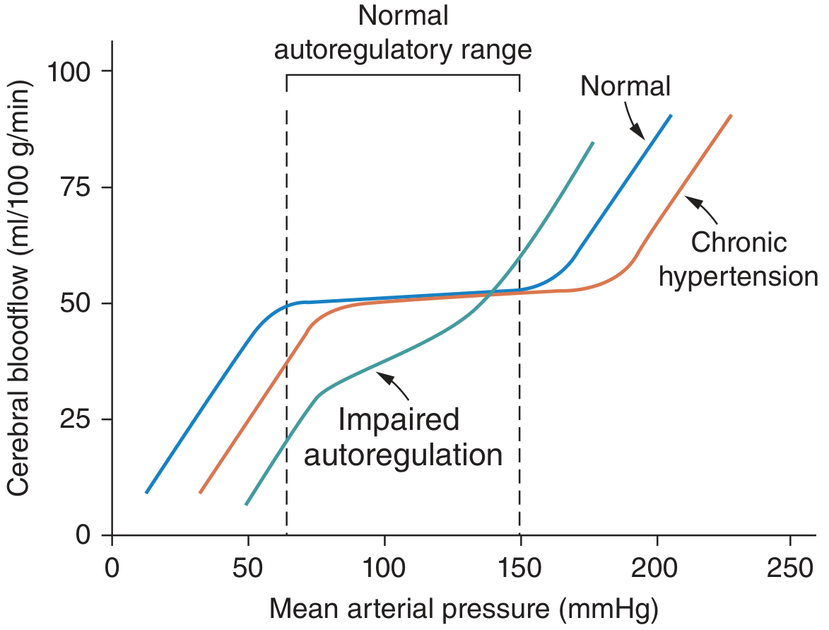
- The autoregulatory curve shifts rightward in chronic hypertension, meaning lower BPs are now required to maintain adequate perfusion - rapid BP lowering can cause ischemia
Endothelin
-
Produced by vascular endothelium and collecting tubules
-
ET-A receptors mediate vasoconstriction; ET-B receptors promote NO release and natriuresis
-
ET-A overactivation contributes to increased SVR, especially in volume-expanded and CKD-related hypertension
-
National Kidney Foundation Primer on Kidney Diseases, 8e; Fuster and Hurst's The Heart, 15th Ed.
6. Blood Flow Autoregulation in Hypertension
The body regulates tissue blood flow acutely (seconds-minutes) via:
- Myogenic response: vessels constrict when stretched by high pressure
- Metabolic response: vasodilation in response to local hypoxia/CO₂ accumulation
In hypertension, this autoregulatory range shifts to higher pressures (see figure below). This protects organs from overperfusion short-term but means BP must be lowered gradually with antihypertensive therapy to avoid ischemia.
7. Hormonal & Humoral Modulators
| Factor | Effect on BP | Mechanism |
|---|---|---|
| Aldosterone | Raises | Sodium retention via ENaC in collecting duct |
| ANP (Atrial Natriuretic Peptide) | Lowers | Increases GFR, inhibits renin, promotes natriuresis |
| Endothelin-1 | Raises (ET-A) | Vasoconstriction, sodium retention |
| Nitric Oxide | Lowers | Vasodilation, natriuresis |
| Prostaglandins (PGI₂) | Lowers | Vasodilation |
| Thromboxane A₂ | Raises | Vasoconstriction, platelet aggregation |
| Reactive Oxygen Species | Raises | Inactivate NO, promote vasoconstriction |
| NSAIDs (COX inhibition) | Raises | Block vasodilatory prostaglandins → sodium retention |
- National Kidney Foundation Primer on Kidney Diseases, 8e
8. Obesity-Hypertension Nexus
Obesity causes hypertension through multiple parallel pathways:
- Renal compression by perirenal fat → impaired pressure natriuresis
- SNS activation via leptin hypersecretion
- RAAS activation (adipose tissue expresses local renin-angiotensin components)
- Insulin resistance/hyperinsulinemia → promotes tubular sodium reabsorption and SNS activity
- Obstructive sleep apnea (common in obesity) → intermittent hypoxia → SNS surges → sustained daytime hypertension
9. Genetic Causes (Secondary/Monogenic)
Rare but well-characterized Mendelian causes illustrate the importance of sodium handling:
| Disorder | Mechanism | Key Features |
|---|---|---|
| Liddle Syndrome | Gain-of-function ENaC mutation | Low aldosterone, low K⁺, responds to amiloride |
| Gordon Syndrome | Excess NaCl reabsorption (WNK kinase mutations) | High K⁺, responds to thiazides |
| Glucocorticoid-remediable aldosteronism (FH-I) | Aldosterone under ACTH control | High aldosterone, responds to dexamethasone |
| AME (Apparent Mineralocorticoid Excess) | 11β-HSD2 deficiency; cortisol acts on MR | Low aldosterone, low K⁺ |
- Fuster and Hurst's The Heart, 15th Ed.
10. Hypertension in CKD (Vicious Cycle)
CKD creates a self-reinforcing cycle:
- Nephron loss → reduced natriuretic capacity → salt sensitivity and volume expansion
- Volume expansion → higher BP → further glomerular hypertension and injury
- Injured glomeruli → additional nephron loss → worsening hypertension
- CKD also: increases endothelin, reduces NO (via ADMA accumulation), amplifies SNS discharge via abnormal afferent renal nerve signals, and promotes oxidative stress
Summary: Integrated Pathophysiology
Genetic predisposition + environmental factors (obesity, excess salt, sedentary lifestyle)
        ↓
Impaired pressure-natriuresis (kidney set-point shifted rightward)
        ↓
Sodium and water retention → ↑ blood volume → ↑ CO
        ↓
↑ Ang II, SNS activation, endothelin
        ↓
Vasoconstriction + vascular remodeling → ↑ TPR
        ↓
Endothelial dysfunction (↓ NO, ↑ ROS) → further ↑ TPR
        ↓
SUSTAINED HYPERTENSION
        ↓
Target-organ damage: LVH, nephrosclerosis, atherosclerosis, stroke
The kidney sits at the center of long-term BP control - ultimately, sustained hypertension requires a defect in renal sodium excretion, whether primary (essential) or secondary (CKD, RAAS excess, structural renal disease).
Xanthochromic CSF seen in
Xanthochromia refers to the yellow discoloration of CSF, classically caused by breakdown of red blood cells after subarachnoid hemorrhage. Other causes include high protein, jaundice, and hypercarotenemia.
Searching sources...
Reading File
Finding Sources
Finding Sources
Found excellent sources. Let me read the detailed content from Henry's Clinical Diagnosis and Quick Compendium now.
Reading File
Reading File
I have comprehensive, authoritative content. Here is the full answer:
Xanthochromic CSF - Causes and Mechanisms
Xanthochromia refers to the pale pink, orange, or yellow discoloration of the supernatant of centrifuged CSF. It is detected by centrifuging the CSF sample and comparing the supernatant against a tube of distilled water.
Pigments Responsible and Their Colors
| Pigment | Color | Origin |
|---|---|---|
| Oxyhemoglobin | Pink to orange | Early RBC lysis (2-4 hours post-bleed) |
| Bilirubin | Yellow | Hemoglobin breakdown (12+ hours post-bleed) |
| Methemoglobin | Brown | Older, encapsulated hematomas |
| Carotenoids | Orange | Dietary hypercarotenemia |
| Melanin | Brownish | Meningeal metastatic melanoma |
| Rifampin | Red-orange | Drug contamination |
Causes of Xanthochromic CSF
1. Subarachnoid Hemorrhage (SAH) - Most Important Cause
- The classic and most clinically significant cause
- RBCs lyse in CSF after a few hours, releasing hemoglobin
- Pink/orange xanthochromia (from oxyhemoglobin): detected as early as 2-4 hours after onset; some cases take up to 12 hours
- Yellow xanthochromia (from bilirubin): develops ~12 hours after the bleed, peaks at 2-4 days (72 hours), persists for 2-4 weeks
- Xanthochromia + erythrophagocytosis + hemosiderin-laden macrophages = confirms true SAH vs traumatic tap
2. Elevated CSF Protein (>150 mg/dL)
Seen in pathological states such as:
- Froin's syndrome (complete spinal block) - CSF may also clot spontaneously
- Guillain-Barré syndrome (polyneuritis) - markedly elevated protein
- Bacterial/tuberculous meningitis
- Acoustic neuroma and other spinal cord tumors causing spinal block
3. Jaundice (Hyperbilirubinemia / Bilirachia)
- Bilirubin crosses the blood-brain barrier and enters CSF in patients with severe jaundice (serum bilirubin typically >10-15 mg/dL)
- Produces yellow discoloration without any hemorrhagic event
4. Hypercarotenemia
- Excess dietary carotene (hypervitaminosis A) or carotenoid pigments in blood enter CSF
- Produces an orange tint
5. Meningeal Melanomatosis
- Metastatic melanoma to the meninges releases melanin into CSF
- Produces a brownish discoloration
6. Drug-Related
- Rifampin therapy - produces red-orange discoloration of CSF (same pigment responsible for orange urine)
7. Artifactual / Spurious Xanthochromia
- Traumatic tap with very high RBC count (>100,000 RBCs/μL): enough plasma protein is introduced to raise CSF protein >150 mg/dL, causing xanthochromia
- Delay >1 hour before examination without refrigeration: in-vitro RBC lysis produces false-positive oxyhemoglobin
- Detergent contamination of the collection needle or tube
Timeline of Xanthochromia in SAH
Hemorrhage occurs
    ↓
2-4 hours   → Pink/orange xanthochromia (oxyhemoglobin) first detectable
    ↓
12 hours    → Yellow xanthochromia (bilirubin) begins
    ↓
24-36 hours → Peak oxyhemoglobin intensity
    ↓
2-4 days    → Peak bilirubin intensity
    ↓
4-8 days    → Oxyhemoglobin disappears
    ↓
2-4 weeks   → Bilirubin clears completely
Key Exam Points
- Xanthochromia is the gold standard for distinguishing true SAH from a traumatic tap - it is present in SAH but absent (or occurs only with massive traumatic bleeding >100,000 RBCs/μL) in a simple traumatic tap
- In a traumatic tap, fluid clears progressively from tube 1 to tube 4; in SAH, color remains uniform
- Spectrophotometry is more sensitive than visual inspection and can differentiate oxyhemoglobin from bilirubin by their different absorption peaks
- A negative CT head + positive xanthochromia = SAH until proven otherwise
Sources:
- Henry's Clinical Diagnosis and Management by Laboratory Methods - CSF Xanthochromia
- Quick Compendium of Clinical Pathology, 5th Ed. - CSF Chemistry
This is a shared conversation. Sign in to Orris to start your own chat.