
Nepphrolithiasis
Reading File
Now let me search the medical library for nephrolithiasis in parallel with a PubMed search.
Finding Sources
Searching PubMed
"nephrolithiasis"[MeSH Terms] AND "management"[MeSH Terms]
Good results. Now let me read the most comprehensive sources in parallel - Campbell-Walsh Urology and Comprehensive Clinical Nephrology, plus the Quick Compendium for a concise overview.
Reading File
Reading File
Reading File
The Brenner & Rector section turned out to be about kidney donors, not nephrolithiasis per se. Let me now search for a comprehensive nephrolithiasis section and also get the Rosen's Emergency Medicine and Campbell-Walsh sections.
Reading File
Finding Sources
Excellent. Now let me get more from Rosen's Emergency Medicine (the clinical presentation, diagnosis, and management sections) and the Robbins Pathology section.
Reading File
Reading File
I now have rich content from multiple sources. Let me read more of the Rosen's clinical features, diagnosis, and management section and the Robbins morphology/treatment section.
Reading File
Reading File
I have plenty of material now. Let me also get the morphology section from Robbins for nephrolithiasis, and fetch the stone impaction diagram.
Reading File
Loading Image
I now have all the material needed for a thorough, well-structured response. Let me compile the full answer.
Nephrolithiasis (Kidney Stones / Urolithiasis)
Epidemiology
Urolithiasis affects 5-12% of the world population over a lifetime. Males are affected about twice as often as females, with peak onset between ages 20 and 30. Up to 50% of patients experience a recurrence within 5 years. Geographic, ethnic, dietary, and genetic factors all influence incidence. - Rosen's Emergency Medicine, p. 1396; Robbins Pathologic Basis of Disease
Types of Stones and Pathogenesis
The fundamental mechanism for all stone types is supersaturation of the urine with a stone-forming substance. A low urine volume is the most universal predisposing factor.
| Stone Type | % of All Stones | Key Conditions / Promoters |
|---|---|---|
| Calcium oxalate (± calcium phosphate) | ~70% | Hypercalciuria, hyperoxaluria, hypocitraturia, hyperuricosuria |
| Struvite (magnesium ammonium phosphate) | 5-15% | UTI with urea-splitting organisms |
| Uric acid | 5-10% | Acidic urine (pH <5.5), gout, hyperuricosuria, leukemia |
| Cystine | 1-2% | Cystinuria (autosomal recessive) |
(Robbins, Table 20.12)
Calcium Stones (Most Common)
- ~5% have hypercalcemia (hyperparathyroidism, sarcoidosis, malignancy)
- ~55% have hypercalciuria without hypercalcemia: absorptive hypercalciuria, renal tubular hypercalciuria, or idiopathic
- ~20% are associated with hyperuricosuria - uric acid crystals nucleate calcium oxalate deposition in the collecting ducts
- ~5% are driven by hyperoxaluria (Crohn disease, small bowel resection, bariatric surgery, radiation enteritis, or rare primary/hereditary oxaluria)
- Hypocitraturia (citrate normally inhibits stone formation) can be idiopathic or due to metabolic acidosis/chronic diarrhea
- Robbins Pathologic Basis of Disease; Rosen's Emergency Medicine
Struvite Stones
- Formed by urea-splitting organisms: Proteus, Providencia, Klebsiella, Pseudomonas, Staphylococcus
- Produce ammonia, raising urinary pH and precipitating MgNH₄PO₄
- Often grow very large - staghorn calculi (75% of staghorn calculi are struvite) that fill much of the renal pelvis
- Common in patients with anatomic abnormalities predisposing to recurrent UTI
- Robbins; Rosen's Emergency Medicine
Uric Acid Stones
- Radiolucent (do not show on plain X-ray) - a classic distinction
- Require persistently acidic urine (pH <5.5) for precipitation
- Associated with gout (~15% of gout patients develop stones), leukemia/rapid cell turnover, uricosuric drugs
- More than 50% of uric acid stone formers have neither hyperuricemia nor hyperuricosuria - urine pH is the primary driver
- Robbins; Rosen's Emergency Medicine
Cystine Stones
- Autosomal recessive defect in renal (and intestinal) reabsorption of dibasic amino acids: Cystine, Ornithine, Lysine, Arginine (mnemonic: COLA)
- Cystine is the least soluble; hexagonal crystals on urine microscopy
- Quick Compendium of Clinical Pathology, 5th Ed.
Urine Crystal Morphology
| Crystal | Shape |
|---|---|
| Calcium oxalate | "Envelopes" |
| Uric acid | Pleomorphic - diamond, square, rod; polarize in multiple colors |
| Struvite | "Coffin lids" (form in alkaline pH) |
| Cystine | Hexagonal |
| Ammonium biurate | "Thorn apples" |
(Quick Compendium of Clinical Pathology, 5th Ed.)
Sites of Impaction
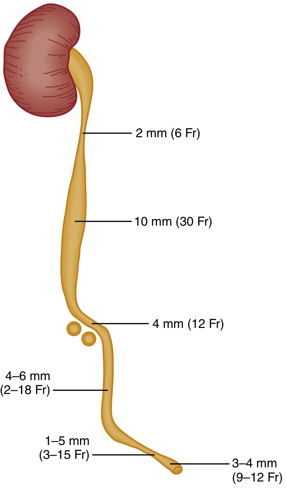
Stones originate in the kidney and migrate. There are five classic sites of impaction along the ureter:
- Calyx / renal pelvis
- Ureteropelvic junction (UPJ) - abrupt narrowing from ~1 cm pelvis to 2-3 mm ureter
- Pelvic brim - where ureter crosses the iliac vessels
- Ureterovesical junction (UVJ) - the most constricted point; most common site of impaction
- Vesical orifice
At diagnosis, up to 75% of stones are in the distal third of the ureter.
Clinical Features
- Renal colic: abrupt onset, crescendo of extreme flank pain radiating laterally around the abdomen and into the groin/labia/testicle
- Hematuria (microscopic or gross) - present in most cases but not universal
- Nausea and vomiting - common
- Pain is typically colicky, not positional (unlike peritonitis)
- Urinary urgency and dysuria with distal ureteral stones
- Fever/chills if there is concomitant infection - this is a urologic emergency
- Rosen's Emergency Medicine
Complications
- Obstruction - causes rapid redistribution of renal blood flow and decreased GFR. Complete obstruction for 1-2 weeks can cause irreversible renal damage
- Infection - obstructed + infected stone = urosepsis risk. Pyelonephritis, perinephric abscess, gram-negative sepsis
- Partial obstruction still risks permanent damage
- Rosen's Emergency Medicine
Diagnosis
Urinalysis
- Hematuria (micro or macro)
- Crystals (see crystal morphology table above)
- Pyuria + bacteriuria suggest concurrent infection
Imaging
Non-contrast CT (NCCT) of abdomen/pelvis is the gold standard outside pregnancy:
- Sensitivity and specificity >90%; detects stones as small as 1 mm
- Identifies hydronephrosis, hydroureter, ureteral edema, and alternative diagnoses (AAA, abscess, malignancy)
- Radiolucent uric acid stones are visible on CT (unlike plain film)
- Low-dose protocols are appropriate for BMI <30 kg/m²
- Patients with known stone history and classic renal colic without fever, infection, solitary kidney, or concern for alternative diagnosis may not need CT
Ultrasound:
- First-line in pregnancy and pediatrics
- Sensitivity ~54%, specificity ~91% for stones; up to 100% sensitive for ureteral obstruction
- Detects hydronephrosis reliably
- Less accurate for stone size and location
Plain X-ray (KUB):
- Detects radiopaque stones (calcium-containing)
- Misses uric acid, cystine, and many small stones
Rosen's Emergency Medicine
Management
Acute Pain Control
- NSAIDs are first-line (ketorolac 30 mg IV or diclofenac 75 mg IM)
- Reduce ureteral spasm and renal capsular pressure by lowering GFR in the obstructed kidney
- Use with caution in renal insufficiency or peptic ulcer disease
- IV opioids (fentanyl 1-2 mcg/kg) are also effective; combination with NSAIDs may reduce ED length of stay
- Antiemetics (ondansetron 4 mg IV) + IV fluids for vomiting
- High-volume IV hydration has NOT been shown to improve stone passage
Medical Expulsive Therapy (MET)
- Alpha-blockers (tamsulosin) facilitate passage of distal ureteral stones by relaxing ureteral smooth muscle
Spontaneous Passage Rates (by stone size)
| Size | Spontaneous Passage Rate |
|---|---|
| <5 mm | ~90% within 4 weeks |
| 5-8 mm | ~15% |
| >8 mm | ~5% (usually requires intervention) |
Rosen's Emergency Medicine
Urologic Emergency Indications
Immediate intervention (ureteral stent or percutaneous nephrostomy) is required for:
- Obstructing stone + infection/sepsis
- Bilateral obstructing stones
- Obstruction of a solitary or transplanted kidney
- Significant renal impairment
Surgical Options
- Extracorporeal shock wave lithotripsy (ESWL)
- Ureteroscopy with laser lithotripsy
- Percutaneous nephrolithotomy (PCNL) - for large/staghorn calculi
- Surgery can generally be outpatient if oral intake is tolerated and pain is controlled
Indications for Admission
- Inability to tolerate oral intake / uncontrolled pain
- High-grade obstruction, large stone unlikely to pass
- Concomitant infection
- Solitary/transplanted kidney obstruction
- Significant renal impairment
Prevention (Recurrence Reduction)
| Stone Type | Prevention Strategy |
|---|---|
| All types | Increase fluid intake (urine output >2 L/day) |
| Calcium oxalate | Thiazide diuretics (reduce hypercalciuria); potassium citrate (raises pH, increases citrate); low-oxalate diet; dietary calcium is NOT restricted (reduces enteric oxalate absorption) |
| Uric acid | Allopurinol; urinary alkalinization (potassium citrate) to pH 6-6.5 |
| Struvite | Eradicate underlying infection; complete stone removal |
| Cystine | High fluid intake; urinary alkalinization; D-penicillamine or tiopronin |
Special Considerations: Pregnancy
- Incidence is similar to non-pregnant women (1 in 188 to 1 in 4600)
- Calcium phosphate stones are most common in pregnancy (vs. calcium oxalate outside pregnancy); this is because pregnancy causes physiologic hypercalciuria balanced by increased citrate and nephrocalcin
- ~64-84% of stones pass spontaneously in pregnancy
- Imaging: ultrasound is first-line; MR urography or limited IVU if needed; CT is reserved for cases where benefits outweigh radiation risk
- Risks: increased rates of preterm labor, pyelonephritis, hypertensive disorders
- Comprehensive Clinical Nephrology, 7th Ed.
Sources: Robbins & Cotran Pathologic Basis of Disease | Rosen's Emergency Medicine | Quick Compendium of Clinical Pathology, 5th Ed. | Comprehensive Clinical Nephrology, 7th Ed.
This is a shared conversation. Sign in to Orris to start your own chat.