
In biochemistry Explain the about Collagen and elastin
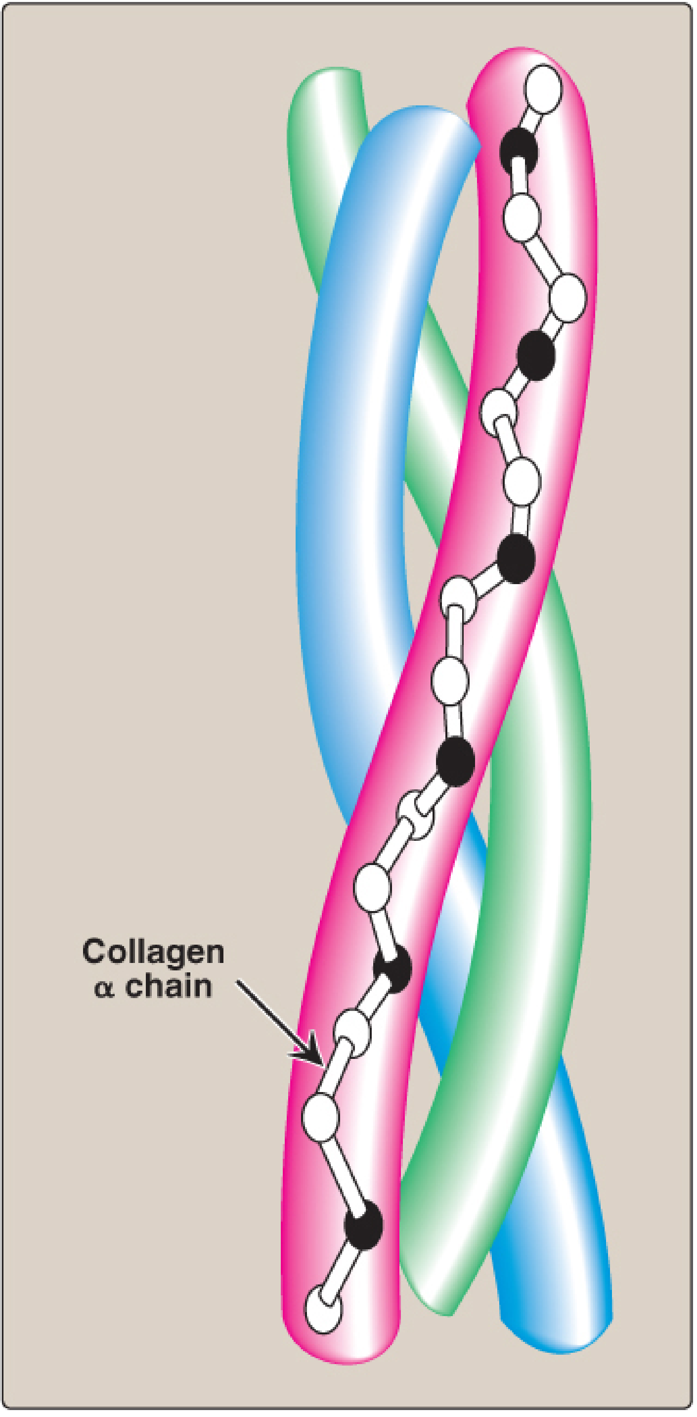
collagen structure triple helix biochemistry diagram
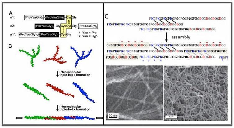
Educational diagram illustrating the self-assembly of collagen mimetic peptides (CMPs). Panel A shows the primary structure of cross-linked CMPs (alpha-1 and alpha-2 chains) stabilized by disulfide bonds between Cys residues. Panel B depicts a two-stage assembly process: intramolecular triple-helix formation of individual colored strands followed by intermolecular association into a super triple helix. Panel C focuses on the charge-pair directed self-assembly of the KOD peptide. The schematic details the alignment of three peptide strands containing PKG (positive), POG (neutral), and DOG (negative) repeating units. An assembly arrow leads to a stabilized structure where directional, interchain Lys-Asp salt bridges (marked by slanting bars) facilitate nucleation. Accompanying Transmission Electron Microscopy (TEM) images confirm these hierarchical structures: the left micrograph (50 nm scale) shows individual filamentous KOD fibrils, while the right micrograph (1 μm scale) displays a dense, interconnected KOD hydrogel network. This demonstrates the transition from molecular sequence to supramolecular biomaterials suitable for hemostatic or tissue engineering applications.
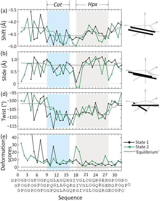
This biophysical data visualization presents the structural parameters of matrix metalloproteinase 1 (MMP1)-bound collagen triple helices. The figure contains four line graphs (a, b, d, e) aligned by amino acid sequence and step number, comparing State 1 (black), State 2 (green), and an 'Equilibrium' state (dotted line). Graph (a) measures 'Shift' in Angstroms (Å), (b) measures 'Slide' in Å, (d) measures 'Twist' in degrees (°), and (e) illustrates 'Deformation scores'. Two critical protein interaction zones are highlighted: the N-terminal catalytic (Cat) domain (shaded light cyan) and the C-terminal hemopexin (Hpx) domain (shaded grey). Schematic diagrams to the right of each graph illustrate the geometric meaning of each translational and rotational parameter. The data demonstrates how MMP1 binding induces conformational changes in the collagen triple helix, specifically showing localized deformations and deviations from equilibrium at the Cat and Hpx contact sites. These parameters are essential for understanding the mechanical and biochemical mechanisms of collagen degradation by matrix metalloproteinases in human connective tissue remodeling.
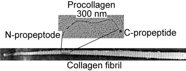
This diagnostic image consists of two electron microscopy (EM) micrographs displaying the ultrastructure of human collagen at different stages of assembly. The top micrograph shows a single procollagen molecule, approximately 300 nm in length. It features a tripartite morphology with a central, wavy triple-helical domain flanked by globular extensions identified as the N-propeptide and C-propeptide. The bottom micrograph illustrates a larger, self-assembled collagen fibril. This mature structure exhibits characteristic periodic striations, appearing as alternating dark and light bands along its longitudinal axis. This visual comparison demonstrates the structural transition from a precursor procollagen molecule to a densely packed, organized collagen fibril following the enzymatic cleavage of the terminal propeptides. The image serves as an educational tool for molecular biology and histology, illustrating the fundamental protein components of connective tissues such as skin, bone, and tendon.
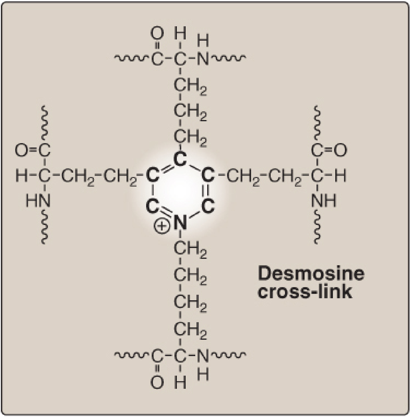
elastin fiber structure cross-linking desmosine
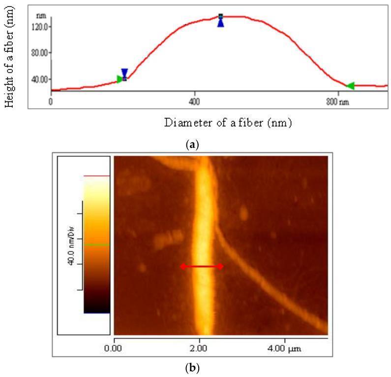
This composite educational graphic details the Atomic Force Microscopy (AFM) analysis of human elastin fiber morphology. Panel (b) presents a 2D topographic AFM image (5 x 5 μm scale) showing a prominent, vertically oriented elastin fiber against a darker background. The fiber appears bright yellow-white, indicating higher elevation on the 40.0 nm/Div color scale. A red horizontal line indicates the cross-sectional measurement path. Panel (a) shows the corresponding quantitative profile plot derived from that measurement. The y-axis represents the height of the fiber in nanometers (reaching a peak of approximately 120 nm), while the x-axis measures the diameter (spanning roughly 800 nm). Blue markers (triangles) define the vertical height measurement, and green markers denote the horizontal diameter width. This content illustrates the self-assembly of elastin from nanofibrils into larger fibers, providing key metrics for connective tissue research and biomedical engineering.
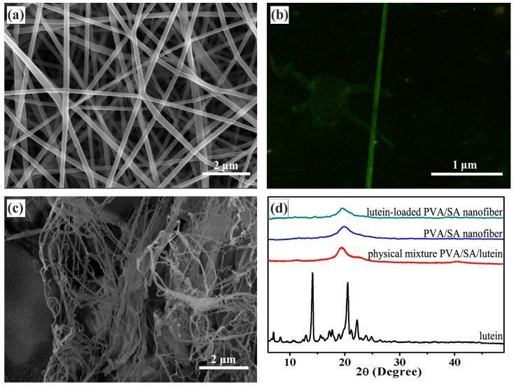
Educational panel illustrating the characterization of a lutein-loaded polyvinyl alcohol/sodium alginate (PVA/SA) drug delivery system. (a) Scanning electron microscopy (SEM) of un-crosslinked nanofibers shows a uniform, smooth, bead-free web-like structure with fiber diameters between 240-340 nm. (b) Fluorescence microscopy demonstrates the uniform distribution of lutein along the axis of a single nanofiber. (c) SEM image following 1-hour of cross-linking shows morphological changes where fibers appear collapsed, swollen, and adhered together, forming an aggregated mass. (d) X-ray diffraction (XRD) patterns compare crystalline free lutein, which exhibits sharp peaks at 2̘ = 14.06° and 20.54°, against the amorphous patterns of PVA/SA nanofibers and the lutein-loaded composite. The absence of crystalline peaks in the loaded nanofibers suggests that lutein is dispersed in an amorphous or molecular state within the polymer matrix, which is favorable for solubility and bioavailability. This content demonstrates materials science techniques used to develop sustained-release ocular or nutritional therapeutic delivery systems.
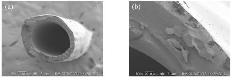
This figure presents Scanning Electron Microscopy (SEM) images detailing the morphology of a C-HF (cation-exchange hollow fiber) membrane, a key component used in medical and industrial separation processes like dialysis. Image (a) is a low-magnification (50x) cross-sectional view showing the global hollow fiber structure. It depicts a cylindrical tube with a central lumen, essential for facilitating fluid flow in diffusion dialysis or electrodialysis systems. The scale bar indicates a 200 µm dimension, illustrating the outer diameter and significant wall thickness of the fiber. Image (b) provides a high-magnification (500x) close-up of the cross-section with a 20.0 µm scale bar. This view reveals a non-porous, relatively dense matrix with irregular granular textures and surface irregularities. These features are indicative of the membrane's composite material properties resulting from the dehydration, coagulation, and chemical cross-linking processes (using glutaraldehyde) intended to optimize water content, mechanical strength, and ionic selectivity for selective ion transport.
Collagen and Elastin - Biochemistry
PART 1 - COLLAGEN
Overview
Triple-Stranded Helix Structure
A. Types of Collagen
| Group | Types | Structure | Location | Function |
|---|---|---|---|---|
| Fibril-forming | I, II, III | Rope-like fibrils with 67-nm banding | Skin, tendon, bone (I); Cartilage (II); Blood vessels, gut (III) | Tensile strength |
| Network-forming | IV, VIII | 3D mesh/sheet | Basement membranes | Structural support, filtration |
| Fibril-associated (FACIT) | IX, XII | Bind fibril surfaces | Links fibrils to ECM components | ECM organization |
- Type I is the most abundant: found in tendon, bone, skin, dentin; extremely high tensile strength
- Type II: restricted to cartilaginous structures
- Type III: found in distensible tissues such as blood vessels (absent in vascular EDS)
- Type IV: forms flexible meshwork in basement membranes (affected in Alport syndrome)
B. Amino Acid Composition and Primary Structure
- Glycine (Gly) appears at every third position - it is the smallest amino acid and fits into the core of the triple helix. Any substitution of glycine disrupts the helix (as in osteogenesis imperfecta).
- X position is frequently Proline - its ring structure causes kinks in the chain, facilitating helix formation. It cannot form an alpha helix.
- Y position is frequently Hydroxyproline or Hydroxylysine - these are formed by post-translational hydroxylation (requiring Vitamin C).
C. Biosynthesis of Collagen (7 Steps)
| Step | Location | Event | Key Enzyme/Factor |
|---|---|---|---|
| 1. Synthesis | Ribosomes (RER) | Alpha chains synthesized as preprocollagen (with signal peptide) | - |
| 2. Hydroxylation | RER lumen | Proline → Hydroxyproline; Lysine → Hydroxylysine | Prolyl hydroxylase, Lysyl hydroxylase (require Vitamin C and Fe²+) |
| 3. Glycosylation | RER lumen | Hydroxylysine residues are glycosylated | Glycosyltransferases |
| 4. Triple helix assembly | RER/Golgi | Three pro-alpha chains associate via C-terminal disulfide bonds → form procollagen triple helix (C-terminus to N-terminus) | Disulfide bond formation |
| 5. Secretion + Cleavage | Extracellular | N- and C-terminal propeptides cleaved by procollagen peptidases → tropocollagen | N- and C-procollagen peptidases (ADAMTS2) |
| 6. Fibril formation | Extracellular | Tropocollagen molecules self-assemble in a staggered parallel array (~3/4 overlap) → collagen fibrils with characteristic banding | Spontaneous |
| 7. Cross-link formation | Extracellular | Lysine/hydroxylysine oxidized to aldehydes; condense to form covalent cross-links → mature collagen fibers | Lysyl oxidase (copper-dependent) |
Vitamin C (ascorbate) is essential as a cofactor for prolyl and lysyl hydroxylases. Deficiency causes scurvy - characterized by weak collagen, poor wound healing, perifollicular hemorrhages, and bleeding gums.
D. Degradation
E. Collagenopathies (Diseases of Defective Collagen)
| Disease | Collagen/Gene Affected | Mechanism | Key Features |
|---|---|---|---|
| Osteogenesis Imperfecta | COL1A1/COL1A2 (Type I) | Gly substitution in triple helix | Brittle bones, blue sclerae, hearing loss |
| Ehlers-Danlos Syndrome (vascular type) | COL3A1 (Type III) | Abnormal type III collagen | Spontaneous arterial/bowel rupture |
| Ehlers-Danlos Syndrome (classical) | COL5A1, COL5A2 | Defective fibril packing | Hyperextensible skin and joints |
| Ehlers-Danlos Syndrome (kyphoscoliotic) | Lysyl hydroxylase deficiency | Deficient hydroxylysine → poor cross-linking | Progressive scoliosis |
| Dermatosparaxis EDS | ADAMTS2 (procollagen N-proteinase) | Abnormal propeptide cleavage | Fragile, sagging skin |
| Alport Syndrome | COL4A3/4/5 (Type IV) | Abnormal basement membrane | Hereditary nephritis, hearing/eye defects |
| Scurvy | Vitamin C deficiency | Prolyl/lysyl hydroxylase failure | Weak collagen, hemorrhage, poor wound healing |
| Menkes Disease | ATP7A (copper ATPase) | Copper deficiency → lysyl oxidase failure | Defective cross-linking, neurological damage |
PART 2 - ELASTIN
Overview
- Lungs (stretch with each breath)
- Walls of large arteries (deform with cardiac pulsation)
- Elastic ligaments (ligamentum flavum, ligamentum nuchae)
- Skin
A. Structure of Elastin
- Rich in small, nonpolar amino acids: glycine, alanine, valine
- Rich in proline and lysine
- Low in hydroxyproline and hydroxylysine (unlike collagen)
- Has two types of alternating domains (~16 regions each):
- Hydrophilic cross-linking domains: rich in lysine and alanine
- Hydrophobic domains: rich in Val-Pro-Gly repeats (VPGVG or VGGVG) - responsible for elastic recoil
B. Cross-Linking: Desmosine
- Tropoelastin is secreted into the ECM
- It interacts with fibrillin-1 and fibrillin-2 microfibrils (scaffold)
- Lysyl oxidase (copper-dependent) oxidatively deaminates lysyl residues → allysine aldehydes
- Three allysine + one unmodified lysine condense to form a desmosine (pyridinium ring) cross-link
- This creates an extensively interconnected, rubbery 3D network
C. Elastic Behavior
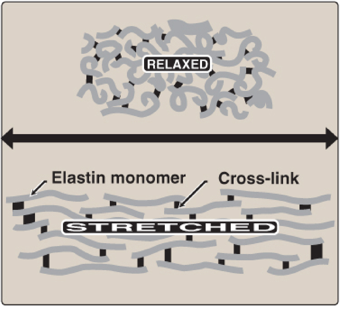
- In the relaxed state: polypeptide chains are randomly coiled and disordered (high entropy)
- Under stretch: chains extend and become ordered (lower entropy)
- On release: entropy-driven recoil restores the random coil state
- The desmosine cross-links prevent the network from falling apart
D. Degradation of Elastin: Alpha-1-Antitrypsin
| Condition | Mechanism | Consequence |
|---|---|---|
| A1AT deficiency | Inherited (PIZZ genotype); mutant protein misfolds in hepatocytes | Reduced elastase inhibition in lungs → excessive elastin degradation → emphysema |
| Cigarette smoking | Oxidizes Met358 of A1AT → inactivates it | Loss of elastase inhibition → lung elastin destruction → emphysema |
This is why smokers with A1AT deficiency have an extremely high risk of early-onset emphysema.
Comparison: Collagen vs. Elastin
| Feature | Collagen | Elastin |
|---|---|---|
| Primary function | Tensile strength | Elastic recoil |
| Structure | Triple helix (Gly-X-Y repeats) | Amorphous, random coil network |
| Precursor | Preprocollagen → Procollagen → Tropocollagen | Tropoelastin |
| Key amino acids | Gly, Pro, Hydroxyproline, Hydroxylysine | Gly, Val, Ala, Pro, Lys (low hydroxyproline) |
| Cross-linking enzyme | Lysyl oxidase (Cu²+) | Lysyl oxidase (Cu²+) |
| Cross-link type | Lysine-derived aldehyde cross-links | Desmosine (unique to elastin) |
| Vitamin C role | Required for hydroxylation | Not required |
| Scaffold protein | - | Fibrillin-1 and -2 |
| Degraded by | Matrix metalloproteinases (collagenases) | Elastase (inhibited by A1AT) |
| Key disease | Scurvy, OI, EDS, Alport syndrome | Emphysema (A1AT deficiency), Marfan syndrome |
| Location | Tendons, bone, skin, basement membranes | Lungs, large arteries, ligaments |
Key Clinical Connections Summary
- Scurvy: Vitamin C deficiency → impaired prolyl/lysyl hydroxylation → unstable collagen
- Osteogenesis Imperfecta: Gly substitution in type I collagen → brittle bones, blue sclerae
- Ehlers-Danlos Syndrome: Various collagen gene/enzyme defects → hypermobility, fragile skin
- Marfan Syndrome: Fibrillin-1 mutation → abnormal elastin scaffold → aortic aneurysm, lens dislocation, tall stature
- Emphysema: A1AT deficiency or smoking → unchecked elastase → alveolar wall destruction
- Menkes Disease: Copper deficiency (ATP7A mutation) → lysyl oxidase failure → defective cross-linking of both collagen and elastin