
Schizophrenia etiology & pathogenesis
"schizophrenia"[MeSH Terms] AND "etiology"[MeSH Terms]
"schizophrenia"[MeSH Terms] AND "pathogenesis"
schizophrenia dopamine hypothesis pathogenesis brain diagram
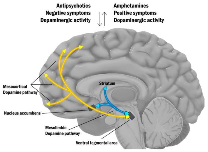
An anatomical diagram of the human brain in a midsagittal view illustrating the dopamine hypothesis of schizophrenia. The illustration highlights two primary dopaminergic pathways originating from the Ventral Tegmental Area (VTA) in the midbrain: the Mesocortical Dopamine Pathway, represented by yellow arrows projecting to the prefrontal cortex, and the Mesolimbic Dopamine Pathway, indicated by yellow arrows projecting to the Nucleus Accumbens. Additionally, blue arrows indicate projections toward the Striatum. At the top, a conceptual comparison lists clinical correlations: 'Antipsychotics' are associated with reduced 'Dopaminergic activity' and 'Negative symptoms,' while 'Amphetamines' are linked to increased 'Dopaminergic activity' and 'Positive symptoms.' Vertical arrows between these lists indicate an inverse relationship between the effects of these substances. This diagram serves as a neurobiological model for medical students and clinicians to visualize the pathophysiology of schizophrenia and the mechanism of action for antipsychotic medications and psychostimulants.
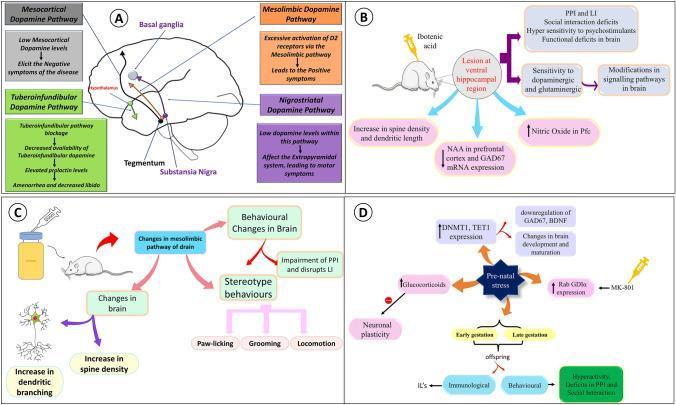
Educational medical infographic illustrating the dopamine hypothesis and neurodevelopmental models of schizophrenia. Panel A (Anatomical Diagram) maps the four major dopaminergic pathways: Mesocortical (linked to negative symptoms), Mesolimbic (excessive D2 activation linked to positive symptoms), Nigrostriatal (motor symptoms via the extrapyramidal system), and Tuberoinfundibular (prolactin dysregulation). Panel B (Flowchart) details the Neonatal Ventral Hippocampal Lesion (NVHL) rodent model using ibotenic acid, showing neurochemical outcomes like decreased NAA and GAD67, and behavioral deficits such as impaired prepulse inhibition (PPI). Panel C (Pathophysiology Diagram) correlates mesolimbic alterations with stereotyped behaviors including paw-licking and grooming. Panel D (Concept Map) illustrates the 'Prenatal Stress' model, highlighting epigenetic changes (increased DNMT1, TET1), downregulation of BDNF, and glucocorticoid-mediated inhibition of neuronal plasticity. The visual utilizes a mix of anatomical mapping and logical flowcharts to explain how neurotransmitter dysregulation, structural brain changes, and prenatal environmental stressors contribute to the complex clinical manifestations of schizophrenia.
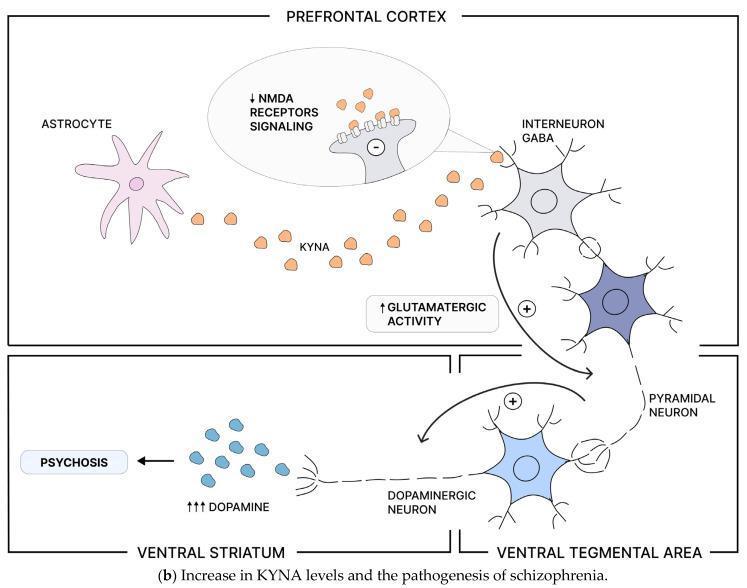
This pathophysiology diagram illustrates the neurobiological mechanism linking increased kynurenic acid (KYNA) levels to the pathogenesis of schizophrenia and psychosis across specific brain regions. In the Prefrontal Cortex (PFC), an astrocyte is shown producing KYNA, which acts as an antagonist at N-methyl-D-aspartate (NMDA) receptors. This interaction results in decreased NMDA receptor signaling on GABAergic interneurons. The subsequent reduction in inhibitory GABAergic output leads to disinhibition and increased glutamatergic activity of cortical pyramidal neurons (indicated by a '+' sign). This pyramidal neuron projects to the Ventral Tegmental Area (VTA), where it provides excitatory stimulation to dopaminergic neurons. This activation of the mesolimbic pathway causes excessive dopamine release in the Ventral Striatum (indicated by '↑↑↑ DOPAMINE'). The final outcome of this increased dopaminergic signaling is labeled as 'PSYCHOSIS'. The diagram highlights the complex interaction between the kynurenine pathway, glutamatergic signaling, and dopaminergic neurotransmission in psychiatric disorders.
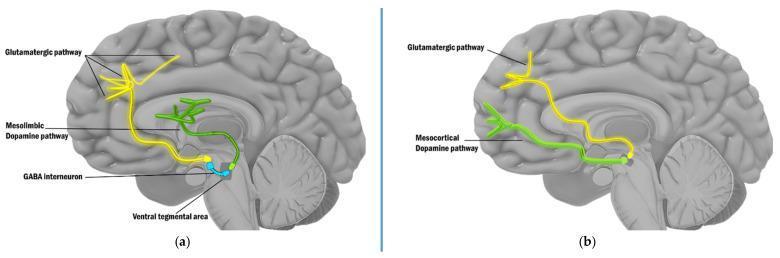
This pathophysiology diagram illustrates the cortical regulation of dopaminergic pathways in the human brain, focusing on the neurobiology of schizophrenia. The image is divided into two panels, (a) and (b), depicting a sagittal view of the brain with color-coded neural circuits. In panel (a), the diagram shows the regulation of the mesolimbic dopamine pathway (green) by a descending glutamatergic pathway (yellow) originating from the prefrontal cortex. A blue GABA interneuron in the ventral tegmental area (VTA) serves as an inhibitory bridge between the glutamate signal and the mesolimbic dopamine neurons; disruption of this 'brake' is linked to psychotic symptoms. In panel (b), the diagram shows the direct excitatory influence of the descending glutamatergic pathway (yellow) on the mesocortical dopamine pathway (green), which projects back to the cortex. This panel illustrates that dysfunction in this direct stimulatory circuit is associated with cognitive deficits. Key anatomical landmarks include the prefrontal cortex, the limbic system, and the midbrain tegmentum. The visual provides a comparison of indirect inhibition versus direct excitation in dopamine modulation.
Schizophrenia: Etiology & Pathogenesis
Overview
I. ETIOLOGY
1. Genetic Factors
| Relationship to proband | Risk of schizophrenia |
|---|---|
| General population | ~1% |
| First-degree relatives | ~6.6-11% |
| Both parents affected | ~40-46% |
| Dizygotic (fraternal) twin | ~10-11% |
| Monozygotic (identical) twin | ~50-68% |
- The concordance rate of ~50-68% in monozygotic twins confirms that genetics alone is insufficient - environmental factors are also required.
- A standard Mendelian (single-gene) inheritance pattern has not been identified. Large-scale genome-wide association studies (GWAS) have identified hundreds of small-effect risk loci plus rare, high-impact copy number variants (CNVs).
- Key implicated genes include: neuregulin (NRG1), dysbindin (DTNBP1), COMT (catechol-O-methyltransferase), DISC1 (Disrupted in Schizophrenia 1), proline dehydrogenase, and rare alleles in NRXN1, SHANK3, CNTNAP2, PRODH - genes also overrepresented in autism spectrum disorder.
- Pathways implicated by GWAS include immunity, inflammation, and cell signaling. There is also recent evidence that brain gene expression in schizophrenia resembles accelerated aging, implicating parallel mechanisms for cognitive deterioration.
- The cumulative use of polygenic risk scores (PRS) is now entering clinical exploration for diagnosis and prognosis.
2. Neurodevelopmental Hypothesis
- Absence of gliosis on neuropathology (gliosis would indicate a postnatal destructive process; its absence points to a prenatal developmental lesion)
- Aberrant distribution of interstitial neurons in frontal white matter, suggesting disrupted neuronal migration from embryologic subplate
- Decreased dendritic spine density in frontal and temporal cortex
- Fewer GABAergic inhibitory interneurons (chandelier cells) in the prefrontal cortex; reduced neurons in layers I and II of the anterior cingulate
- Children who later develop schizophrenia show delays in motor milestones, speech, social withdrawal, and lower scholastic achievement - subtle signs preceding psychosis by decades
3. Obstetric and Perinatal Risk Factors
- Obstetric complications (fetal hypoxia, intrauterine growth restriction, poor neonatal condition) are modestly but consistently associated with later schizophrenia.
- Fetal hypoxia is associated with increased structural brain abnormalities (reduced gray matter, enlarged ventricles) in patients with psychotic disorders and their nonaffected siblings - demonstrating gene-environment interaction.
- Winter/spring birth in northern latitudes confers elevated risk, with speculation that maternal influenza infection during mid-pregnancy may cause viral or immune-mediated fetal brain injury.
- A gene-environment correlation may also operate: parents with psychotic disorders have increased risk of social adversity that predisposes to obstetric complications.
4. Environmental Risk Factors
- Over 20 studies document a consistent ~2-fold increase in schizophrenia risk with urban vs. rural residence, with a dose-response gradient.
- Urban exposure prior to illness onset appears more pathogenic than exposure at the time of presentation.
- Advanced paternal age
- Cannabis use (particularly high-potency THC products)
- Social adversity, migration, and minority group status (thought to act via chronic social defeat and sensitization of dopaminergic pathways)
- Childhood trauma and maltreatment
II. PATHOGENESIS
1. Dopamine Hypothesis
- All clinically effective antipsychotics have high affinity for D2 receptors; clinical potency directly parallels D2 receptor affinity
- Psychostimulants (amphetamine, cocaine) that increase extracellular dopamine can induce or worsen psychotic symptoms
- PET neuroimaging demonstrates increased striatal dopamine synthesis and release in schizophrenic patients
| Brain Region | Dopamine Status | Clinical Correlate |
|---|---|---|
| Striatum / Mesolimbic pathway | Hyperfunction (excess D2 stimulation) | Positive symptoms (hallucinations, delusions) |
| Prefrontal Cortex / Mesocortical pathway | Hypofunction | Negative symptoms and cognitive deficits |
2. Glutamate / NMDA Receptor Hypofunction Hypothesis
- Phencyclidine (PCP) and ketamine (NMDA receptor antagonists) produce a syndrome in healthy subjects that mimics the full spectrum of schizophrenia - both positive and negative symptoms, plus cognitive deficits - unlike dopaminergic stimulants which produce only positive symptoms.
- NMDA receptors are found especially on GABAergic interneurons in the prefrontal cortex. NMDA hypofunction on these inhibitory cells leads to:
- Disinhibition of pyramidal neurons → increased cortical glutamate release → downstream dopamine overactivation in subcortical regions
- This explains the bridge between cortical glutamate hypofunction and subcortical dopamine hyperfunction
- Astrocytes produce KYNA, which antagonizes NMDA receptors on GABAergic interneurons
- Elevated KYNA in schizophrenia → reduced GABA interneuron activity → disinhibition of pyramidal glutamate neurons → VTA dopamine overactivation → psychosis
3. Serotonin Hypothesis
- Serotonin (5-HT) modulates dopaminergic activity, particularly via 5-HT2A receptors on prefrontal pyramidal neurons.
- Atypical ("second-generation") antipsychotics (e.g., clozapine, olanzapine, risperidone) act by combining D2 antagonism with 5-HT2A receptor antagonism. 5-HT2A blockade in the prefrontal cortex increases mesocortical DA release, which may explain their superior efficacy for negative and cognitive symptoms vs. first-generation agents.
- The development of newer agents targets D3, 5-HT1A, mGlu2/3, and muscarinic (M1, M4) receptors to further address cognitive and negative symptom domains.
4. GABA Hypothesis
- Postmortem studies consistently show a reduction in GABAergic interneurons (especially parvalbumin-positive chandelier and basket cells) in the prefrontal cortex and hippocampus.
- These inhibitory interneurons normally provide synchronized, rhythmic inhibition to pyramidal neurons. Their loss impairs gamma oscillations - neural synchrony patterns essential for working memory and cognitive function.
- This explains the characteristic cognitive impairment in schizophrenia independent of positive symptom severity.
5. Structural Brain Abnormalities
- Enlarged lateral and third ventricles (CT/MRI) - a robust, replicated finding
- Reduction in gray matter volume, especially in temporal lobes (including hippocampus), prefrontal cortex, and anterior cingulate cortex
- Thinning of prefrontal cortical laminae II and III with smaller, more densely packed pyramidal cells
- Reduced dendritic spine density in frontal and temporal cortex
- No gliosis (pointing to a prenatal, not acquired, insult)
- EEG abnormalities in ~1/3 of patients; more frequent in those with positive family history and enlarged ventricles
6. Integrated Neurodevelopmental Model
- Genetic susceptibility genes (NRG1, DISC1, COMT, etc.) establish a biological vulnerability
- Early environmental insults (obstetric hypoxia, prenatal infection, stress) interact with this vulnerability during fetal brain development
- Disrupted neuronal migration and synaptic connectivity produce subtle abnormalities in hippocampus and prefrontal cortex
- These changes are clinically silent through childhood, but during adolescence, synaptic pruning (a normal developmental process - excessive in schizophrenia) unmasks the deficit
- Stress-related activation of the HPA axis and sensitization of dopaminergic systems precipitates the first psychotic break
Summary Table: Etiology & Pathogenesis
| Domain | Key Facts |
|---|---|
| Heritability | ~80%; MZ twin concordance 50-68% |
| Genetics | Polygenic; NRG1, DISC1, COMT, CNVs at 22q11, 15q13, 1q21 |
| Neurotransmitters | Dopamine: subcortical hyperfunction; cortical hypofunction. Glutamate: NMDA hypofunction. GABA: interneuron deficit. Serotonin: 5-HT2A dysregulation |
| Structural changes | Enlarged ventricles, reduced gray matter, dendritic spine loss, no gliosis |
| Neurodevelopment | Prenatal insult → aberrant neuronal migration → PFC/hippocampal connectivity deficits |
| Environmental risks | Urban birth, winter birth, obstetric complications, cannabis, migration, social adversity |
| Timing | Symptoms emerge adolescence/early adulthood during synaptic pruning |
- Adams and Victor's Principles of Neurology, 12th Ed. - Theories of Causation and Mechanism
- Harrison's Principles of Internal Medicine 22E - Epidemiology and Pathophysiology of Schizophrenia
- Goodman & Gilman's Pharmacological Basis of Therapeutics - Dopamine receptor antagonists and schizophrenia
- Kaplan & Sadock's Comprehensive Textbook of Psychiatry - Risk factors and neurodevelopmental model