Mycotoc aneurysm imaging

Reading File
Finding Sources
Searching PubMed

mycotic aneurysm imaging diagnosis

Finding Sources
Finding Sources
Reading File
Reading File
Reading File
Reading File
Loading Image
Loading Image
Finding Sources
Loading Image
Reading File
Finding Sources
Finding Sources
Finding Sources
Finding Sources
Reading File
Searching the Web

mycotic aneurysm imaging CT MRI DSA features radiology 2024 2025

Now I have comprehensive data. Here is the full synthesis:

Mycotic Aneurysm - Imaging

Background

A mycotic aneurysm (MA) is an infected aneurysm caused by bacterial or fungal invasion of an arterial wall. William Osler coined the term (misnomer - these are not fungal by nature). They arise most commonly as a complication of infective endocarditis (IE), affecting 1-5% of IE patients with emboli, and are also seen in IV drug users and immunocompromised hosts. Streptococcus viridans is the most frequent pathogen; Staphylococcus spp. are increasing. Fungal MAs (Aspergillus, Candida) also occur. The most common locations are intracranial (66%) - particularly MCA and branches - followed by visceral and peripheral arteries.

Imaging Modalities

1. CT Angiography (CTA) - First-Line for Most Locations

CT angiography is the initial imaging modality of choice for most mycotic aneurysms, especially aortic and peripheral. It provides excellent spatial resolution and demonstrates both the aneurysm anatomy and surrounding soft tissue changes.
CT findings highly suggestive of mycotic etiology:
  • Saccular and lobulated vessel contours - eccentric or multilobulated shape, rather than smooth fusiform
  • Perianeurysmal soft tissue inflammation - manifested as perivascular contrast enhancement / inflammatory mass
  • Intramural or perivascular air - pathognomonic when present
  • Perianeurysmal fluid collection
  • Eccentric, rapidly enlarging aneurysm in the context of fever/infection
These features are well illustrated in AJR studies on mycotic aneurysm CT/MRI, which confirmed moderately high sensitivity and high specificity for CT/MRI detection.
"Features pointing to an infectious etiology of an aneurysm include saccular, eccentric, or multi-lobulated shape surrounded by inflammatory mass, intramural or perivascular air, or fluid. These features are well demonstrated with CT angiography."
  • Fuster and Hurst's The Heart, 15th Edition

2. MR Angiography (MRA) / MRI

MRA is an alternative to CTA, especially when avoiding ionizing radiation or iodinated contrast. CE-MRA has moderately high sensitivity/specificity comparable to CTA.
  • MRI with gadolinium enhancement: higher sensitivity for small lesions, microbleeds, cerebritis, and brain abscesses (critical in IE workup)
  • MRI detects microbleeds (round T2* hypointensities ≤10 mm) in up to 80% of IE patients on imaging
  • For intracranial MAs specifically: MRI/MRA has approximately 40% sensitivity - significantly lower than DSA for small distal aneurysms
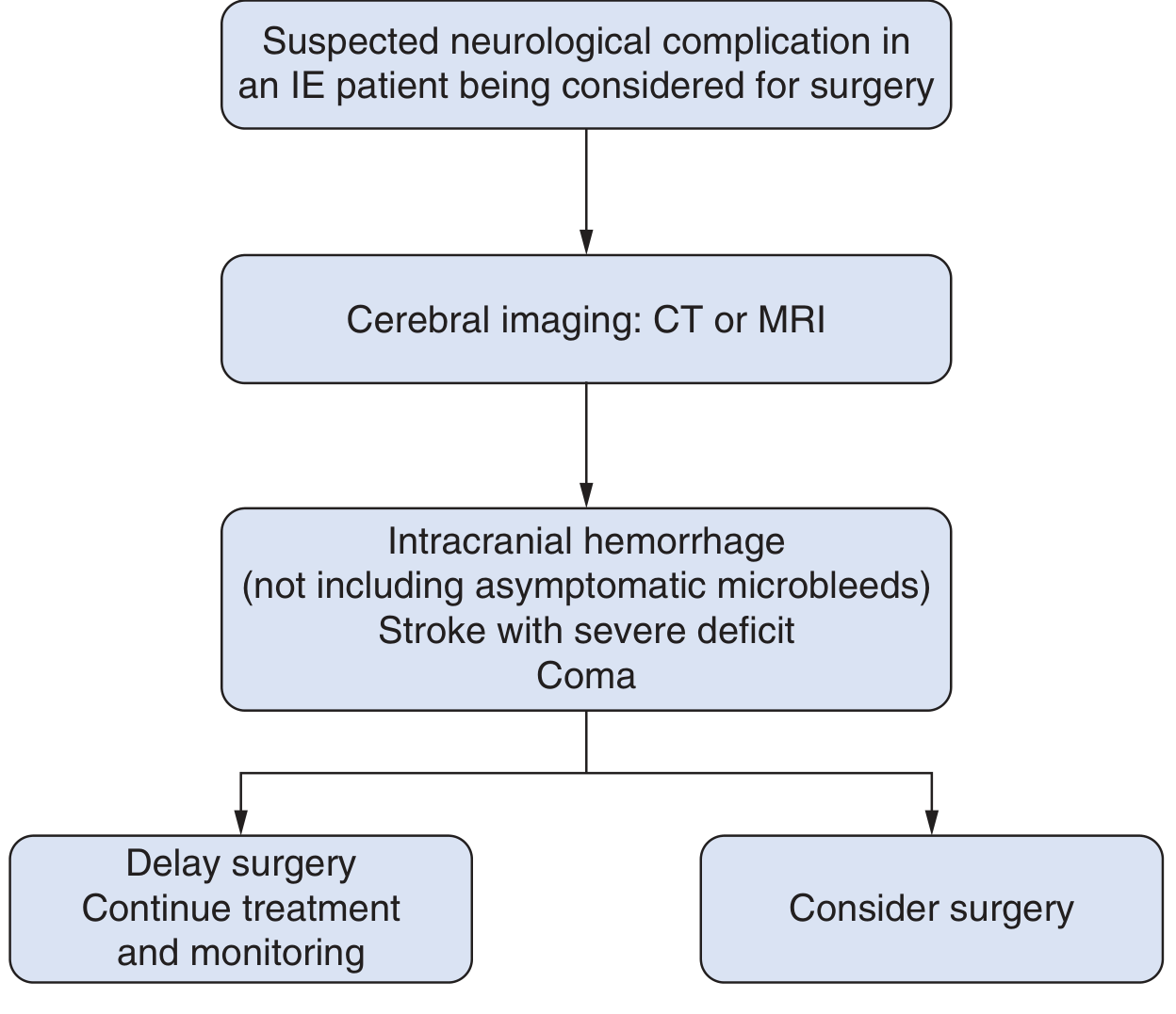
  • Cerebral imaging is mandatory (CT or MRI) for any suspicion of neurological complication in IE patients
IE neurological complication imaging decision flowchart
Neurological complication imaging algorithm in IE patients (Fuster & Hurst's The Heart, 15th Ed., adapted from 2015 ESC Guidelines)

3. Digital Subtraction Angiography (DSA) - Gold Standard for Intracranial MAs

DSA is the gold standard for intracranial mycotic aneurysms due to the small size of these lesions and the inadequate sensitivity of CTA/MRA (~40% sensitivity for small MAs).
Classical DSA hallmarks of intracranial mycotic aneurysms:
  • Distal location - beyond M2 (MCA), P1 (PCA), or equivalent; peripheral rather than at the circle of Willis
  • Multiplicity - multiple aneurysms are characteristic (vs. saccular berry aneurysms)
  • Fusiform or irregular shape
  • Change in size on follow-up - new aneurysm appearance or growth on repeat DSA during antibiotic treatment
  • Located at arterial bifurcations (sites of flow turbulence where emboli lodge in the vasa vasorum)
Conventional angiography is reasonable even after negative non-invasive imaging when clinical suspicion for intracranial MA remains high.
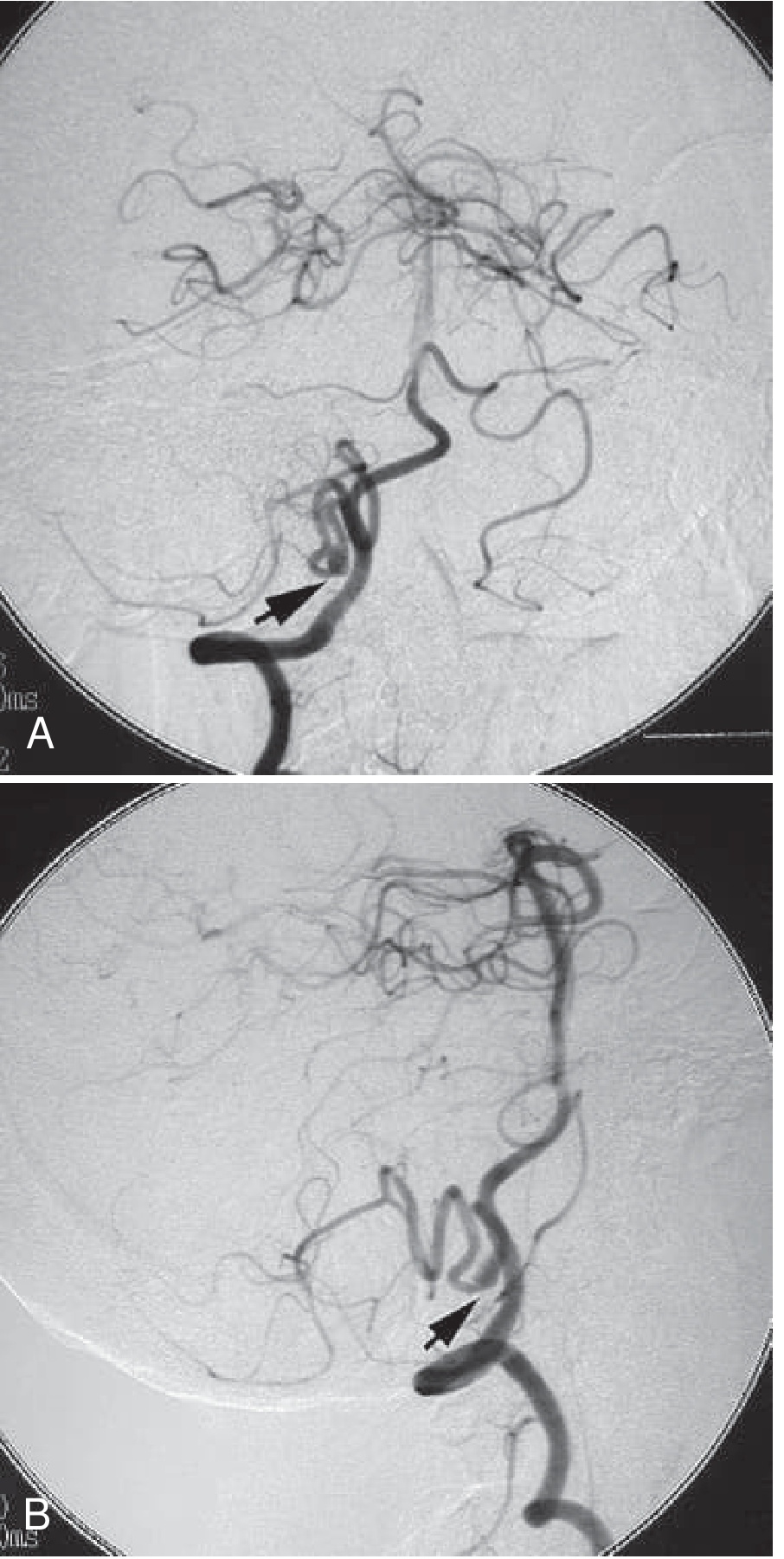
DSA of a ruptured dissecting PICA aneurysm - AP and lateral views showing fusiform dilatation
Digital subtraction angiograms (AP and lateral) of the right vertebral artery demonstrating a small fusiform dilatation of the PICA (arrows) - characteristic peripheral location of a mycotic aneurysm (Bradley and Daroff's Neurology in Clinical Practice)
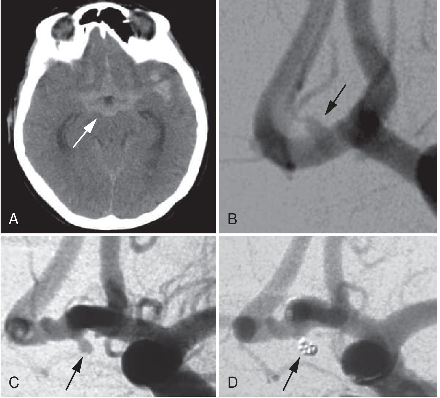
CT-SAH and serial DSA showing mycotic aneurysm evolution
Sequence: (A) CT brain with aneurysmal SAH; (B) Initial DSA - no definite aneurysm; (C) Repeat DSA at day 7 - new 2mm aneurysm (arrow); (D) Post-coil embolization with complete obliteration. Classic mycotic aneurysm pattern of delayed appearance on serial DSA. (Bradley and Daroff's Neurology in Clinical Practice)

4. Duplex Ultrasound

  • Useful for peripheral artery mycotic aneurysms (femoral, popliteal, radial/ulnar)
  • Can detect pulsatile mass, confirm flow within aneurysm sac
  • Color Doppler demonstrates swirling "yin-yang" pattern
  • Limited for deep visceral or intracranial locations

5. Nuclear Medicine / FDG-PET

  • FDG-PET (often combined as PET-CT) is primarily used to detect graft infection and aortic/large vessel mycotic aneurysms
  • Useful in cases where CT findings are equivocal - metabolic activity in the aneurysm wall suggests active infection
  • Particularly helpful for mycotic aortic aneurysms and post-endovascular graft infection surveillance

6. Echocardiography

  • Transthoracic echocardiography (TTE) is the preferred modality for coronary artery mycotic aneurysms
  • Transesophageal echocardiography (TEE) is essential for the underlying IE diagnosis and source identification

Imaging by Location

LocationFirst-LineSupplementaryNotes
IntracranialCT brain (hemorrhage screen) then DSAMRI/MRADSA gold standard; CTA/MRA ~40% sensitivity
Aorta/large vesselsCTAFDG-PETPerivascular air/fluid on CT suggestive
Visceral arteriesCTAUltrasound, MRACT most commonly establishes diagnosis
Peripheral arteriesDuplex ultrasound, CTAMRAHand/finger: doppler first
Coronary arteriesTTECTA coronaryCoronary MA rare

Key Imaging Differentiators from Non-Infectious Aneurysms

FeatureMycotic AneurysmAtherosclerotic Aneurysm
ShapeSaccular, eccentric, lobulatedFusiform, smooth
LocationDistal/peripheral branchesProximal, major vessels
Perivascular changesAir, fluid, inflammationCalcification, mural thrombus
MultiplicityCommonLess common
Rate of growthRapidSlow
Clinical contextFever, bacteremia, IEElderly, cardiovascular risk factors

Serial Imaging - Critical Point

Because mycotic aneurysms can appear on an initially negative DSA, repeat angiography is essential in patients with IE who develop neurological symptoms or unexplained SAH. The natural history includes both spontaneous resolution (in ~30% with antibiotics alone) and rapid enlargement with rupture. Serial angiography guides treatment decisions.

Recent Evidence

A 2024 meta-analysis (PMID 38064880) on endovascular treatment of mycotic aneurysms confirms neuroendovascular intervention as preferred first-line therapy with lower mortality, easier access to distal aneurysms, and ability to treat multiple aneurysms vs. microsurgical craniotomy - all supported by imaging-guided planning.

Sources: Fuster and Hurst's The Heart 15th Ed.; Bradley and Daroff's Neurology in Clinical Practice; Adams and Victor's Principles of Neurology 12th Ed.; Sabiston Textbook of Surgery; StatPearls - Mycotic Aneurysm (2025); AJR mycotic aneurysm CT/MRI study; Cerrahpasa Medical J 2024 endovascular series

Explain the pathophysiology: how do infected emboli from endocarditis weaken the arterial wall?

Finding Sources
Finding Sources
Searching PubMed

mycotic aneurysm pathophysiology vasa vasorum endocarditis mechanism

Finding Sources
Finding Sources
Finding Sources
Finding Sources
Finding Sources
Finding Sources
Reading File
Searching the Web

mycotic aneurysm pathophysiology mechanism vasa vasorum septic emboli wall destruction proteases 2023 2024

Reading File
Reading Web Page

mycotic aneurysm pathophysiology vasa vasorum infected emboli arterial wall mechanism matrix metalloproteinase

Now I have a rich multi-source picture. Here is the full synthesis:

Pathophysiology of Mycotic Aneurysm Formation: How Infected Emboli Weaken the Arterial Wall

Overview of the Sequence

The arterial wall destruction follows a stereotyped cascade: embolus lodgment → bacterial seeding of the wall → inflammatory cell recruitment → proteolytic enzyme release → structural protein degradation → wall thinning and dilation → aneurysm formation (or rupture).

Step 1 - The Source: Vegetations on the Valve

In infective endocarditis (IE), bacteria colonize the cardiac valves, forming vegetations - friable masses of fibrin, platelets, and organisms. These vegetations are mechanically fragile. Turbulent blood flow across the infected valve continuously sheds fragments into the systemic circulation as septic emboli - microthrombi laden with live bacteria (most commonly Streptococcus viridans, Staphylococcus aureus, or enterococci).

Step 2 - Two Routes of Entry into the Arterial Wall

Once in the circulation, infected emboli reach arteries by one of two mechanisms:

Route A: Occlusion of the Vasa Vasorum (primary and most likely)

The vasa vasorum are the tiny nutrient vessels that supply the outer layers (adventitia and outer media) of large and medium-sized arteries from the outside. Septic microemboli become lodged in the vasa vasorum, blocking its lumen - causing a focal infarction of the arterial wall. This ischemic injury then allows bacteria to establish themselves in the avascular, necrotic adventitia. Experimental canine models (Molinari et al., 1973) confirmed that the first histological changes appear in the adventitial layer, spreading inward to involve the media.
"The most likely cause is impaction of infected material in the vasa vasorum of the artery, with resulting destruction of the arterial wall."
  • Bradley and Daroff's Neurology in Clinical Practice

Route B: Intraluminal Impaction

Less commonly, a larger infected embolus occludes the lumen of a smaller artery (particularly distal intracranial branches). Bacteria in the embolus then invade the intima directly, spreading outward through the layers. Subsequent infection weakens the wall at the occlusion site.

Route C: Contiguous Spread

A third mechanism involves direct extension from an adjacent infectious focus (e.g., meningitis, vertebral osteomyelitis, abscess) spreading to the adjacent arterial adventitia from the outside in.

Step 3 - Bacterial Infection of the Wall Layers

Once bacteria establish in the adventitia or intima:
  1. Bacterial toxins and enzymes (particularly from Staphylococcus spp.) are released directly, degrading the extracellular matrix of the vessel wall.
  2. Pattern recognition by toll-like receptors on resident vascular cells triggers a local inflammatory response.
  3. Pro-inflammatory cytokines (IL-1β, TNF-α, IL-6) are released by endothelial cells, smooth muscle cells, and macrophages - recruiting neutrophils and monocytes into the wall.

Step 4 - The Central Destructive Mechanism: Matrix Metalloproteinases (MMPs)

This is the core molecular event:
  • Recruited neutrophils and macrophages, activated by cytokine signaling, release matrix metalloproteinases (MMPs) - particularly MMP-2, MMP-3, MMP-9, and MMP-12.
  • MMPs are zinc-dependent proteolytic enzymes that degrade the structural proteins of the arterial wall: elastin (the elastic fiber backbone of the media) and collagen (the tensile-strength scaffold of all three layers).
  • Normally, MMP activity is controlled by TIMPs (tissue inhibitors of metalloproteinases). In an infected arterial wall, the inflammatory state upregulates MMPs while reducing TIMPs, creating an unchecked proteolytic environment.
  • The PGE2-cAMP signaling pathway, activated by inflammatory cytokines, amplifies MMP production.
"Bacterial infection triggers the release of proinflammatory cytokines, which attract neutrophils. These cells activate matrix metalloproteinases, which contribute to the focal breakdown of the blood vessel wall. The degree of metalloproteinase activity correlates with the risk of future aneurysm rupture."
  • StatPearls: Mycotic Aneurysm (2025)

Step 5 - Structural Failure of the Arterial Wall

The three layers of the artery are progressively destroyed:
LayerNormal FunctionWhat Infection Destroys
IntimaSmooth barrier, anti-thromboticEndothelial integrity, platelet activation
MediaSmooth muscle + elastic laminae - provides tensile strength and elasticityElastin fibers (MMP-12), smooth muscle cells (apoptosis from cytotoxins)
AdventitiaCollagen scaffold, houses vasa vasorumCollagen (MMP-1, MMP-9), vasa vasorum itself infarcted first
The result is transmural inflammation with abscess formation, necrosis, and thrombosis - what pathologists find in resected specimens.
With the media and adventitia both compromised, the arterial wall can no longer withstand normal pulsatile blood pressure. The vessel progressively dilates - aneurysmal enlargement occurs by arterial pulsation against the weakened wall (Bradley and Daroff).

Step 6 - Immune Complex Deposition (Additional Mechanism)

In IE-associated mycotic aneurysms, there is also a type III hypersensitivity component: circulating antigen-antibody immune complexes deposit in the vessel wall. This triggers complement activation, further recruiting inflammatory cells and amplifying local tissue destruction - independent of direct bacterial invasion.

Step 7 - Why Intracranial Arteries Are Particularly Vulnerable

Intracranial arteries have structural features that make them especially susceptible:
  • No external elastic lamina (absent or poorly developed)
  • Thin adventitia compared to systemic vessels
  • No vasa vasorum in the smallest branches - meaning emboli lodge directly in the arterial lumen and invade the intima
  • Located at bifurcation points where flow turbulence and eddy currents cause endothelial shear stress, facilitating embolus impaction and bacterial adherence

Summary Diagram (Conceptual)

Valve vegetation (IE)
        ↓
Septic embolus shed into circulation
        ↓
    ┌──────────────────────────────┐
    │ Lodges in vasa vasorum       │  ← Primary route
    │ (infarcts wall from outside) │
    └──────────────────────────────┘
    ┌──────────────────────────────┐
    │ Occludes arterial lumen      │  ← Secondary route
    │ (bacteria invade intima)     │
    └──────────────────────────────┘
        ↓
Bacteria establish in adventitia/media
        ↓
Cytokines (IL-1β, TNF-α) → recruit neutrophils & macrophages
        ↓
MMP release (MMP-2, -3, -9, -12) + TIMP suppression
        ↓
Elastin + collagen degradation → transmural necrosis/abscess
        ↓
Wall cannot withstand arterial pulsation
        ↓
Aneurysmal dilation → RUPTURE (SAH, intracerebral hemorrhage, or hemorrhage into the relevant territory)

Why Some Resolve and Others Rupture

This is not fully understood, but:
  • Effective antibiotics can sterilize the wall before irreversible structural damage occurs - explaining why ~30% of intracranial MAs resolve on antibiotic therapy alone with serial imaging.
  • MMP activity level may be the key prognostic variable - higher activity correlates with rupture risk.
  • Aneurysm growth on follow-up imaging is the clearest predictor of imminent rupture, which is why serial DSA during antibiotic treatment is standard practice.

Sources: Bradley and Daroff's Neurology in Clinical Practice; Adams and Victor's Principles of Neurology 12th Ed.; StatPearls: Mycotic Aneurysm (2025); Diagnostics: New Insights into the Pathophysiology of Coronary Artery Aneurysms (MDPI 2024)

Viva question toigh

Here are some tough viva questions on mycotic aneurysms that an examiner might throw at you — with model answers:

Viva: Mycotic Aneurysms


Q1. Why are mycotic aneurysms typically located distally rather than at the circle of Willis?
Expected answer: Septic emboli from IE are small microthrombi that travel distally until they lodge in smaller-caliber vessels - particularly at bifurcation points of distal MCA, PCA branches, etc. These are sites of flow turbulence and eddy currents that facilitate impaction. The circle of Willis vessels are larger and proximal; emboli pass through them. This is the reverse of saccular (berry) aneurysms, which classically occur at proximal bifurcations due to hemodynamic stress on congenitally weak walls.

Q2. A patient with known IE develops sudden headache. CT brain is normal. What do you do next and why?
Expected answer: Normal CT does not exclude mycotic aneurysm. CT/MRA has only ~40% sensitivity for small MAs. The next step is lumbar puncture to look for xanthochromia (SAH). If LP is positive or clinical suspicion remains high, catheter-based DSA is mandatory - it is the gold standard. Every IE patient with SAH requires catheter angiography regardless of CT findings.

Q3. You find a 4mm unruptured intracranial mycotic aneurysm on DSA. The patient is on IV antibiotics. What is your management and why?
Expected answer: Conservative management with antibiotics and serial DSA (or MRA) at 1-2 week intervals is the initial approach for unruptured MAs. Approximately 30% resolve on antibiotic therapy alone. Indications to intervene (surgical clipping or endovascular coiling/trapping) include: aneurysm growth on serial imaging, rupture, failure to resolve, or the patient needing cardiac surgery. Endovascular treatment is now preferred over microsurgery - lower mortality, better access to distal aneurysms, can treat multiple simultaneously.

Q4. Why does a mycotic aneurysm appear on a repeat DSA 7 days after the first DSA was negative?
Expected answer: This is a classic hallmark of mycotic aneurysms and reflects the time course of the pathophysiology. The initial embolic event causes vasa vasorum occlusion and wall ischemia, but MMP-mediated collagen/elastin degradation takes days to produce enough wall thinning to be angiographically visible. The aneurysm can literally form within days of bacterial seeding. This is why a negative initial DSA does not exclude MA, and repeat DSA at 1-2 weeks is mandatory if suspicion persists.

Q5. Why is anticoagulation withheld in IE patients with cerebral embolism?
Expected answer: Two risks: (1) hemorrhagic transformation of septic embolic infarcts - the blood-brain barrier is disrupted around infarcted tissue; anticoagulation converts ischemic infarcts into hemorrhagic ones. (2) Rupture of an unrecognized mycotic aneurysm - a patient may harbour an MA that has not yet been imaged; anticoagulation dramatically increases rupture risk and bleeding severity. Antibiotics must first sterilize the source and serial imaging must exclude MA before anticoagulation is considered.

Q6. What is the MMP-TIMP imbalance and why does it matter clinically?
Expected answer: Matrix metalloproteinases (MMP-2, -3, -9, -12) degrade structural proteins - elastin and collagen - in the arterial wall. Normally their activity is checked by tissue inhibitors (TIMPs 1-4). Bacterial infection + cytokine signaling upregulates MMPs while suppressing TIMPs. This unchecked proteolysis destroys the media and adventitia. Clinically, the degree of MMP activity may correlate with rupture risk and could serve as a prognostic biomarker - though this is not yet in routine clinical use.

Q7. What is the difference between a mycotic aneurysm and septic arteritis, and why does it matter?
Expected answer: Both result from bacterial infection of the arterial wall in IE. In septic arteritis, the vessel wall is destroyed without aneurysm formation - the wall is weakened but the vessel does not dilate; instead it ruptures directly or causes hemorrhagic infarction. In mycotic aneurysm, the wall weakens and dilates before rupture. The distinction matters because angiography can differentiate them - MA shows a definable aneurysmal outpouching; septic arteritis may show only vessel irregularity or beading. Management implications are the same (antibiotics + intervention if rupture), but MA is amenable to targeted endovascular treatment.

Q8. Why does Staphylococcus aureus endocarditis carry a higher risk of mycotic aneurysm rupture than Streptococcus viridans?
Expected answer: S. aureus is a far more virulent organism - it produces a broader array of exotoxins, proteases (including its own metalloproteinases), coagulase, and panton-valentine leukocidin. These directly destroy tissue and drive a more intense neutrophilic response with higher MMP release. S. aureus IE causes acute endocarditis with rapid valve destruction and large, friable vegetations that shed emboli early and frequently. Streptococcus viridans causes subacute endocarditis with smaller vegetations and a less aggressive inflammatory response - MAs can still form but the wall destruction is slower, allowing more time for antibiotic resolution.

Good luck - if you want me to drill you with rapid-fire questions or go deeper on any of these, just say so.

Suprasellar mass and craniopharngioma imaging

Finding Sources
Finding Sources
Searching PubMed

craniopharyngioma MRI imaging diagnosis

Finding Sources
Finding Sources
Finding Sources
Reading File
Reading File
Finding Sources
Finding Sources
Searching the Web

craniopharyngioma suprasellar mass imaging MRI CT differential diagnosis radiology features 2024

Finding Sources
Finding Sources
Loading Image
Reading Web Page

craniopharyngioma MRI CT imaging features suprasellar adamantinomatous papillary differential diagnosis

Here is the comprehensive answer:

Suprasellar Mass and Craniopharyngioma - Imaging

Anatomy of the Suprasellar Region - Why It Matters

The suprasellar cistern sits directly above the pituitary fossa and is bounded by the optic chiasm anteriorly, the hypothalamus superiorly, the third ventricle, and the circle of Willis. Any mass here can compress the optic chiasm (→ bitemporal hemianopia), obstruct the third ventricle (→ hydrocephalus), or invade the hypothalamus/pituitary stalk (→ endocrine dysfunction). This anatomical complexity means the differential diagnosis is broad - and imaging must both characterize the lesion AND map its relationships to surrounding structures.

Suprasellar Mass - Differential Diagnosis Framework

A useful memory aid is SATCHMO or simply thinking by tissue of origin:
OriginLesion
PituitaryMacroadenoma extending superiorly, pituitary apoplexy
Rathke's pouch remnantCraniopharyngioma, Rathke's cleft cyst
MeningesSuprasellar/tuberculum sellae meningioma
Germ cellsGerminoma (most common suprasellar germ cell tumor)
Optic/hypothalamicOptic pathway/hypothalamic glioma
VascularSuprasellar aneurysm (ICA, ACoA)
Inflammatory/infiltrativeHypophysitis (lymphocytic, IgG4), sarcoidosis, Langerhans cell histiocytosis
InfectionTuberculoma, abscess
Epidermoid/dermoidMidline epidermoid cyst
The most common suprasellar masses by age:
  • Children: Craniopharyngioma, optic/hypothalamic glioma, germinoma
  • Adults: Pituitary macroadenoma, meningioma, craniopharyngioma (papillary type)

Craniopharyngioma

Origin and Types

Craniopharyngiomas arise from epithelial remnants of Rathke's pouch - the embryological precursor of the anterior pituitary. They are WHO Grade 1 (benign histologically) but locally aggressive due to their critical location. There are two distinct subtypes with different imaging, molecular, and clinical profiles:
FeatureAdamantinomatous (ACP)Papillary (PCP)
AgeChildren (peak 5-15 yrs)Adults (peak 40-55 yrs)
MutationCTNNB1 (β-catenin/Wnt pathway)BRAF V600E
Calcification~90% - prominent, "eggshell"Rare (<10%)
CystsMultilocular, "machine oil" fluidUsually solid or simple cyst
LocationSellar + suprasellarPurely suprasellar (3rd ventricle)
HistologyWet keratin, peripheral palisadingPapillary squamous epithelium

CT Imaging

CT is the gold standard for detecting calcification (found in 90% of ACPs).

Adamantinomatous CP on CT:

  • Hypodense cystic component in the suprasellar cistern - often multilocular
  • Calcification - present in ~90%; can be peripheral "eggshell" calcification of the cyst wall, or irregular nodular foci within the solid component
  • Solid enhancing nodule after contrast
  • The classic CT triad: cyst + solid nodule + calcification
  • May extend into the sella inferiorly or the third ventricle superiorly
  • Obstructive hydrocephalus when third ventricle is compressed

Papillary CP on CT:

  • Isodense solid suprasellar mass - lacks the cystic/calcified appearance
  • Typically no calcification
  • Homogeneous enhancement after contrast
  • Often located in the third ventricle floor

MRI Imaging - Gold Standard for Characterization

MRI is the gold standard for craniopharyngioma characterization - it defines the relationship to the optic chiasm, hypothalamus, pituitary stalk, and third ventricle.

Signal characteristics of Adamantinomatous CP:

SequenceAppearanceExplanation
T1 pre-contrastHyperintense cysts (bright)High cholesterol, protein, and old blood in "machine oil" fluid
T2Hyperintense cysts; mixed solidFluid + variable solid matrix
FLAIRCysts remain bright (not suppressed)Protein-rich fluid doesn't suppress like CSF
T1 + GdRim/wall enhancement of cysts + solid nodule enhancesBreakdown of blood-brain barrier in solid component
SWI/GREBlooming artifact from calcificationSusceptibility effect of Ca²⁺
"Cystic components are typically hyperintense and less commonly isointense to CSF on T1-weighted images. Multiple cysts with varying signal intensities are characteristic. Solid components have variable signal intensities and usually enhance with contrast."
  • Brain Imaging with MRI and CT (Cambridge)

Signal characteristics of Papillary CP:

  • T1: Iso- to slightly hypointense (no cholesterol/blood content)
  • T2: Hyperintense solid mass
  • T1 + Gd: Solid, homogeneous enhancement
  • No blooming on SWI (no calcification)
  • Often appears as a well-demarcated solid mass in the third ventricle floor

MRI Sequences to Request:

  1. Sagittal T1 thin-slice (3mm) - best for pituitary stalk, optic chiasm, hypothalamus relationships
  2. Coronal T2 - cyst characterization, third ventricle involvement
  3. Coronal + sagittal T1 post-Gd - enhancement pattern
  4. Axial FLAIR - perilesional edema, hydrocephalus
  5. SWI - calcification, hemorrhage
  6. DWI - helps distinguish epidermoid (restricted diffusion) from craniopharyngioma (no restriction)

Histopathology Correlate

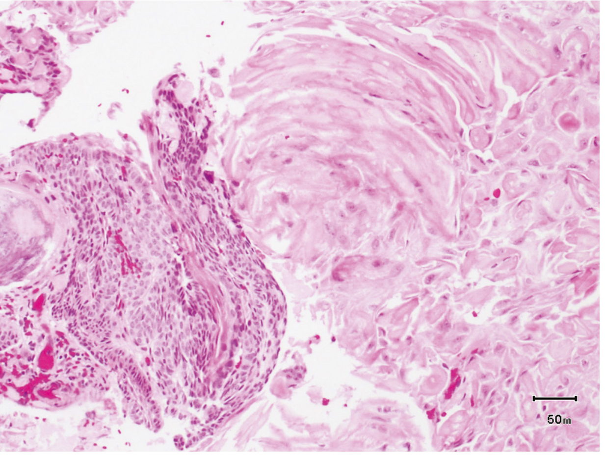
Adamantinomatous craniopharyngioma histology - compact lamellar "wet" keratin (right) with cords of squamous epithelium showing peripheral palisading (left)
Adamantinomatous craniopharyngioma: compact lamellar "wet keratin" (right) and squamous epithelium cords with peripheral palisading (left) - the calcifications and cyst wall that produce the CT and MRI findings originate from this tissue. (Robbins Pathologic Basis of Disease)

Differential Diagnosis on Imaging

How to distinguish key suprasellar lesions:

Pituitary Macroadenoma vs Craniopharyngioma:
  • Adenoma originates from the sella (snowman sign - figure-of-8 expansion through diaphragma sellae); craniopharyngioma is primarily suprasellar
  • Adenoma is isointense to grey matter, solid, delayed homogeneous enhancement; rarely calcifies
  • Adenoma does NOT have the T1-bright cysts of ACP
  • Adenoma may invade the cavernous sinus laterally (Knosp grading)
Rathke's Cleft Cyst (RCC) vs Craniopharyngioma:
  • RCC is a single ovoid cyst - no enhancing solid nodule, no calcification (or rare thin peripheral calcification)
  • T1 signal variable (mucoid: T1 bright; serous: T1 dark)
  • No multilocularity; thin or absent wall enhancement
  • Key: RCC has no solid nodule and no calcification - the hallmarks of craniopharyngioma
Suprasellar Meningioma vs Craniopharyngioma:
  • Meningioma: dural tail sign, dense homogeneous enhancement, isointense T1/T2 to brain, rarely cystic, may show calcification but of different character
  • Meningioma arises from tuberculum sellae/planum sphenoidale - not from Rathke's pouch
Germinoma vs Craniopharyngioma:
  • Germinoma: isointense, solid, no calcification, homogeneous enhancement
  • Germinoma may have elevated CSF/serum β-hCG, AFP
  • Germinoma is highly radiosensitive - distinguishing it from craniopharyngioma changes management completely
  • DWI: germinoma may show mildly restricted diffusion (hypercellular)
Hypothalamic/Optic Glioma vs Craniopharyngioma:
  • Glioma: T2 hyperintense without the cystic-calcified morphology of ACP
  • Strong association with NF1
  • Enhancement variable; may be absent in low-grade glioma
Suprasellar Aneurysm:
  • Pulsation artifact on MRI
  • Flow void on T2; MRA/CTA shows connection to ICA or ACoA
  • Never calcifies in the craniopharyngioma pattern

Summary: Imaging Signature by Lesion

LesionCTT1T2EnhancementKey Clue
ACP CraniopharyngiomaCyst + Ca²⁺ + noduleBright cystsBright cystsRim + nodule"Machine oil" T1 bright cysts + calcification
PCP CraniopharyngiomaSolid, no Ca²⁺Iso/hypoHyperSolid, homogeneousAdult, 3rd ventricle, BRAF+
Pituitary adenomaIso, sella-basedIsoIso/hyperDelayed, solidSnowman, cavernous sinus
Rathke's cleft cystCyst, no Ca²⁺VariableVariableNone/thin rimNo nodule, no Ca²⁺
MeningiomaDense, ± Ca²⁺IsoIsoIntense + dural tailDural tail, en plaque
GerminomaSolid, isoIsoIsoHomogeneousYouth, DI, β-hCG/AFP
Optic gliomaIsoIsoBrightVariableNF1, optic nerve enlargement
AneurysmCalcified rim (thrombosed)Flow voidFlow voidNone/rimMRA confirms
EpidermoidHypo (similar to CSF)HypoHyperNoneDWI restricted (key)

Clinical Correlations

  • Bitemporal hemianopia → optic chiasm compression from above → suprasellar (not sellar) mass
  • Diabetes insipidus at presentation → suggests germinoma or Langerhans cell histiocytosis first (pituitary stalk involvement); craniopharyngioma causes DI more often post-operatively
  • Growth failure + visual loss in a child → craniopharyngioma until proven otherwise; CT first for calcification
  • Pituitary stalk thickening → germinoma, LCH, hypophysitis, TB, sarcoid

Sources: Adams and Victor's Principles of Neurology 12th Ed.; Robbins Pathologic Basis of Disease; Localization in Clinical Neurology 8th Ed.; PMC comprehensive craniopharyngioma review (2024); Brain Imaging with MRI and CT (Cambridge); StatPearls Craniopharyngioma (2025)

Acutr ischemic stroke imaging and management

Finding Sources
Finding Sources
Searching PubMed

"ischemic stroke" AND "thrombectomy"

Finding Sources
Finding Sources
Reading File
Finding Sources
Reading File
Finding Sources
Reading File
Reading File
Reading File
Reading File
Reading File
Searching the Web

acute ischemic stroke imaging CT perfusion MRI DWI ASPECTS score management guidelines 2024 2025

Reading File
Loading Image
Excellent material gathered. Here is the full synthesis:

Acute Ischemic Stroke: Imaging and Management


Pathophysiology in Brief

Ischemic stroke results from vessel occlusion by thromboembolism or atherosclerotic plaque, causing focal brain ischemia. Two zones form:
  • Ischemic core: irreversibly infarcted (CBF <10 mL/100g/min) - dead within minutes
  • Penumbra: ischemic but not yet infarcted (CBF 10-20 mL/100g/min) - potentially salvageable with reperfusion
The entire premise of imaging and management is: identify and save the penumbra before it joins the core.

IMAGING

The "Code Stroke" CT Protocol

The standard imaging triage for acute stroke is a 3-step CT protocol performed simultaneously as a single rapid acquisition:
1. Non-contrast CT (NCCT) → exclude hemorrhage, assess ASPECTS
2. CT Angiography (CTA)   → identify LVO, assess collaterals
3. CT Perfusion (CTP)     → core vs penumbra mismatch (selected patients)
MRI (particularly DWI) is an alternative when available without delaying treatment.

1. Non-Contrast CT (NCCT) - First and Always

Primary purpose: Exclude hemorrhagic stroke (absolute contraindication to thrombolysis and thrombectomy).
Early ischemic signs on NCCT (appear within 3-6 hours, may be subtle):
SignAppearanceSignificance
Hyperdense MCA signBright white MCA on unenhanced CTAcute thrombus in MCA - suggests LVO
Hyperdense basilar signBright basilar arteryBasilar artery occlusion
Loss of grey-white differentiationBlurring of insular ribbon, lentiform nucleusEarly cytotoxic edema
Sulcal effacementLoss of cortical sulciEarly cerebral swelling
"Dot sign"Hyperdense dot in sylvian fissure branchesDistal MCA branch thrombus
"Large cortical infarcts might not be detected by NCCT up to 3 hours from onset, and around 40% of infarcts can be missed by 24 hours."

ASPECTS Score (Alberta Stroke Program Early CT Score)

The ASPECTS system scores early ischemic changes in the MCA territory on NCCT. The brain is divided into 10 regions (M1-M6 cortical + C caudate, L lentiform, IC internal capsule, I insula):
  • Each region scores 1 point if normal
  • Subtract 1 point for each region showing early ischemic change
  • Score 10 = no ischemic change (ideal)
  • Score ≤7 = poor prognosis for independent recovery
  • Score ≥6 traditionally required for thrombectomy eligibility (now reassessed - see below)
Posterior circulation ASPECTS (pc-ASPECTS) is also increasingly used but requires further validation.

2. CT Angiography (CTA) - Identify the Occlusion

CTA from aortic arch to vertex is performed immediately after NCCT. This is now considered standard in all acute stroke patients.
What CTA provides:
  • Identifies the large vessel occlusion (LVO) site: ICA terminus, M1, M2, basilar artery, etc.
  • Collateral circulation grading - good collaterals = larger salvageable penumbra = more time for treatment
  • Rules out underlying vascular lesions (dissection, stenosis)
  • CTA source images (CTA-ASPECTS) are superior predictors of final infarct size compared to NCCT-ASPECTS, because they directly show the degree of collateral perfusion

3. CT Perfusion (CTP) - Core vs Penumbra Mismatch

CTP measures four parameters:
ParameterWhat it measuresCore/Penumbra
CBF (Cerebral Blood Flow)ml/100g/minCore = severely reduced (<30% contralateral)
CBV (Cerebral Blood Volume)ml/100gCore = CBV markedly reduced
MTT (Mean Transit Time)SecondsPenumbra = prolonged
Tmax (Time to peak)SecondsPenumbra = Tmax >6 seconds
Mismatch = Tmax >6s region (at-risk territory) MINUS CBF core (infarcted territory). A large mismatch means large salvageable penumbra - the patient benefits from reperfusion.
Used for patient selection in:
  • Extended thrombolysis window (4.5-9 hours) - EXTEND trial
  • Late-window thrombectomy (6-24 hours) - DAWN and DEFUSE-3 trials

4. MRI - More Sensitive, Used When Available

MRI is more sensitive than NCCT for early infarct detection, but logistics (time, availability, contraindications) often make CT the primary tool.

DWI (Diffusion-Weighted Imaging) - Most Important Sequence

  • Detects ischemic core within minutes of onset - the earliest sequence to show infarction
  • Restricted diffusion (bright on DWI, dark on ADC map) = cytotoxic edema from Na-K ATPase failure
  • Sensitivity ~80-95% for acute infarction (note: false negatives exist, especially in posterior fossa, small lacunes, and hyperacute <6 hours)
  • DWI-FLAIR mismatch: DWI bright + FLAIR normal = stroke likely within 4.5 hours (used in WAKE-UP trial for unknown-onset stroke treatment)

MRI Sequences in Acute Stroke

SequenceRole
DWIIdentify ischemic core (minutes to hours)
ADC mapConfirm restricted diffusion (dark = true infarct)
FLAIREdema, old infarcts; DWI-FLAIR mismatch for timing
GRE/SWIHemorrhage (microbleeds, hemorrhagic transformation), thrombus
MRANon-contrast vessel imaging - identifies LVO
PWI (perfusion)Core-penumbra mismatch (DWI-PWI mismatch)
T2Chronic infarcts, white matter disease

5. Imaging Sequence Summary by Time Window

Time from onsetImaging ProtocolPurpose
0-6 hours ("early window")NCCT + CTA ± CTPRule out ICH, identify LVO for thrombectomy
4.5-9 hours (extended tPA)NCCT + CTP or DWI-FLAIR MRIMismatch for IV tPA eligibility
6-24 hours (late thrombectomy)NCCT + CTA + CTP or DWI MRIClinical-imaging or perfusion mismatch for thrombectomy (DAWN/DEFUSE-3 criteria)
Unknown onset ("wake-up stroke")MRI DWI-FLAIR mismatchProxy for <4.5h onset (WAKE-UP trial)

MANAGEMENT

Emergency Assessment - "Time is Brain"

Every 1 minute of untreated LVO = ~1.9 million neurons lost. Key targets:
  • Door-to-needle time (IV tPA): <60 minutes
  • Door-to-groin puncture (thrombectomy): <90 minutes
Immediate steps:
  1. Airway, breathing, circulation - stabilize
  2. Blood glucose (hypoglycemia mimics stroke - treat if <60 mg/dL)
  3. NCCT + CTA (CT protocol as above)
  4. 12-lead ECG (AF is common stroke cause)
  5. IV access, bloods: FBC, coagulation, metabolic panel
  6. NIHSS scoring

A. Intravenous Thrombolysis (IV tPA / Alteplase)

Mechanism: Alteplase (rt-PA) activates plasminogen to plasmin, dissolving the fibrin clot systemically.
Dose: 0.9 mg/kg (max 90 mg); 10% as IV bolus, remainder over 60 minutes.
Time windows:
WindowEvidenceNotes
0-3 hoursNINDS (1995) - landmark trialFDA approved; maximum benefit
3-4.5 hoursECASS-III (2008)Extended benefit; some additional exclusions apply
4.5-9 hoursEXTEND trial (2019)Requires CTP mismatch showing salvageable tissue
Unknown onsetWAKE-UP trial (2018)Requires DWI+ / FLAIR- MRI mismatch
"The EXTEND study randomized patients presenting between 4.5 and 9 hours after onset - favorable outcome at 90 days was present in more patients in the alteplase group (35.4% vs 29.5%)." - Fuster and Hurst's The Heart, 15th Ed.

Key tPA Contraindications

Absolute ContraindicationsRelative Contraindications
Intracranial hemorrhage on CTRecent surgery (<14 days)
Ischemic stroke within 3 monthsMajor trauma (<14 days)
Active internal bleedingAnticoagulant use (INR >1.7, heparin with elevated PTT)
Severe head trauma (<3 months)Blood glucose <50 or >400 mg/dL
Platelet count <100,000Large infarct (ASPECTS ≤3)
BP >185/110 mmHg (uncontrolled)Recent MI
Note on Tenecteplase: Emerging as an alternative to alteplase - single IV bolus (0.25 mg/kg), similar efficacy, more convenient; gaining traction in guidelines (2024 ESO, AHA/ASA updates).

B. Mechanical Thrombectomy (MT) - LVO Stroke

Thrombectomy is now the standard of care for LVO stroke - one of the most effective interventions in modern medicine with an NNT of 2.6 to reduce disability by one mRS level (HERMES meta-analysis).
Devices:
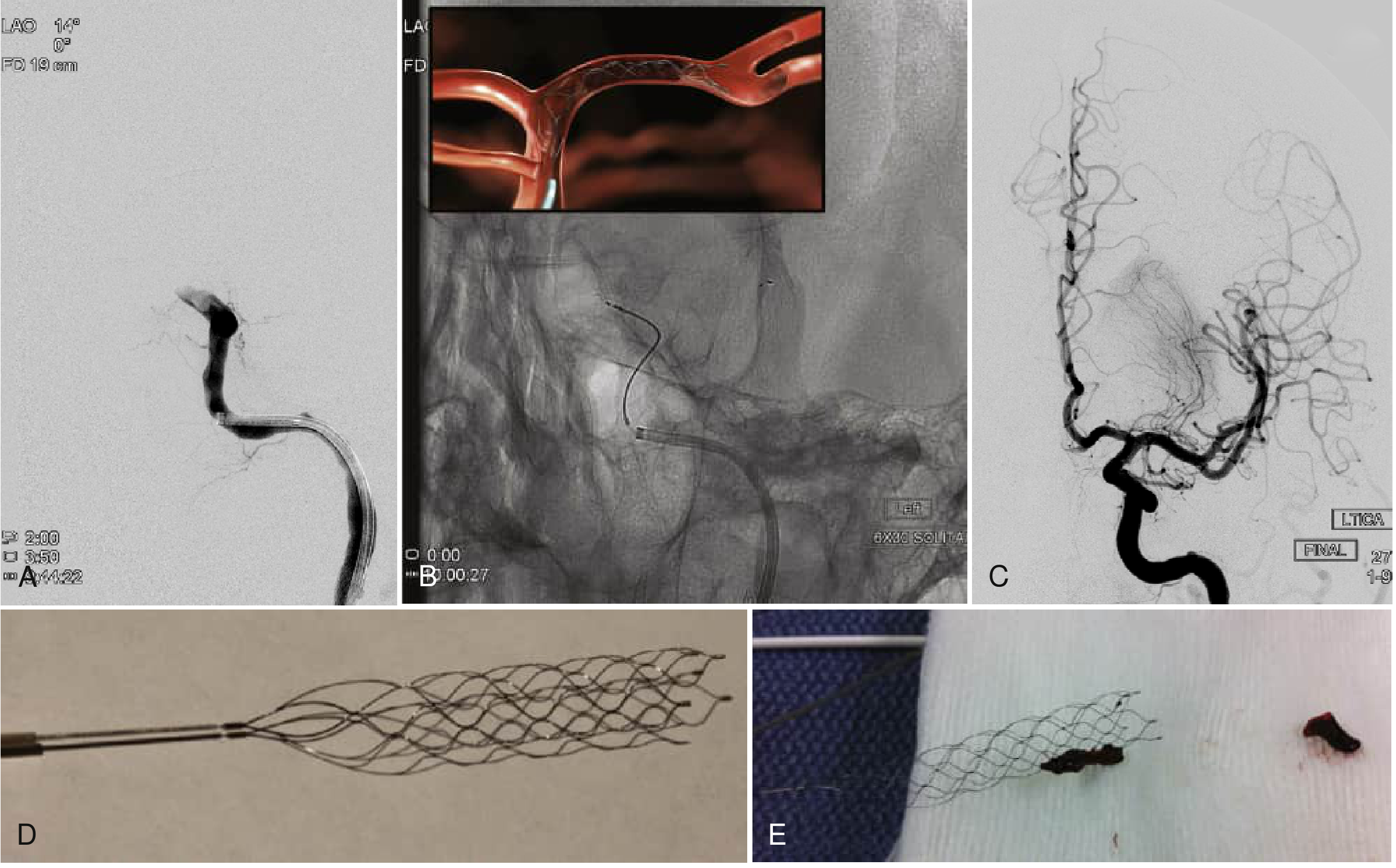
  • Stent retrievers (Solitaire, Trevo): deployed beyond the clot, then withdrawn with clot engaged in mesh
  • Large-bore aspiration catheters (Penumbra): direct suction thrombectomy - equivalent to stent retrievers in ASTER and COMPASS trials
  • Often combined: aspiration + stent retriever ("Solumbra technique")
Mechanical thrombectomy procedure: (A) DSA showing left ICA terminal occlusion; (B) Solitaire stent retriever deployed in LCA; (C) Complete recanalization after thrombectomy; (D) Solitaire FR device; (E) Clot adherent to device after retrieval
Endovascular mechanical thrombectomy: (A) Left ICA terminal occlusion on DSA; (B) Solitaire stent retriever deployed in LCA under fluoroscopy; (C) Complete recanalization of ICA and MCA post-thrombectomy; (D-E) Solitaire FR device with retrieved clot fragments. (Fuster & Hurst's The Heart, 15th Ed.)

Thrombectomy Time Windows and Criteria

Early window (0-6 hours): Five landmark 2015 trials (MR CLEAN, ESCAPE, REVASCAT, SWIFT PRIME, EXTEND-IA) established benefit. Selection: NIHSS ≥6, confirmed LVO on CTA, ASPECTS ≥6, good functional baseline (mRS 0-2).
Late window (6-24 hours) - DAWN and DEFUSE-3:
TrialWindowSelectionKey Result
DAWN (2018)6-24 hClinical-infarct mismatch (NIHSS vs core volume by CTP/DWI); ICA or M1 occlusionmRS 0-2: 49% vs 13% (NNT=2.8); stopped early for efficacy
DEFUSE-3 (2018)6-16 hPerfusion mismatch: core <70mL, Tmax>6s:core ratio ≥1.8; NIHSS ≥6mRS 0-2: 45% vs 17% (P<0.001)
"Patients who present in this delayed window are thought to be slow progressors, with leptomeningeal collaterals that help sustain salvageable brain tissue." - Fuster and Hurst's The Heart, 15th Ed.
Large core thrombectomy (new 2023-2024 evidence): Previously, ASPECTS ≤5 or large infarct core was considered a contraindication. Four recent trials (TENSION, SELECT-2, ANGEL-ASPECTS, LASTE, all 2023) demonstrated benefit of thrombectomy even with ASPECTS 3-5 and core volumes up to 100 mL - expanding eligibility significantly. Guidelines are now being updated to reflect this.
"First pass effect" - achieving complete recanalization in a single device pass - is strongly associated with better clinical outcomes and is now a benchmark for device performance.

C. Blood Pressure Management

  • Pre-tPA: Must lower BP to <185/110 mmHg before administration (IV labetalol or nicardipine); maintain <180/105 for 24h after
  • No thrombolysis/thrombectomy candidate: Permissive hypertension up to 220/120 mmHg for 24-48h (allows autoregulation in penumbra)
  • Post-thrombectomy (successful recanalization): Target <180/105 mmHg to reduce risk of reperfusion hemorrhage
  • 2025 ESO update (PMID 42095756): Reaffirms cautious approach to BP lowering in acute phase

D. Antiplatelet Therapy

  • No LVO/thrombolysis: Aspirin 300 mg loading dose within 48 hours (IST and CAST trials)
  • Post-tPA: Hold antiplatelet and anticoagulants for 24 hours (re-image first to exclude hemorrhagic transformation)
  • TIA/minor stroke: Dual antiplatelet therapy (aspirin + clopidogrel) for 21 days (POINT and CHANCE trials)

E. Neuroprotective / Supportive Measures

InterventionTargetRationale
Glucose control140-180 mg/dLBoth hyper- and hypoglycemia worsen outcome
TemperatureNormothermia (<37.5°C)Hyperthermia expands infarct
OxygenSpO2 >94%; avoid hyperoxiaSupplemental O2 only if hypoxic
PositioningHead of bed 0-30° (flat preferred acutely)Maximize CBF to penumbra
AnticoagulationAvoid in first 24-48hHemorrhagic transformation risk
DVT prophylaxisCompression stockings; LMWH after 24-48hImmobility risk

F. Functional Outcomes (Modified Rankin Scale)

mRSDescription
0No symptoms
1Minimal symptoms, no disability
2Slight disability but independent
3Moderate disability - walks independently, needs some help
4Moderately severe - unable to walk, needs assistance
5Severe - bedridden, incontinent
6Dead
mRS 0-2 = "good functional outcome" is the primary endpoint in all stroke trials.

Summary Algorithm

Suspected AIS
      ↓
NCCT (immediate) → Hemorrhage? → YES → Treat hemorrhagic stroke
      ↓ NO
ASPECTS score + CTA
      ↓
LVO identified?
   ├─ YES → IV tPA (if eligible, no contraindications) + Thrombectomy
   │                 ↑ 0-24h depending on mismatch imaging
   └─ NO  → IV tPA alone (if <4.5h, or 4.5-9h with CTP mismatch)
            Aspirin 300mg if no tPA/thrombectomy
      ↓
Supportive care: BP, glucose, temp, O2
Early rehab: physio, OT, speech therapy
Secondary prevention: antiplatelet/anticoagulant, statin, risk factor control

Sources: Bradley and Daroff's Neurology in Clinical Practice; Fuster and Hurst's The Heart 15th Ed.; Canadian Stroke Best Practices 2024; AHA/ASA Guidelines; PMC: Current state of imaging in acute ischemic stroke 2025; DAWN trial (Nogueira et al. 2018); DEFUSE-3; HERMES meta-analysis; ESO 2025 BP guidelines (PMID 42095756)
This is a shared conversation. Sign in to Orris to start your own chat.