
Gastric carcinoma
I'll start by reading the medical library skill, then search for comprehensive information on gastric carcinoma.
Reading File
Now I'll run parallel searches across headings and paragraphs for gastric carcinoma.
Finding Sources
Finding Sources
Searching PubMed
"gastric cancer"[MeSH Terms] AND "treatment"[MeSH Terms]
Excellent results. Now I'll read the most comprehensive sources in parallel - Sabiston (surgical), Yamada's (gastroenterology), and Grainger & Allison (radiology).
Reading File
Reading File
Reading File
Great content so far. Let me now read more from Sabiston (the most comprehensive surgical text) covering staging, classification, treatment and prognosis.
Reading File
Reading File
Excellent content. Let me continue reading more of the Sabiston text for classification, staging table, treatment, and prognosis sections.
Reading File
I now have comprehensive information. Let me fetch a couple of the key images to include in the answer.
Loading Image
Loading Image
Finding Sources
Now let me read the Lauren classification and molecular classification sections, plus the treatment/chemotherapy/prognosis section from Sabiston.
Reading File
I now have rich, comprehensive information from multiple authoritative textbooks. Let me compile the full answer.
Gastric Carcinoma
Epidemiology
Gastric carcinoma is the fifth most common malignancy and the third most common cause of cancer-related death worldwide. Its incidence is significantly higher in Eastern Asia (Japan, China, Korea) and Latin America than in Western countries. Peak age of onset is between 50 and 70 years. Males are more commonly affected than females (2:1 ratio for intestinal type). About 90-95% of malignant gastric tumors are adenocarcinomas; the remainder comprise lymphoma, gastrointestinal stromal tumors (GISTs), and neuroendocrine tumors.
Overall, the incidence of distal (antral) gastric cancer has been declining globally with aggressive treatment of H. pylori, but proximal (cardia) cancers have been increasing in incidence, particularly among young adults in the West.
Etiology and Risk Factors
Infectious / Medical
- Helicobacter pylori - the most important risk factor. H. pylori induces body-predominant gastritis with atrophy, driving the Correa cascade: nonatrophic gastritis → atrophic gastritis → intestinal metaplasia → dysplasia → adenocarcinoma. Virulence factors cagA and vacA co-opt the host inflammatory response to promote oncogenesis; high-risk H. pylori genotypes acting synergistically with proinflammatory IL-1 gene cluster variants can increase gastric cancer risk up to 87-fold.
- Epstein-Barr virus (EBV) - latent EBV infection in tumor cells alters host gene expression via encoded microRNAs and inducing hypermethylation of host DNA. EBV-associated tumors constitute a distinct molecular subtype (~10% of all gastric cancers).
- Atrophic gastritis and intestinal metaplasia
- Pernicious anemia (achlorhydria from autoimmune destruction of parietal cells)
- Adenomatous polyps
- Prior gastric surgery (increases risk ~3-fold after >15 years)
- Long-term PPI use
- Prior abdominal radiation
Nutritional / Dietary
- High nitrate and N-nitroso compound consumption (smoked, cured, pickled, and salted foods)
- Low fresh fruit and vegetable intake (ascorbic acid inhibits conversion of nitrites to N-nitroso compounds)
- High salt intake
Environmental / Social
- Lack of refrigeration (historically linked to preserved foods)
- Contaminated drinking water
- Smoking (~1.5-fold increased risk)
- Low socioeconomic status
Hereditary
- Hereditary Diffuse Gastric Cancer (HDGC): germline mutation in CDH1 (E-cadherin gene). Lifetime risk: 37-42% in males, 25-33% in females for gastric cancer; 39-55% risk of lobular breast cancer in females. Prophylactic total gastrectomy is recommended between ages 20-30 for fit individuals with significant risk.
- Lynch syndrome, familial adenomatous polyposis (FAP), Peutz-Jeghers syndrome, juvenile polyposis, Li-Fraumeni syndrome are also associated with increased risk.
- Blood type A is associated with diffuse-type gastric cancer.
Pathology
Gross Classification (Borrmann, 1926)
| Type | Appearance |
|---|---|
| I | Polypoid |
| II | Fungating |
| III | Ulcerating |
| IV | Diffusely infiltrating (linitis plastica) |
Histologic Classification: Lauren (1965)
| Feature | Intestinal Type | Diffuse Type |
|---|---|---|
| Differentiation | Well differentiated, forms glands | Poorly differentiated, signet ring cells |
| Precondition | Atrophic gastritis, intestinal metaplasia | Not associated with chronic gastritis |
| Sex | Male predominant | Equal sex distribution |
| Age | Older | Slightly younger |
| Spread | Hematogenous (liver) | Peritoneal (carcinomatosis) |
| Association | H. pylori endemic areas | Blood group A |
| Prognosis | Better | Worse |
| Mixed type | Both components; intermediate to poor prognosis | - |
The modified Lauren classification (combining Lauren type + tumor location as diffuse, proximal nondiffuse, or distal nondiffuse) is a better predictor of survival than histology alone: distal nondiffuse has the best prognosis; diffuse has the worst.
WHO Classification
- Tubular (corresponds to Lauren intestinal type)
- Papillary (corresponds to Lauren intestinal type)
- Mucinous
- Poorly cohesive, including signet ring cell carcinoma (corresponds to Lauren diffuse type)
- Mixed carcinoma
Molecular Subtypes (TCGA/ACRG)
The Cancer Genome Atlas (TCGA) defined four molecular subtypes:
- EBV-positive - frequent PIK3CA mutations, extreme DNA hypermethylation, amplification of JAK2/PD-L1/PD-L2
- Microsatellite Instability (MSI) - elevated mutation rate, hypermethylation of MLH1; best prognosis; responds to immune checkpoint inhibitors
- Genomically Stable (GS) - enriched for diffuse histology, CDH1 and RHOA mutations
- Chromosomal Instability (CIN) - most common in proximal stomach/EGJ; TP53 mutations, RTK-RAS amplifications (EGFR, VEGFR, FGFR, HER2); worst prognosis
Clinical Features
Symptoms are often non-specific in early disease (which is why most Western patients present at an advanced stage):
- Early: dyspepsia, epigastric discomfort, nausea
- Late: weight loss, anorexia, early satiety, dysphagia (proximal tumors), vomiting (antral obstruction), hematemesis/melena, abdominal mass
Paraneoplastic signs:
- Virchow's node - left supraclavicular lymph node (Troisier's sign)
- Sister Mary Joseph nodule - periumbilical metastasis
- Krukenberg tumor - ovarian metastasis via transcoelomic spread
- Blumer's shelf - rectal shelf on digital rectal exam (pouch of Douglas metastasis)
- Acanthosis nigricans
- Dermatomyositis
Early Gastric Cancer (EGC)
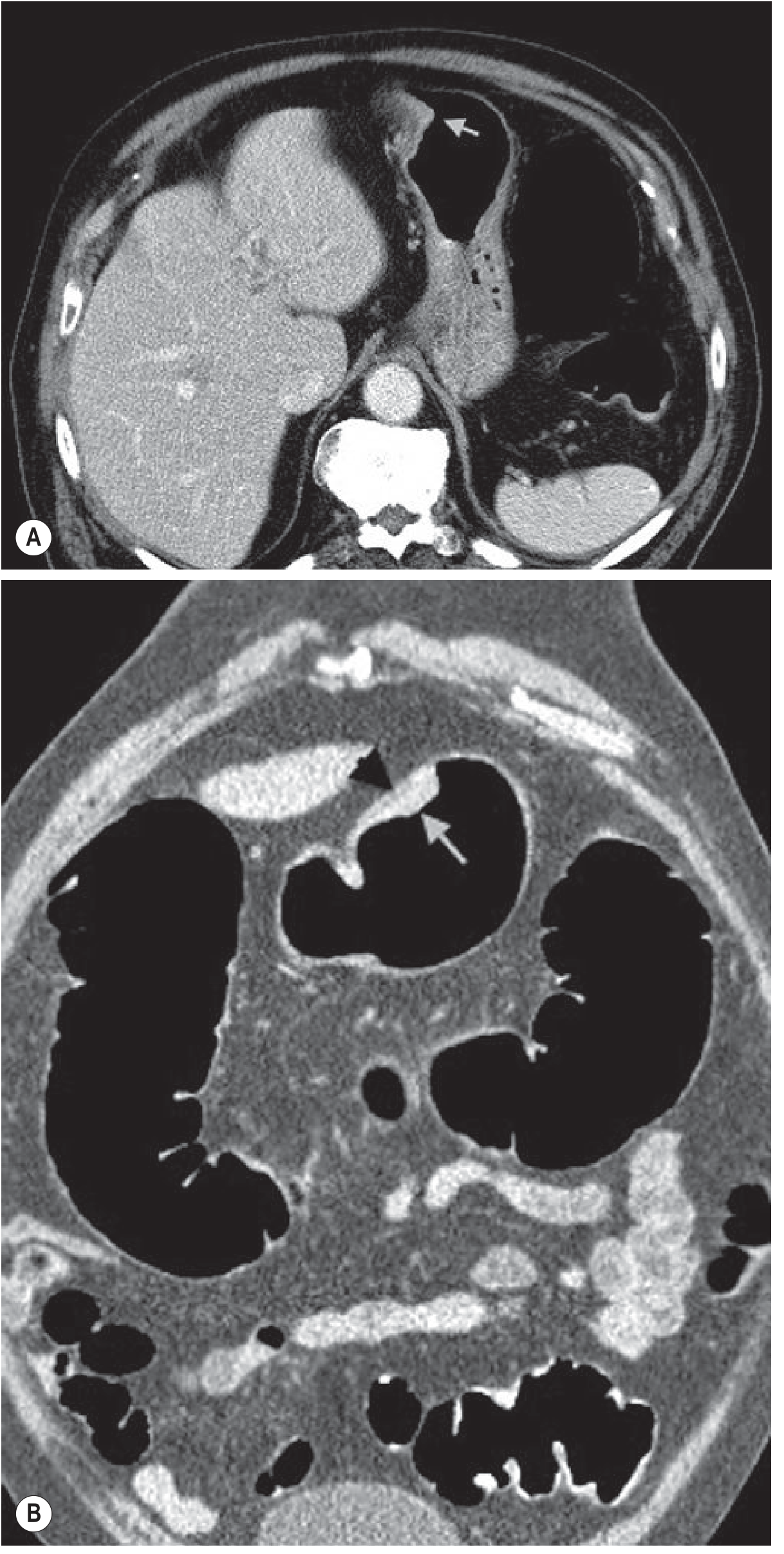
EGC is defined as tumor confined to the mucosa and submucosa, regardless of nodal status (T1, any N). It is curable, with 5-year survival rates up to 95%. The Japanese classification system:
- Type I - Polypoid (>5 mm, protruding)
- Type IIa - Superficial elevated (<5 mm)
- Type IIb - Flat
- Type IIc - Depressed
- Type III - Excavated
- Type IV - Lateral spreading
CT appearance of T1 lesion: focal thickening of the inner layer with preserved low-attenuation outer layer and clear perigastric fat plane.
Staging (AJCC/UICC TNM, 8th Edition)
Key update in the 8th edition: tumors at the esophagogastric junction (EGJ) with epicenter ≤2 cm into the proximal stomach are staged as esophageal cancer; those with epicenter >2 cm into the proximal stomach are staged as gastric cancer.
| T Stage | Description |
|---|---|
| T1a | Invades lamina propria or muscularis mucosae |
| T1b | Invades submucosa |
| T2 | Invades muscularis propria |
| T3 | Invades subserosa |
| T4a | Perforates serosa |
| T4b | Invades adjacent structures |
N staging: N1 (1-2 nodes), N2 (3-6 nodes), N3a (7-15 nodes), N3b (>15 nodes)
The most important prognostic factors in resectable disease are depth of invasion (T stage) and presence/absence of nodal metastases.
Imaging
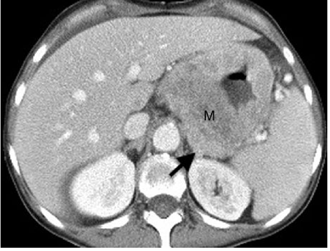
CT Scan
- Modality of choice for staging and surgical planning
- Assessment of depth of invasion (T stage), nodal involvement (N stage), and distant metastases (M stage)
- Perigastric fat stranding, loss of fat planes, and direct organ invasion indicate advanced T4 disease
Barium Studies
- Double-contrast barium: sensitivity ~96% for gastric cancer when performed carefully
- Linitis plastica (leather bottle stomach): diffuse scirrhous tumor causing rigid, narrowed stomach - better appreciated on fluoroscopy than on endoscopy
Endoscopy
- Diagnostic procedure of choice; allows biopsy
- Sensitivity of double-contrast barium can approach endoscopy when performed expertly
Treatment
Endoscopic Resection (Early Gastric Cancer)
Standard criteria for endoscopic resection:
- Intestinal-type adenocarcinoma
- Confined to mucosa (Tis or T1a)
- Absence of lymphovascular invasion
- Non-ulcerated tumor
- < 2 cm in diameter
Two methods:
- EMR (Endoscopic Mucosal Resection): uses saline injection + snare; ~15% bleeding rate; perforation is rare
- ESD (Endoscopic Submucosal Dissection): allows larger tumors and limited submucosal involvement; more technically challenging; higher perforation risk; widely used in East Asia. Expanded criteria (>2 cm without ulceration, ≤3 cm with ulceration, or non-intestinal <2 cm without ulceration) have ~0.7% lymph node involvement risk.
Patients with positive vertical margins, lymphovascular invasion, or submucosal invasion after endoscopic resection should be referred for gastrectomy with lymphadenectomy.
Surgery
Resection margins:
- cT1: proximal margin ≥2 cm
- cT2-T4 (non-infiltrative): ≥3 cm
- cT2-T4 (infiltrative): ≥5 cm
Extent of resection by location:
- Distal stomach (body/antrum): distal gastrectomy
- Proximal/fundus or diffuse: total gastrectomy
- T4 tumors: en-bloc resection of invaded organs
Lymphadenectomy:
- D1: perigastric nodes
- D2: additionally includes celiac axis, left gastric, common hepatic, and splenic nodes (standard in Japan; recommended for resectable disease in Western guidelines for curative intent)
Palliative surgery: for outlet obstruction, hemorrhage, or perforation in unresectable disease.
Perioperative Chemotherapy / Multimodal Treatment
- Perioperative chemotherapy (FLOT regimen - 5-FU, leucovorin, oxaliplatin, docetaxel) is current standard of care for resectable locally advanced gastric cancer in Western countries, based on the FLOT4 trial (improved OS vs. ECF/ECX).
- Postoperative chemoradiotherapy (MacDonald regimen: 5-FU/leucovorin + radiation) - used in the US after D0/D1 resection.
- Adjuvant chemotherapy with S-1 or capecitabine + oxaliplatin is standard in East Asian patients (CLASSIC, ACTS-GC trials).
Targeted Therapy and Immunotherapy
- HER2 overexpression (~15-20% of gastric cancers): trastuzumab (anti-HER2) + chemotherapy improves OS in HER2+ metastatic disease (ToGA trial).
- Anti-VEGFR2: ramucirumab - second-line treatment.
- PD-L1 / MSI-H tumors: immune checkpoint inhibitors (nivolumab, pembrolizumab); particularly effective in MSI-H/EBV-positive subtypes.
- FGFR2b amplification: futibatinib / bemarituzumab (investigational).
Palliative Chemotherapy (Metastatic Disease)
- First-line: FOLFOX, FLOT, or cisplatin/5-FU-based regimens
- HER2+: add trastuzumab
- Second-line: ramucirumab ± paclitaxel, irinotecan, docetaxel
Prophylactic Surgery (HDGC)
For germline CDH1 mutation carriers with significant risk, prophylactic total gastrectomy between ages 20-30 years is recommended. In those who refuse or wish to delay, annual endoscopy with Cambridge protocol biopsies (≥6 random biopsies from 5 anatomic regions) is recommended.
Prognosis
- Overall 5-year survival: 10-30% for advanced disease
- Early gastric cancer (EGC): 5-year survival up to 95% after curative resection
- Key prognostic factors:
- T stage (depth of invasion)
- N stage (number of positive lymph nodes - the more nodes resected, the better the staging accuracy; D2 lymphadenectomy retrieves ≥16 nodes as per guidelines)
- M stage (liver metastases from intestinal type; peritoneal carcinomatosis from diffuse type)
- Lauren/molecular subtype (MSI-H best; diffuse/GS worst)
- Tumor location (distal nondiffuse best, diffuse worst by modified Lauren)
- Most Western patients present at advanced stage due to lack of screening programs, explaining the poor overall survival.
Screening
- Japan/Korea: national mass screening programs using photofluorography (barium), endoscopy, H. pylori antibody testing, and serum pepsinogen - responsible for detection of >60% of tumors as EGC in Japan.
- Western countries: No population-based screening; upper endoscopy or double-contrast barium for symptomatic patients.
- Surveillance recommended for high-risk groups: known H. pylori gastritis with atrophy/metaplasia, hereditary syndromes, prior gastric surgery.
Sources:
- Sabiston Textbook of Surgery, 21st ed., Chapter 87
- Grainger & Allison's Diagnostic Radiology, 7th ed., Chapter 20
- Yamada's Textbook of Gastroenterology, 7th ed., Chapter 130
- Park's Textbook of Preventive and Social Medicine
This is a shared conversation. Sign in to Orris to start your own chat.