
Circle of willis
Reading File
The Circle of Willis is the arterial anastomotic ring at the base of the brain formed by interconnected cerebral and communicating arteries. It supplies blood to the cerebral hemispheres and acts as a collateral network when one vessel is compromised.
Searching sources...
Finding Sources
Finding Sources
Good results. Now let me read the dedicated neuroanatomy section and grab the diagram image from Clinical Anesthesia.
Reading File
Reading File
Reading File
Excellent content gathered. Now let me fetch the key diagram images and read the remaining content on branches/clinical correlations.
Loading Image
Loading Image
Reading File
I now have comprehensive, well-sourced content with excellent diagrams. Here is the full answer:
Circle of Willis
Overview
The Circle of Willis is an anastomotic arterial ring located at the base of the brain in the subarachnoid space. It connects the anterior circulation (from the internal carotid arteries) with the posterior circulation (from the vertebrobasilar system), forming a collateral network that can redistribute blood if one vessel is occluded. A complete, full-caliber ring is present in only ~34% of individuals; anatomical variants and hypoplasia are very common. - Neuroanatomy through Clinical Cases, 3rd ed.
Diagrams
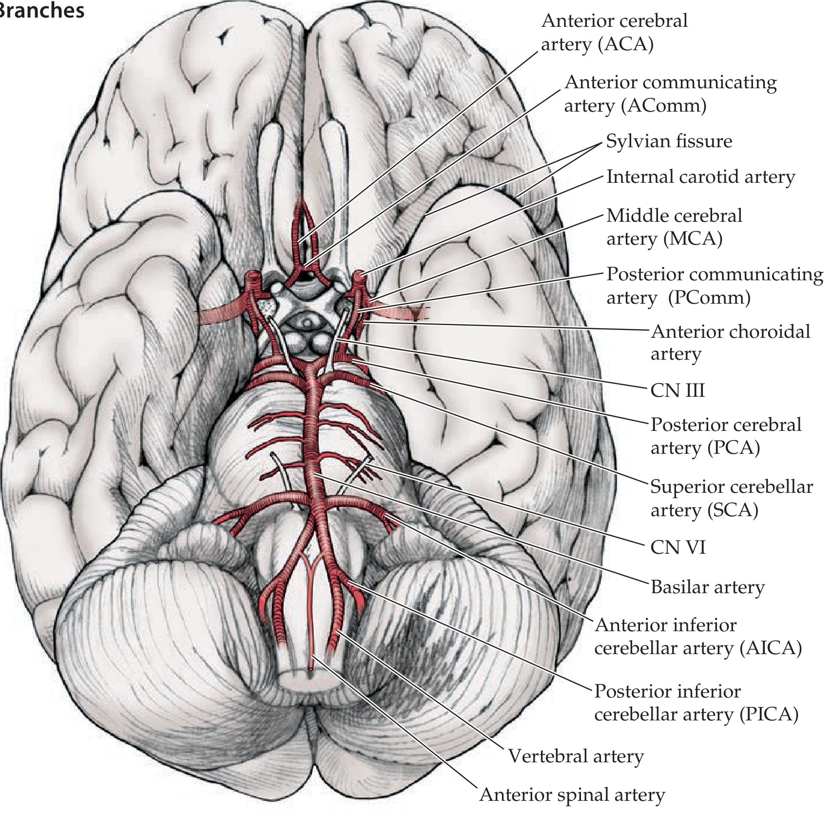
Circle of Willis and its main branches (view from below the brain):
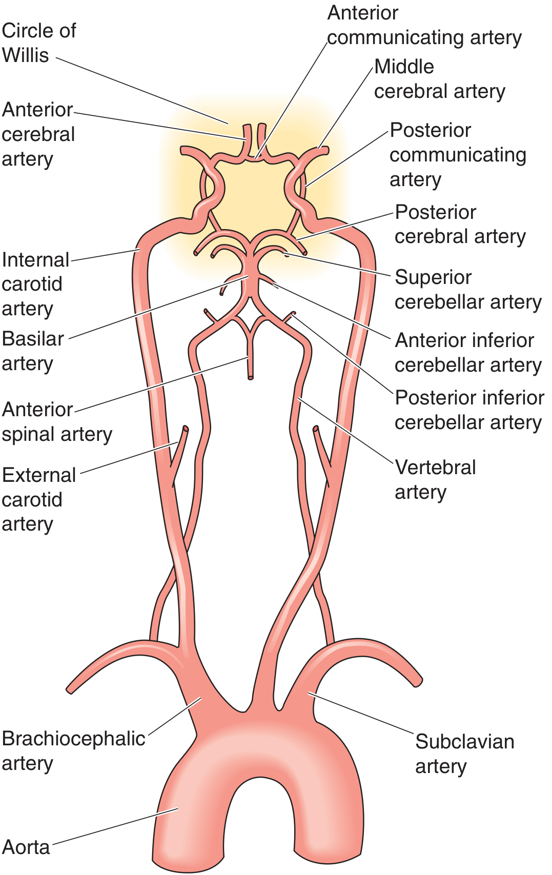
Full arterial supply from aorta to circle (schematic):
Arterial Inputs
The circle receives blood from two sources:
| Source | Arteries | Origin |
|---|---|---|
| Anterior circulation | Bilateral internal carotid arteries (ICA) | Common carotid arteries (from aorta/brachiocephalic trunk) |
| Posterior circulation | Bilateral vertebral arteries | Subclavian arteries - ascend through cervical vertebral foramina transversaria - join at pontomedullary junction to form the basilar artery |
Components of the Ring
The circle itself is formed by 7 vessels (bilaterally paired where applicable):
| Vessel | Abbreviation | Part of Ring |
|---|---|---|
| Anterior communicating artery | AComm | Anterior - connects the two ACAs |
| Anterior cerebral artery (x2) | ACA | Anterolateral |
| Internal carotid artery (x2) | ICA | Lateral inflow |
| Posterior communicating artery (x2) | PComm | Connects ICA to PCA - links anterior and posterior circulations |
| Posterior cerebral artery (x2) | PCA | Posterior - arise from the top of the basilar artery |
Major Branches Arising from the Ring
From the circle and its immediate feeders, the three main cerebral arteries supply the hemispheres:
1. Anterior Cerebral Artery (ACA)
- Passes anteriorly into the interhemispheric fissure, sweeps back over the corpus callosum
- Supplies: medial frontal and parietal cortex, medial sensorimotor cortex (legs/feet representation)
- Deep branches: medial lenticulostriate arteries (basal ganglia)
2. Middle Cerebral Artery (MCA)
- Largest branch of the ICA; turns laterally into the Sylvian fissure
- Bifurcates into superior and inferior divisions
- Supplies: lateral frontal, parietal, temporal cortex - includes primary motor/sensory cortex for the face and arm, Broca's area, Wernicke's area
- Deep branches: lateral lenticulostriate arteries (internal capsule, putamen, caudate)
3. Posterior Cerebral Artery (PCA)
- Arises from the top of the basilar artery; also receives contribution via PComm
- Supplies: occipital lobe (primary visual cortex), medial temporal lobe (hippocampus), thalamus
- Deep branches: thalamoperforating arteries
Other branches from the ICA (mnemonic OPAAM):
- Ophthalmic artery - retinal supply
- Posterior communicating artery (PComm)
- Anterior choroidal artery - supplies posterior limb of internal capsule, optic tract, choroid plexus
- Anterior cerebral artery (ACA)
- Middle cerebral artery (MCA) - Neuroanatomy through Clinical Cases, 3rd ed.
Vascular Territories at a Glance
| Artery | Cortical territory | Deep territory |
|---|---|---|
| ACA | Medial frontal/parietal (leg area) | Medial basal ganglia |
| MCA | Lateral frontal, parietal, temporal | Internal capsule, lateral BG |
| PCA | Occipital lobe, medial temporal | Thalamus |
Collateral Function
The circle provides collateral flow in three ways:
- Circle of Willis itself - connects carotid and vertebrobasilar systems
- Extracranial-intracranial anastomoses - e.g., ophthalmic artery collaterals
- Leptomeningeal anastomoses - between terminal branches of ACA, MCA, and PCA at watershed zones
Patients with complete proximal carotid occlusion can sometimes tolerate it if the circle is intact, as blood reaches the hemisphere via the contralateral ICA through the AComm, or from the vertebrobasilar system through the PComm. - Schwartz's Principles of Surgery, 11th ed. | Localization in Clinical Neurology, 8th ed.
Clinical Correlations
| Condition | Mechanism |
|---|---|
| Berry (saccular) aneurysm | Most common at arterial junctions of the circle - AComm is the single most frequent site, followed by PComm-ICA junction. Rupture causes subarachnoid hemorrhage. |
| MCA stroke | Contralateral face/arm weakness, aphasia (dominant hemisphere), neglect (non-dominant) |
| ACA stroke | Contralateral leg weakness, abulia, incontinence |
| PCA stroke | Contralateral homonymous hemianopia, thalamic pain, memory deficits |
| PComm aneurysm | Compresses CN III (runs adjacent) - ipsilateral pupil dilation, ptosis, "down and out" eye |
| Carotid endarterectomy | Brain depends on collateral flow from an intact circle when the ICA is cross-clamped. Autopsy studies show majority of specimens have anatomical anomalies in the circle. - Clinical Anesthesia, 9th ed. |
Imaging
The circle is best visualized with MR angiography (MRA) or CT angiography (CTA). Both reliably demonstrate the ring and any anatomical variants such as hypoplastic vessels or absent segments. - Grainger & Allison's Diagnostic Radiology
Images of all transverse section at different level in neuroanatomy 1st year
Reading File
This is a visual/anatomy query - image retrieval is the priority. Let me search for transverse sections of the brainstem and spinal cord across the library simultaneously.
Finding Sources
Finding Sources
Finding Sources
Finding Sources
Finding Sources
Finding Sources
Gray's Anatomy for Students has dedicated internal/external medulla sections. Let me read those plus search for pons, midbrain, and spinal cord in parallel.
Reading File
Finding Sources
Finding Sources
Excellent - all sections are in the same block file. Let me read the internal midbrain, pons, medulla, and spinal cord sections to harvest all the diagram image URLs.
Reading File
Reading File
I have the image URLs now. Let me read more to get all the remaining section images (caudal midbrain, medulla sections, and spinal cord cross-sections).
Reading File
Reading File
Now I have a comprehensive set of image URLs from the sections. Let me now fetch all the key transverse section diagrams in parallel - midbrain, pons, medulla, and spinal cord levels.
Loading Image
Loading Image
Reading File
I have all the image URLs. Let me now fetch all remaining section images in parallel - pons levels, caudal midbrain, medulla levels, and spinal cord cross-section.
Loading Image
Loading Image
Loading Image
Reading File
Now let me fetch the remaining images - caudal pons, rostral & mid medulla, and spinal cord cross-section.
Loading Image
Loading Image
Loading Image
Reading File
I now have all the key transverse section images. Let me get the spinal cord cross-section image too.
Reading File
Loading Image
I now have all images needed. Here is the complete, image-rich answer:
Transverse Sections of the CNS - Neuroanatomy (1st Year)
All sections below are from Gray's Anatomy for Students and Neuroanatomy through Clinical Cases, 3rd ed. (Crossman/Nolte), arranged rostral to caudal.
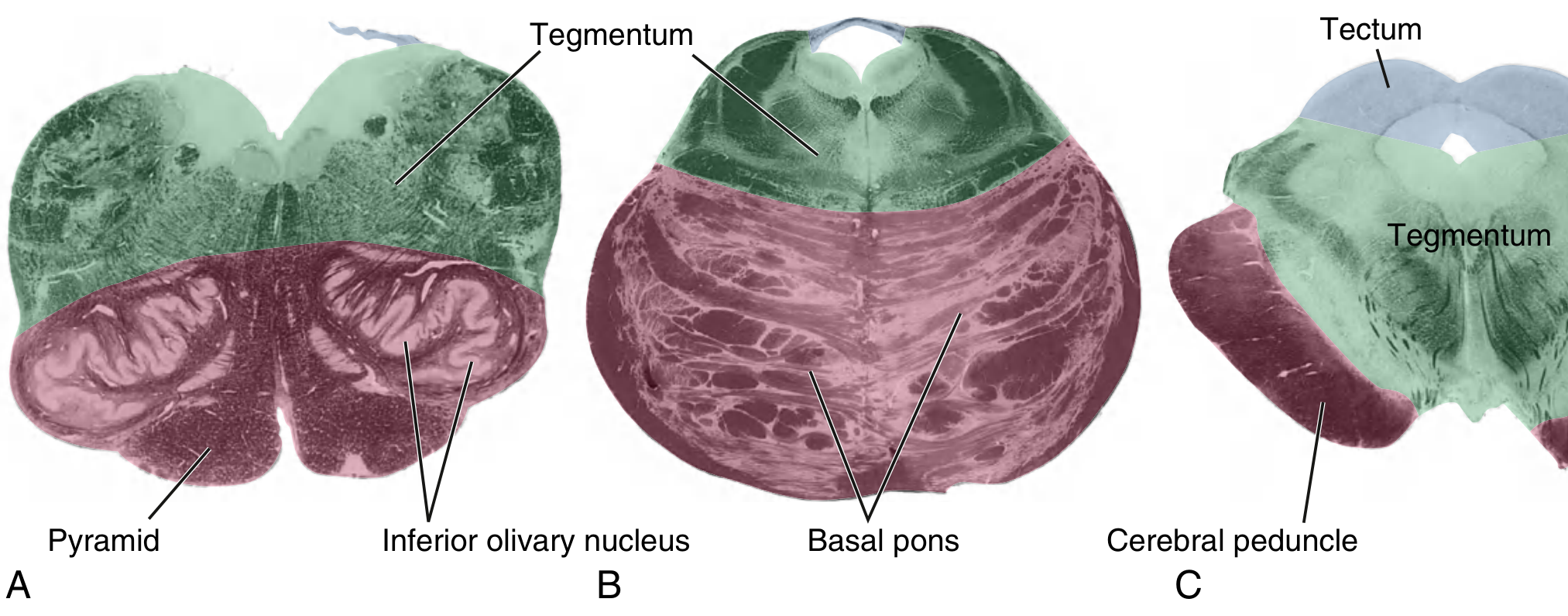
Overview: Brainstem Compartments
Every brainstem level is divided into three zones:
| Zone | Location | Contents |
|---|---|---|
| Tectum ("roof") | Dorsal - midbrain only | Superior/inferior colliculi |
| Tegmentum ("covering") | Dorsal - all levels | Cranial nerve nuclei, reticular formation, tracts |
| Basis | Ventral | Descending motor fibers (corticospinal, corticobulbar) |
1. Rostral Midbrain (Level of Superior Colliculus)
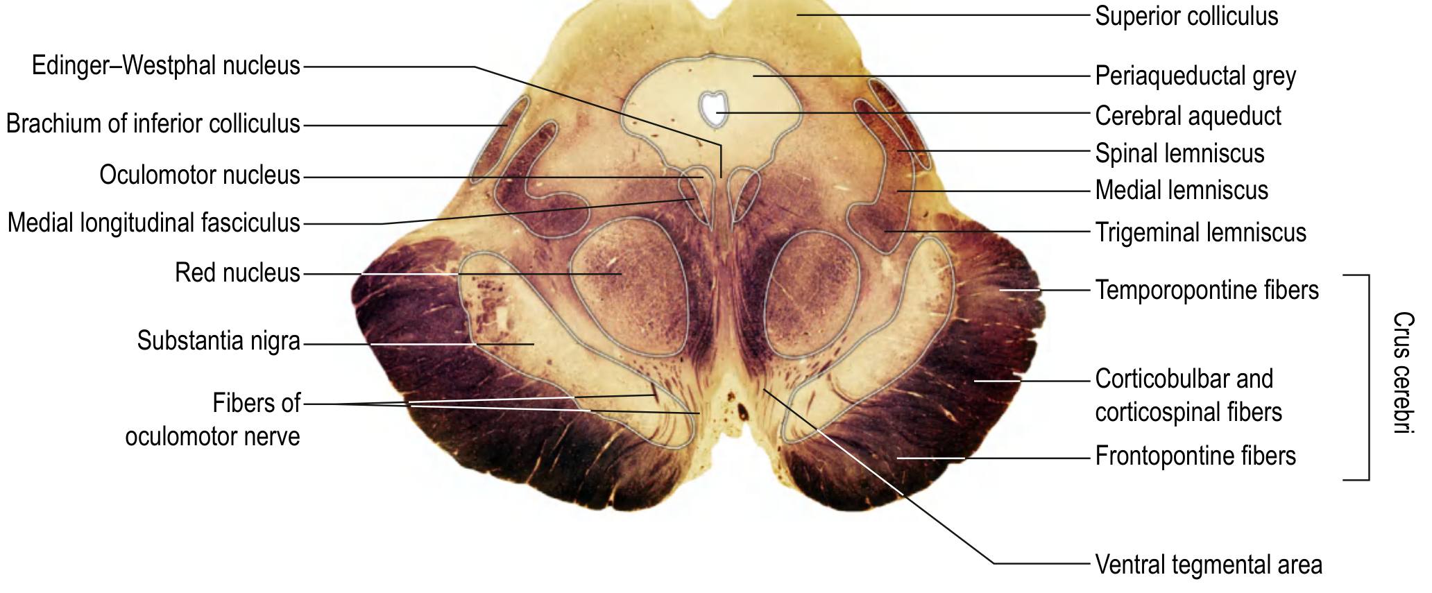
Key structures to identify:
| Structure | Notes |
|---|---|
| Superior colliculus | Tectum - visual reflex |
| Cerebral aqueduct | CSF passage |
| Periaqueductal grey (PAG) | Pain modulation |
| Oculomotor nucleus (CN III) | Near midline, ventral to PAG |
| Edinger-Westphal nucleus | Parasympathetic to pupil |
| Red nucleus | Rubrospinal tract origin |
| Substantia nigra | Dopaminergic; basal ganglia circuit |
| Crus cerebri (cerebral peduncle) | Corticospinal + corticobulbar fibers |
| Medial lemniscus | Fine touch/proprioception ascending |
| MLF (medial longitudinal fasciculus) | Coordinates eye movements |
2. Pontine-Mesencephalic Junction (Level of Inferior Colliculus)
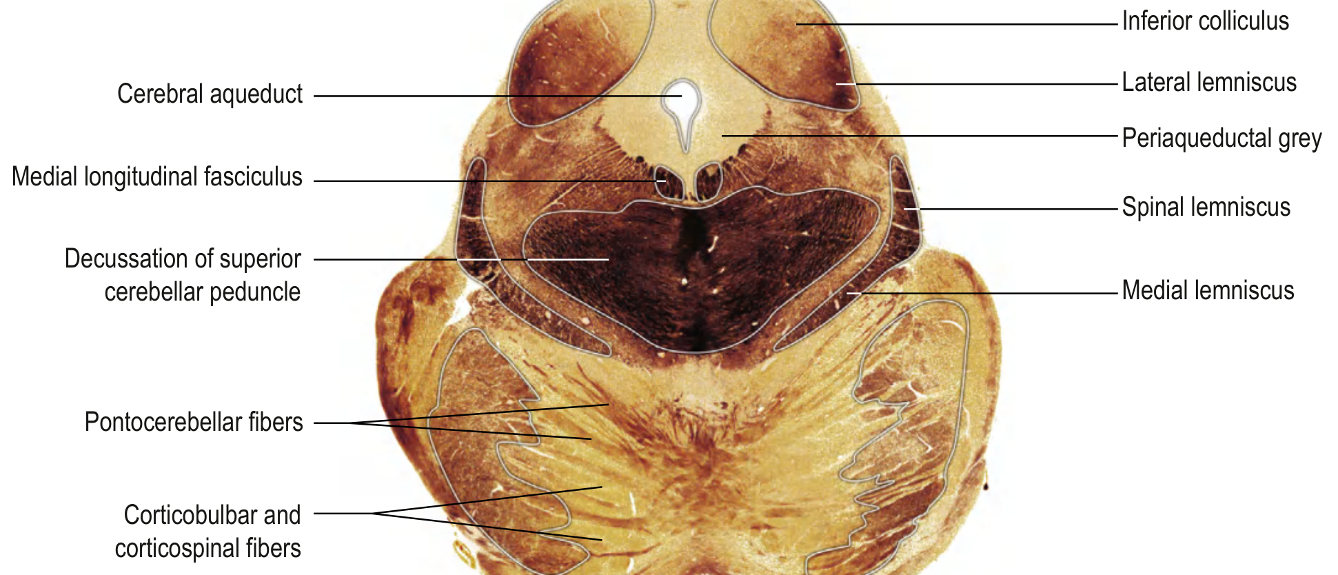
Key structures:
- Inferior colliculus (auditory reflex)
- Decussation of superior cerebellar peduncle (dentatorubrothalamic fibers crossing)
- Lateral lemniscus (auditory pathway)
- Cerebral aqueduct + periaqueductal grey
- Medial lemniscus and spinal lemniscus
- Pontocerebellar fibers
3. Rostral Pons
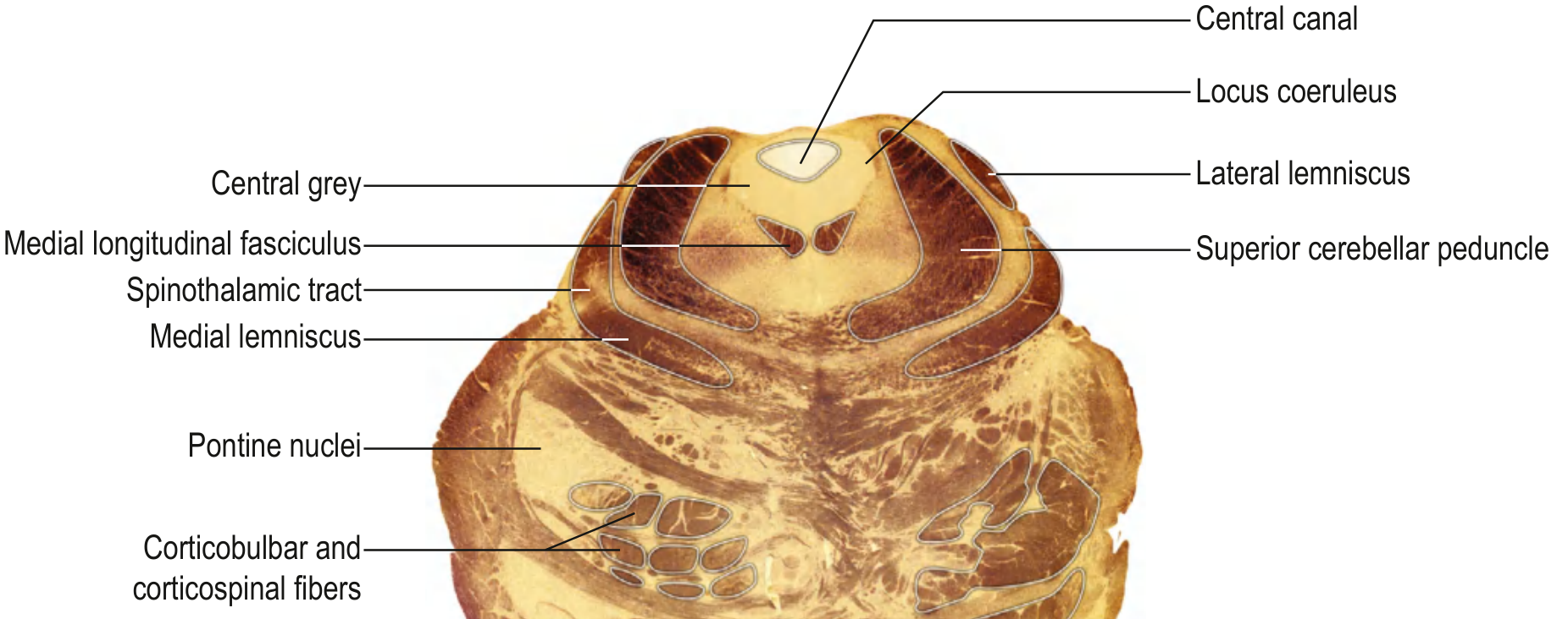
Key structures:
| Structure | Notes |
|---|---|
| Central grey | Around 4th ventricle |
| Locus coeruleus | Noradrenergic nucleus |
| Superior cerebellar peduncle | Lateral wall of 4th ventricle |
| MLF | Near midline |
| Medial lemniscus | Horizontal slab between tegmentum and basis |
| Spinothalamic tract | Lateral to medial lemniscus |
| Pontine nuclei | Relay cortex → cerebellum |
| Corticospinal fibers | Scattered in basal pons |
4. Mid-Pons (Level of CN V - Trigeminal)
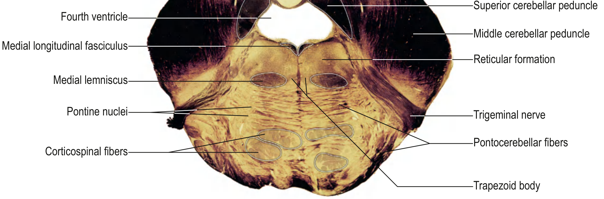
Key structures:
- Fourth ventricle expanding
- Middle cerebellar peduncle (largest peduncle - bridges to cerebellum)
- Motor nucleus of CN V + Principal sensory nucleus of CN V
- Trigeminal nerve exiting laterally
- MLF near midline
- Medial lemniscus
- Pontine nuclei + Pontocerebellar fibers
- Trapezoid body (auditory)
- Reticular formation
5. Caudal Pons (Level of CN VI - Abducens)
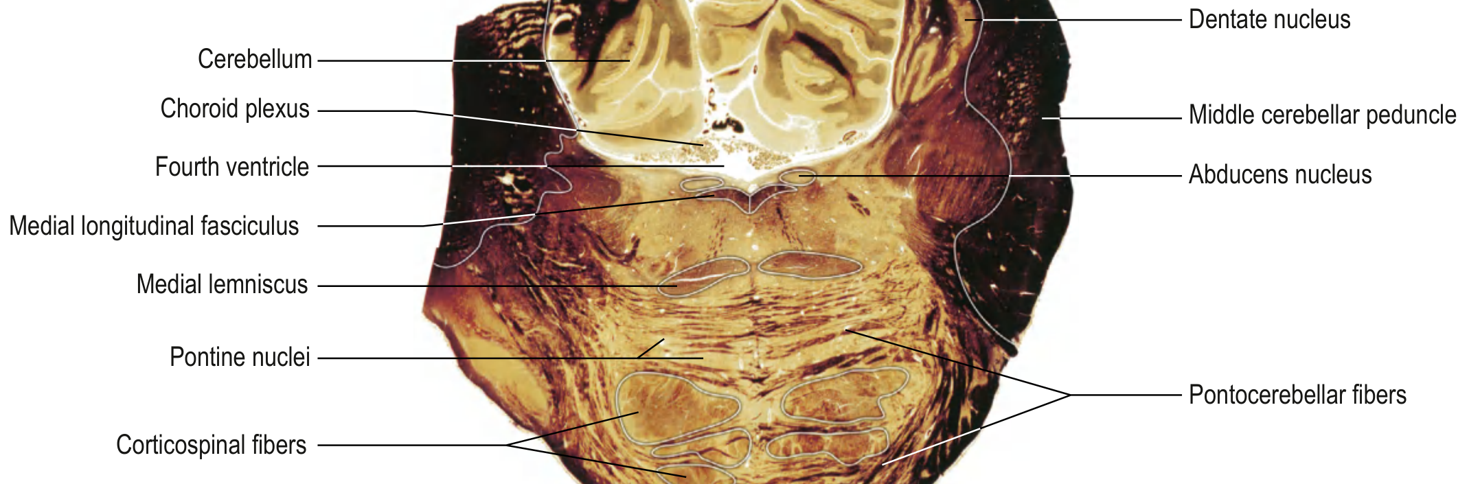
Key structures:
- Fourth ventricle with cerebellum and choroid plexus dorsal
- Abducens nucleus (CN VI) - just lateral to MLF
- Middle cerebellar peduncle
- Dentate nucleus of cerebellum visible dorsally
- Medial vestibular nucleus
- MLF and Medial lemniscus
- Corticospinal fibers + Pontocerebellar fibers
6. Rostral Medulla (Level of Inferior Olivary Nucleus)
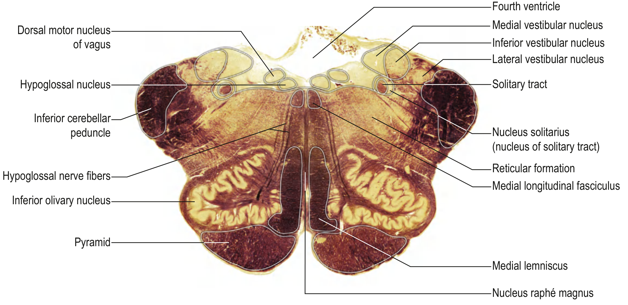
Key structures:
| Structure | Notes |
|---|---|
| Inferior olivary nucleus | Large, convoluted - landmark of rostral medulla |
| Pyramid | Corticospinal fibers anteriorly |
| Hypoglossal nucleus (CN XII) | Near midline |
| Dorsal motor nucleus of vagus (CN X) | Lateral to CN XII nucleus |
| Solitary tract + nucleus solitarius | Taste and visceral afferents |
| Vestibular nuclei (medial, inferior, lateral) | CN VIII |
| Inferior cerebellar peduncle | Lateral |
| Medial lemniscus | Vertical, near midline |
| MLF | |
| Nucleus raphé magnus | Serotonergic pain modulation |
| Reticular formation |
7. Mid-Medulla (Level of Internal Arcuate Fibers / Sensory Decussation)
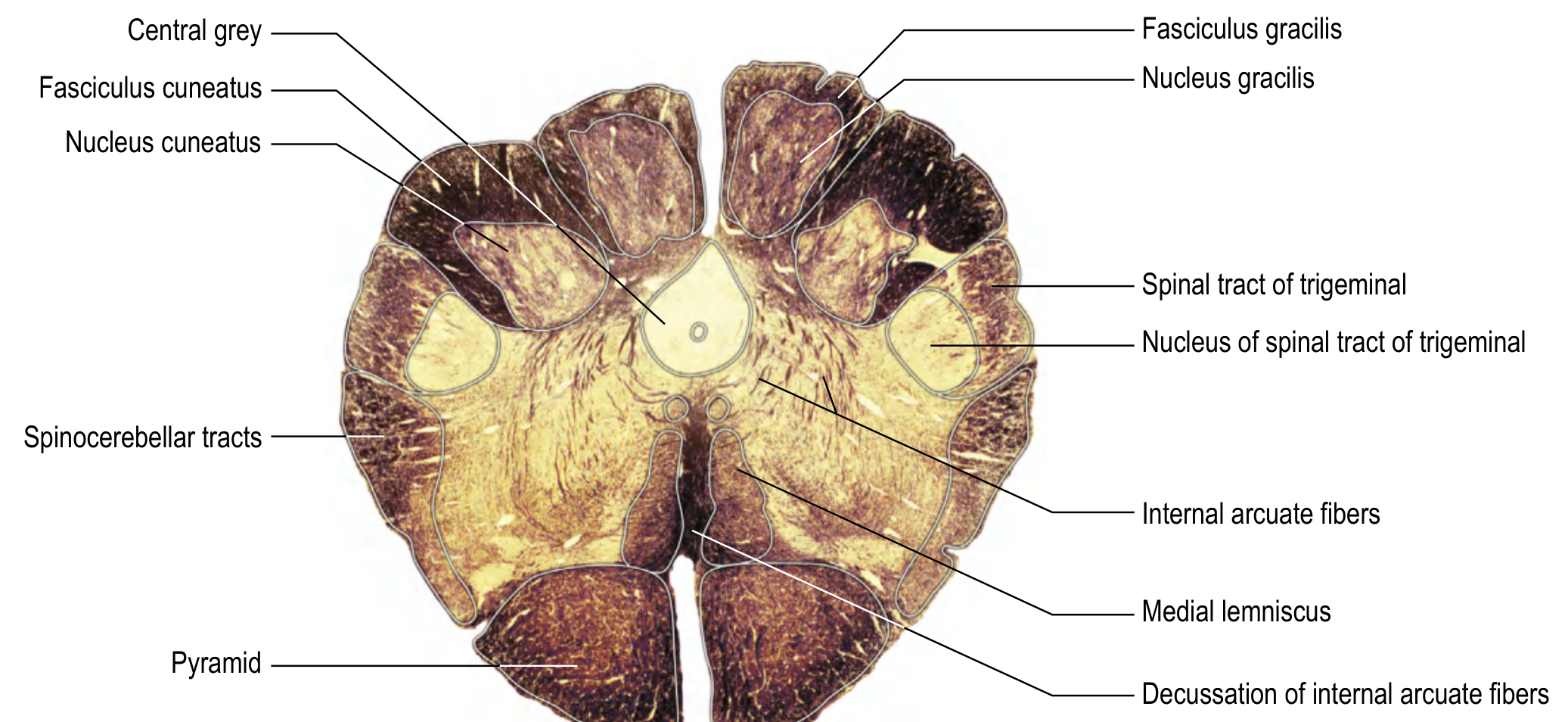
Key structures:
- Nucleus gracilis (medial) and Nucleus cuneatus (lateral) - 2nd-order neurons of posterior column pathway
- Fasciculus gracilis and Fasciculus cuneatus - ascending dorsal column fibers
- Internal arcuate fibers - axons of gracile/cuneate nuclei decussating to form the medial lemniscus
- Decussation of internal arcuate fibers - sensory decussation (fine touch crosses here)
- Spinal tract and nucleus of trigeminal (CN V)
- Spinocerebellar tracts laterally
- Pyramid anteriorly
8. Spinal Cord - Cross Section (C8 Cervical Segment)
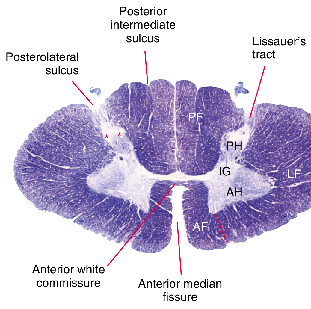
Key structures:
| Structure | Abbreviation | Location |
|---|---|---|
| Posterior funiculus | PF | Dorsal white matter (fine touch, proprioception) |
| Posterior horn | PH | Dorsal grey - receives sensory input |
| Intermediate grey | IG | Contains lateral horn at T1-L2 (sympathetic) |
| Anterior horn | AH | Motor neurons |
| Anterior funiculus | AF | Ventral white matter |
| Lateral funiculus | LF | Mixed motor (corticospinal) + sensory (spinothalamic) |
| Anterior white commissure | - | Crossing of spinothalamic fibers |
| Anterior median fissure | - | Deep midline cleft anteriorly |
| Lissauer's tract | - | Posterolateral; pain fibers ascend 1-2 levels before synapsing |
| Substantia gelatinosa (*) | - | Apex of posterior horn; pain gate |
The internal grey matter follows an H-shape, further subdivided into Rexed's laminae I-X (I = superficial dorsal horn, IX = motor neurons, X = central canal zone).
Quick Summary: What to Look for at Each Level
| Level | Landmark Structure | CN present |
|---|---|---|
| Rostral midbrain | Superior colliculus, Red nucleus, Substantia nigra | CN III |
| Caudal midbrain | Inferior colliculus, SCP decussation | CN IV (exits dorsally) |
| Rostral pons | Locus coeruleus, SCP | - |
| Mid-pons | CN V nuclei, MCP | CN V |
| Caudal pons | Abducens nucleus, Dentate | CN VI, VII |
| Rostral medulla | Inferior olivary nucleus | CN VIII-XII |
| Mid-medulla | Sensory decussation, Gracile/Cuneate nuclei | - |
| Caudal medulla | Pyramidal decussation (motor cross here) | - |
| Spinal cord | H-shaped grey, Rexed laminae | - |
Source: Gray's Anatomy for Students (Crossman), Neuroanatomy through Clinical Cases 3rd ed. (Blumenfeld)
This is a shared conversation. Sign in to Orris to start your own chat.