
Kaisay Pata Chalay Ga Left Or Right Ventricle Or Atria Mein Issue Hai
Reading File
Yeh ek broad cardiac diagnostic question hai. Pehle quick answer deta hoon, phir medical library se detailed information lata hoon.
Quick Answer: Dil ke chambers (Left/Right Ventricle aur Atria) mein issue identify karne ke liye doctors ECG, Echocardiography, chest X-ray, aur clinical symptoms dekhtay hain. Har chamber ki apni distinct features hoti hain - jaise left ventricle hypertrophy high blood pressure mein hoti hai, right ventricle problems pulmonary issues mein.
Searching sources...
Finding Sources
Finding Sources
Finding Sources
Finding Sources
Reading File
Finding Sources
Finding Sources
Reading File
Reading File
Reading File
Loading Image
Loading Image
Loading Image
Ab main aapko ek complete aur detailed jawab deta hoon, Harrison's Principles of Internal Medicine aur doosri textbooks ke hawale se.
Dil Ke Chambers Mein Issue Kaise Pata Chalta Hai?
Dil ke chaar chambers hain - Left Atrium (LA), Right Atrium (RA), Left Ventricle (LV), aur Right Ventricle (RV). Har chamber mein problem alag tareekon se detect hoti hai. Doctors teen main tools use karte hain:
- ECG (Electrocardiogram)
- Echocardiography (Echo)
- Clinical Symptoms + Chest X-Ray
1. ATRIA (Auricles) Mein Issue
Right Atrium (RA) Enlargement
ECG par signs:
- Lead II mein P-wave ki height barh jaati hai - normal se zyada (≥2.5 mm se zyada)
- Yeh "P-pulmonale" kehlataa tha
- Wajah: Lungs ki bimari (COPD, pulmonary hypertension), ya right side valve problems
Left Atrium (LA) Enlargement
ECG par signs:
- P-wave chauri aur notched ho jaati hai (≥120 ms)
- Lead V1 mein biphasic P-wave - pehle upward phir deep negative component
- Yeh "P-mitrale" kehlataa tha
- Wajah: Mitral valve stenosis, hypertension, atrial fibrillation
Atrion ki ECG waveforms:
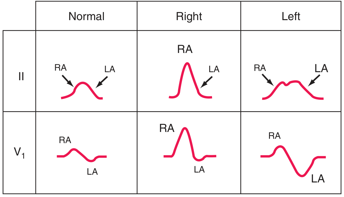
Upar ki image mein:
- Normal P wave - dono RA aur LA components normal size
- Right (RA enlargement) - Lead II mein tall, peaked P wave
- Left (LA enlargement) - Lead V1 mein deep negative component, lead II mein broad notched wave
2. VENTRICLES Mein Issue
Left Ventricular Hypertrophy (LVH)
ECG par signs:
- SV1 + RV5 ya RV6 > 35 mm (Sokolow-Lyon criterion)
- V5-V6 mein tall R waves
- Left axis deviation
- ST depression + T-wave inversion (left ventricular "strain" pattern)
- RaVL > 20 mm (women), > 28 mm (men)
Wajah: High blood pressure (hypertension), aortic stenosis, athletic heart
Right Ventricular Hypertrophy (RVH)
ECG par signs:
- Lead V1 mein tall R wave (R ≥ S wave)
- Right axis deviation (QRS axis right side shift)
- V1 mein qR pattern ya RSR' pattern
- V1-V4 mein ST depression aur T-wave inversion
Wajah: Pulmonary hypertension, pulmonary stenosis, Atrial Septal Defect (ASD)
Ventricular Hypertrophy ECG comparison:
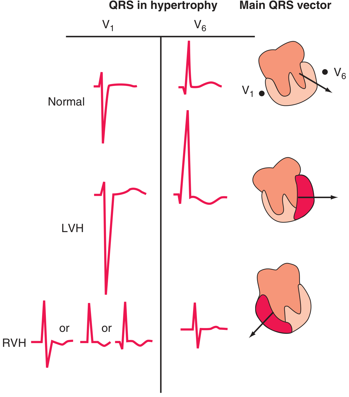
Is image mein:
- Normal - V1 mein small r, V6 mein normal R
- LVH - V1 mein deep S wave, V6 mein very tall R wave - electrical axis left ki taraf
- RVH - V1 mein tall R wave - electrical axis right ki taraf shift
3. ECHOCARDIOGRAPHY (Echo) - Sabse Behtar Test
ECG sirf electrical signals batata hai, lekin Echo seedha dil ka ultrasound hai. Yeh batata hai:
| Chamber | Echo mein kya dekha jaata hai |
|---|---|
| Left Ventricle | Wall thickness (>11 mm = hypertrophy), ejection fraction (EF <55% = problem), dilation |
| Right Ventricle | Size comparison LV ke saath (normally RV < LV), free wall thickness, pressure estimate via tricuspid jet |
| Left Atrium | Size measurement (>4 cm = enlarged), clot check in appendage |
| Right Atrium | Size, IVC size (fluid overload check) |
Important: Harrison's notes that "more definitive anatomic and functional information is provided by echocardiographic and cardiac magnetic resonance imaging studies" - ECG ka akela use limited hai.
4. Symptoms se Kaise Pata Chalein
| Chamber | Common Symptoms |
|---|---|
| Left Ventricle fail | Saanns phoolna (dyspnea), pulmonary edema, raat ko saaans ki takleef |
| Right Ventricle fail | Pair soojhna (leg edema), liver barhna, jugular veins phoolna (JVD) |
| Left Atrium | Atrial fibrillation, stroke, mitral valve issues |
| Right Atrium | Pericardial disease, ASD, tricuspid valve issues |
5. Aur Tests
- Chest X-Ray: Heart ka size aur shape (cardiomegaly, pulmonary congestion)
- Cardiac MRI: Sab se precise - tissue characterization bhi hoti hai
- Cardiac Catheterization: Pressures directly measure karta hai har chamber mein
- BNP / NT-proBNP blood test: Heart failure confirm karta hai (LV dysfunction ki marker)
Summary Table
| Test | LV Issue | RV Issue | LA Issue | RA Issue |
|---|---|---|---|---|
| ECG | Tall R V5-V6, deep S V1, left axis | Tall R V1, right axis deviation | Broad/biphasic P wave | Tall peaked P wave |
| Echo | Low EF, wall thickening, dilation | RV > LV size, free wall thick | LA diameter > 4 cm | RA dilation |
| Symptoms | Breathlessness, pulmonary edema | Leg swelling, JVD, liver enlargement | Atrial fibrillation | Pericardial/valvular disease |
Agar aap ya kisi patient ko yeh symptoms hain, toh sabse pehle ECG + Echo karwana chahiye - yeh dono saath mein dil ke kissi bhi chamber ki problem clearly batate hain.
(Sources: Harrison's Principles of Internal Medicine 22nd Edition, Textbook of Clinical Echocardiography, Tintinalli's Emergency Medicine)
This is a shared conversation. Sign in to Orris to start your own chat.