
Greenstick fracture in detail
Reading File
A greenstick fracture is an incomplete fracture seen almost exclusively in children, where one cortex of the bone breaks while the other bends (like a green, flexible twig). The periosteum remains intact on the concave side.
Searching sources...
Finding Sources
Searching PubMed
greenstick fracture children treatment
Finding Sources
Searching PubMed
greenstick fracture pediatric
Good - I have several relevant sources. Let me read the key sections in parallel.
Reading File
Reading File
Reading File
Now let me get the radiology section and Bailey & Love section for imaging details, management, and the figure images.
Reading File
Finding Sources
Finding Sources
Loading Image
Loading Image
Finding Sources
Searching the Web
greenstick fracture pathophysiology management clinical features 2024
Reading File
Reading Web Page
https://www.ncbi.nlm.nih.gov/books/NBK513279
I now have comprehensive information. Let me compile the full detailed answer.
Greenstick Fracture - Detailed Overview
Definition
A greenstick fracture is an incomplete fracture unique to pediatric bone, where one cortex and its overlying periosteum are disrupted (the tension/convex side), while the opposite cortex (the compression/concave side) remains intact but bends. The name comes from the behavior of a young, green tree branch: it cracks on the outer arc but does not break clean through.
- Tintinalli's Emergency Medicine describes it as "cortical disruption and periosteal tearing on the convex side of the bone, with an intact periosteum on the concave side of the fracture."
- Pfenninger and Fowler's Procedures for Primary Care notes it is "a level worse than a torus fracture - it involves an actual crack or disruption of one cortex and buckling of the opposite cortex."
Pathophysiology
The greenstick pattern is only possible because of specific properties of pediatric bone:
| Property | Effect |
|---|---|
| Lower mineral density | Bone is softer and more pliable |
| Thicker, biologically active periosteum | Maintains cortical continuity on the concave side; limits displacement |
| Greater elasticity | Bone absorbs more energy before complete failure |
| Open physes | Growth plates are mechanically weaker but also alter fracture propagation |
When a bending force is applied:
- Tensile stress develops on the convex (outer) side - this side fails first
- The cortex cracks and periosteum tears on the tension (convex) side
- Compressive stress on the concave (inner) side causes buckling without complete disruption
- The intact periosteal hinge on the concave side stabilizes the fracture
As children age and bone mineralizes, this energy-absorbing capacity is lost, and identical forces produce complete fractures in adults. - Sabiston Textbook of Surgery (Fracture Types table: "Incomplete cortical disruption, plastic deformation")
Epidemiology & Risk Factors
- Affects almost exclusively children under 10 years (peak age 4-8 years)
- Boys slightly more affected than girls (more active/risk-taking behavior)
- Most common bones: radius, ulna (forearm), followed by tibia, fibula, humerus, clavicle
- Forearm is by far the most common site because children brace falls with an outstretched arm (FOOSH - Fall On OutStretched Hand)
- Risk factors:
- Vitamin D deficiency / rickets (weakens cortical bone disproportionately)
- Malnutrition
- Physical abuse (non-accidental trauma - must be considered with inconsistent history)
Etiology / Mechanism of Injury
| Mechanism | Notes |
|---|---|
| FOOSH (fall on outstretched hand) | Most common; produces forearm fractures |
| Sports injuries | Contact sports, gymnastics |
| Motor vehicle collision | Higher energy |
| Direct blow / non-accidental trauma | Child abuse - consider if mechanism is inconsistent |
Clinical Features
Symptoms:
- Localized pain (often mild-moderate, less than a complete fracture)
- Swelling and soft tissue edema at the fracture site
- Point tenderness over the bone
- Refusal to use the affected limb (especially in younger children who cannot verbalize pain)
- Visible angulation or deformity when bending is substantial
Signs:
- Mild deformity - may not be dramatically apparent
- Pain on passive movement
- Neurovascular status usually intact (rare compromise given the stable nature)
- Less swelling and bruising than complete fractures
Note: Greenstick fractures are more stable and somewhat less painful than complete fractures because the intact periosteum limits bony displacement. - Tintinalli's Emergency Medicine
Radiology
X-ray Findings
- Fracture line visible through one cortex only (typically the convex/tension side)
- Opposite cortex shows buckling or bowing without a discrete fracture line
- No complete cortical disruption across both cortices
- Angulation of the bone at the fracture level
- Two views (AP + lateral) are mandatory - the fracture may only be visible on one projection
AP and lateral radiographs of greenstick fractures of the distal radius and ulna in a child:
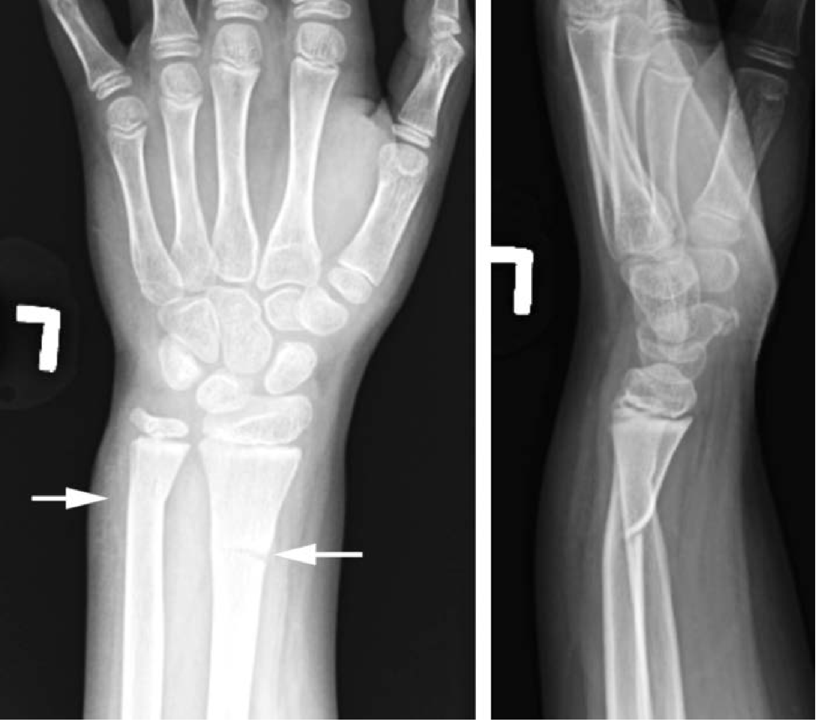 Fig: AP and lateral radiographs demonstrating greenstick fractures of the distal radius and ulna. Arrows indicate the fracture lines. - Tintinalli's Emergency Medicine
Lateral radiograph of greenstick fracture of the radius after FOOSH:
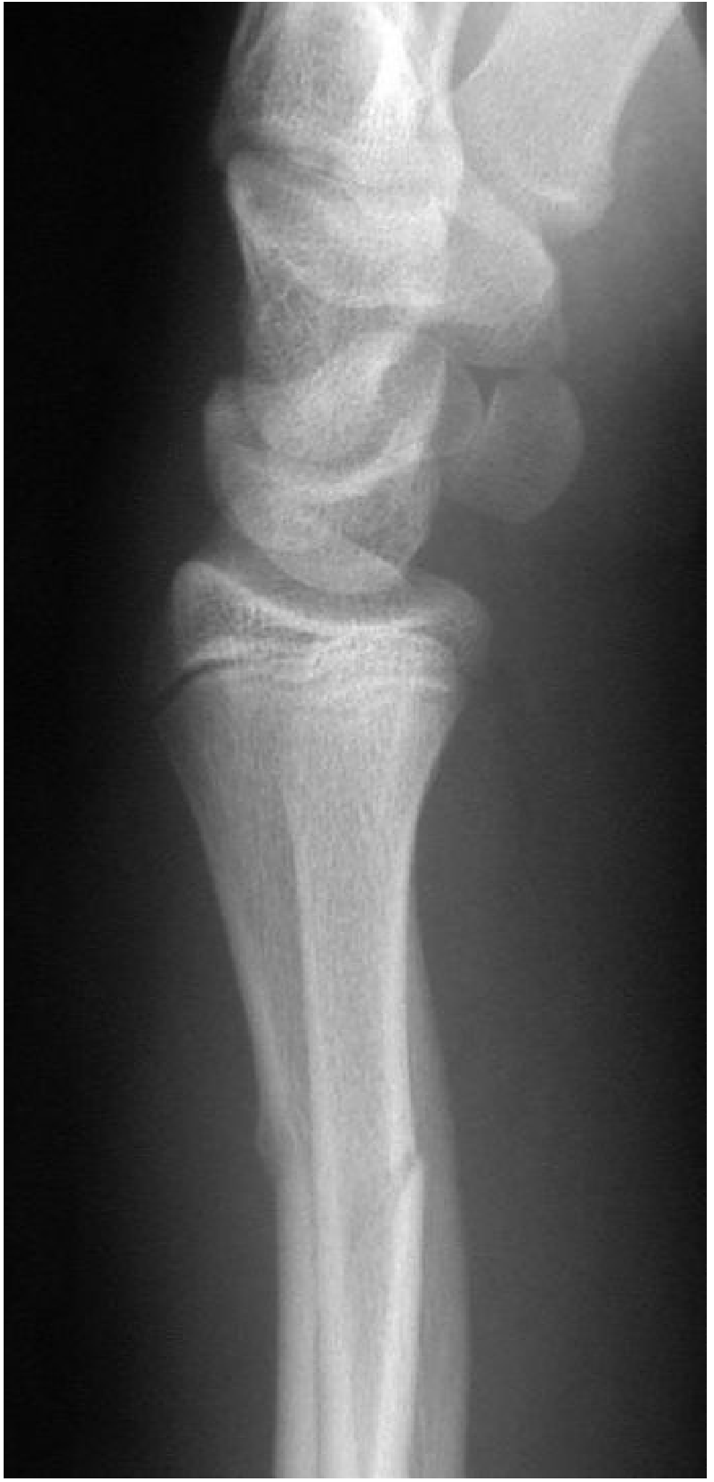 Fig: Lateral radiograph following a fall on outstretched hand, demonstrating an oblique cortical fracture on the volar aspect of the radius and buckling of the dorsal cortex. - Grainger & Allison's Diagnostic Radiology
Subtle Cases
- Comparison views of the contralateral, uninjured limb are helpful in mild cases where angulation is subtle
- MRI or bone scan can detect occult greenstick fractures in the rare case where X-ray is negative but clinical suspicion is high
Differential Diagnosis
| Condition | Key Distinguishing Feature |
|---|---|
| Torus (buckle) fracture | Cortex buckles/bulges but does not crack - compression only, no tension-side disruption; even more stable |
| Plastic bowing fracture | Entire diaphysis deforms with NO visible fracture line; only in forearm and lower leg |
| Complete transverse fracture | Both cortices broken, full displacement possible |
| Salter-Harris fracture | Involves the physis; Salter-Harris classification applies |
| Normal physiologic bowing | No mechanism, no tenderness, bilateral, symmetric |
The spectrum from least to most severe: torus fracture → greenstick fracture → complete fracture.
Treatment and Management
General Principles
All greenstick fractures require immobilization. The approach depends on the degree of angulation:
Mild Angulation (<15°)
- Short-arm splint or cast - typically for 4-6 weeks
- Casting a few days after injury (once swelling has stabilized) reduces need for recasting due to edema
- Analgesia (paracetamol ± ibuprofen)
- Elevation of the limb initially
Significant Angulation (>15°-20°)
- Closed reduction under procedural sedation or general anaesthesia (in children)
- After reduction, a long-arm cast is applied (above elbow for forearm fractures)
- The intact periosteum on the concave side acts as a hinge and aids reduction
Reduction Technique
A key point: because the concave-side periosteum is intact and intact, there is a risk the fracture will spring back to its angulated position after reduction. The technique involves:
- Applying traction along the long axis of the bone
- Completing the fracture by breaking through the intact cortex (intentionally completing it) - this prevents the "spring-back" tendency
- Then reducing and casting
Cast Immobilization Duration
- Distal radius/forearm: ~4-6 weeks
- Tibia/fibula: 6-8 weeks
- Follow-up X-rays at 1-2 weeks post-reduction to confirm maintained alignment
Surgical Management
Rare - only if:
- Closed reduction fails or cannot maintain alignment
- Severe angulation with unacceptable deformity
- Open fracture (uncommon for greenstick mechanism)
Orthopedic Referral
- Simple greenstick without angulation: orthopedic referral at follow-up is recommended but may not be emergent
- Any significant angulation: orthopedic referral at initial visit is generally recommended
Prognosis and Remodeling
Pediatric bone has exceptional remodeling capacity:
- Children's bones remodel angulation over time, especially in younger children with more growth remaining
- Remodeling is most effective when deformity is in the plane of joint movement (e.g., volar-dorsal angulation at the wrist remodels well)
- Rotational deformity does NOT remodel - this must be corrected at the time of reduction
- Outcomes are generally excellent with prompt treatment
Complications
| Complication | Notes |
|---|---|
| Refracture | ~6.7% risk within ~49 days of plaster removal; bone is mechanically weakened until fully consolidated |
| Malunion | Residual angulation if reduction inadequate or not performed; may remodel in young children |
| Redisplacement | Occurs if reduction is not maintained; usually detected at 1-2 week check X-ray |
| Compartment syndrome | Rare but can occur with swelling; monitor for the 5 P's post-casting |
| Cast complications | Pressure sores, skin maceration, tight cast from post-injury swelling |
| Non-union | Extremely rare in children due to high bone turnover and excellent blood supply |
| Growth disturbance | If physis is involved (technically then a Salter-Harris fracture, not pure greenstick) |
Comparison with Related Fracture Types (Pediatric Spectrum)
TORUS / BUCKLE         GREENSTICK            PLASTIC BOWING        COMPLETE FRACTURE
Axial compression      Bending force          Pure bending           High energy
Cortex buckles,        Tension side cracks,   No fracture line,      Both cortices
no crack               compression side       entire shaft bows      disrupted
                       buckles
Most stable            Stable                 Stable (but hard       Unstable
                                              to diagnose)
Immobilize 3-4 wks     Cast 4-6 wks          Reduction often        Reduction + cast
                       ± reduction            needed                 ± surgery
Summary Points
- Greenstick = incomplete fracture exclusive to children due to pliable bone and thick periosteum
- Mechanism: tension side cracks, compression side bends/buckles - periosteal hinge intact on concave side
- Most common site: distal radius and ulna (FOOSH mechanism)
- Diagnosis by 2-view X-ray - look for one-sided cortical break with angulation
- Treatment: immobilization ± closed reduction depending on angulation degree
- Prognosis: excellent - remodeling capacity in children corrects residual angulation (not rotation)
- Key risk: refracture - avoid high-risk activities and ensure adequate immobilization period
- Always consider non-accidental trauma when mechanism is inconsistent with injury
Sources: Tintinalli's Emergency Medicine (p. 949), Pfenninger & Fowler's Procedures for Primary Care, Pye's Surgical Handicraft, Grainger & Allison's Diagnostic Radiology (p. 1148), Sabiston Textbook of Surgery, StatPearls - Greenstick Fracture
This is a shared conversation. Sign in to Orris to start your own chat.