I now have comprehensive information from both embryology textbooks. Let me compile the full answer.
Pharyngeal (Branchial) Arches
Overview
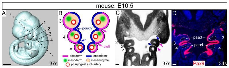
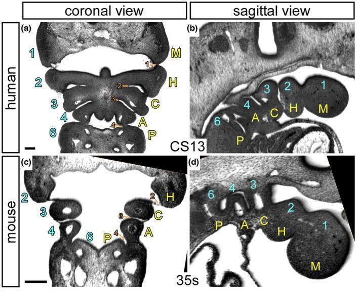
The pharyngeal arches develop during the 4th and 5th weeks of embryonic life as a series of bilateral mesodermal swellings in the head and neck region, separated by pharyngeal clefts (grooves) externally and pharyngeal pouches internally.
There are 5 arches in humans (1, 2, 3, 4, and 6 - the 5th is rudimentary or absent).
Structure of Each Arch
Each arch has four components (CAME mnemonic):
- Cartilage (skeletal element)
- Artery (aortic arch artery)
- Muscle (with its own myotome)
- Endothelium + nerve supply
Tissue of origin:
- Ectoderm - covers externally
- Endoderm - lines internally
- Mesoderm - forms central core (muscle primordium) - paraxial mesoderm migrates in
- Neural crest cells - migrate in during week 4 and form most of the mesenchyme, including cartilage, dermis, and smooth muscle
"During the fourth week, most of the mesenchyme is derived from neural crest cells that migrate into the arches... Migration of the multipotent neural crest stem cells into the arches and their differentiation into mesenchyme produce the maxillary and mandibular prominences." - The Developing Human
Derivatives by Arch
Arch 1 - Mandibular Arch (CN V - Trigeminal)
| Component | Derivatives |
|---|
| Cartilage (Meckel's) | Malleus, incus (middle ear ossicles); anterior ligament of malleus; sphenomandibular ligament |
| Bone (membranous ossification) | Mandible, maxilla, premaxilla, zygomatic bone, part of temporal bone |
| Muscles | Muscles of mastication (temporalis, masseter, pterygoids), mylohyoid, anterior belly of digastric, tensor tympani, tensor veli palatini |
| Nerve | CN V - trigeminal (V1 ophthalmic, V2 maxillary, V3 mandibular) |
| Artery | Terminal branches of maxillary artery |
The middle part of Meckel's cartilage regresses; its perichondrium forms the anterior ligament of malleus and sphenomandibular ligament.
Arch 2 - Hyoid Arch (CN VII - Facial)
| Component | Derivatives |
|---|
| Cartilage (Reichert's) | Stapes, styloid process, stylohyoid ligament, lesser horn and upper body of hyoid bone |
| Muscles | Muscles of facial expression, stapedius, stylohyoid, posterior belly of digastric, auricular muscles |
| Nerve | CN VII - facial |
| Artery | Stapedial artery (transient); contributes to corticotympanic artery |
Arch 3 (CN IX - Glossopharyngeal)
| Component | Derivatives |
|---|
| Cartilage | Greater horn and lower body of hyoid bone |
| Muscles | Stylopharyngeus (the only muscle from arch 3) |
| Nerve | CN IX - glossopharyngeal |
| Artery | Common carotid artery, proximal internal carotid artery |
Arch 4 (CN X superior laryngeal branch)
| Component | Derivatives |
|---|
| Cartilage | Superior cornu of thyroid cartilage, thyroid cartilage |
| Muscles | Cricothyroid, levator veli palatini, constrictors of pharynx |
| Nerve | Superior laryngeal branch of CN X (vagus) |
| Artery | Right: right subclavian artery; Left: arch of aorta |
Arch 6 (CN X recurrent laryngeal branch)
| Component | Derivatives |
|---|
| Cartilage | Cricoid, arytenoid, corniculate, and cuneiform cartilages |
| Muscles | Intrinsic muscles of larynx, striated muscles of esophagus |
| Nerve | Recurrent laryngeal branch of CN X (vagus) |
| Artery | Right: right pulmonary artery; Left: ductus arteriosus + left pulmonary artery |
Complete Summary Table
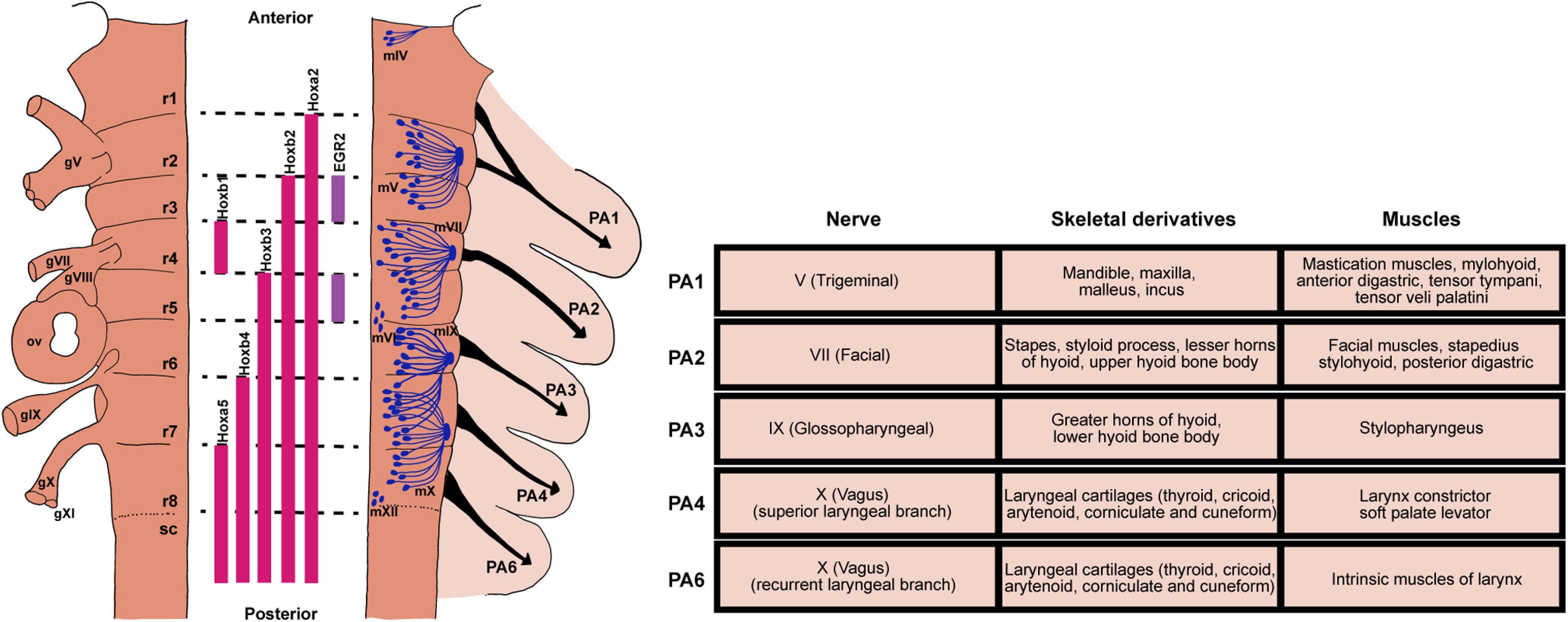
From The Developing Human (Table 9.1):
| Arch | Nerve | Muscles | Skeletal Structures | Ligaments |
|---|
| 1st (mandibular) | CN V (trigeminal) | Mastication muscles, mylohyoid, ant. digastric, tensor tympani, tensor veli palatini | Malleus, incus | Ant. ligament of malleus, sphenomandibular lig. |
| 2nd (hyoid) | CN VII (facial) | Facial expression, stapedius, stylohyoid, post. digastric | Stapes, styloid process, lesser horn + upper body of hyoid | Stylohyoid ligament |
| 3rd | CN IX (glossopharyngeal) | Stylopharyngeus | Greater horn + lower body of hyoid | - |
| 4th | CN X - superior laryngeal | Cricothyroid, levator veli palatini, pharyngeal constrictors | Superior cornu + thyroid cartilage | - |
| 6th | CN X - recurrent laryngeal | Intrinsic laryngeal muscles, esophageal striated muscle | Cricoid, arytenoid, corniculate, cuneiform cartilages | - |
Pharyngeal Pouches (Endodermal)
The 5th is rudimentary. The pouches give rise to vital organs:
| Pouch | Derivatives |
|---|
| 1st | Tubotympanic recess → middle ear cavity + auditory (Eustachian) tube |
| 2nd | Tonsillar fossa → palatine tonsil (surface epithelium) |
| 3rd | Inferior parathyroid glands (III); thymus |
| 4th | Superior parathyroid glands (IV); ultimobranchial body (parafollicular C-cells of thyroid) |
| 5th | Rudimentary (sometimes fused with 4th pouch) |
Note: The parathyroid glands from the 3rd pouch (inferior) actually migrate further down than those from the 4th pouch (superior) - hence the counterintuitive naming (3rd pouch → inferior parathyroids).
Pharyngeal Clefts (Ectodermal)
| Cleft | Derivative |
|---|
| 1st | External acoustic meatus |
| 2nd, 3rd, 4th | Normally obliterated (buried by 2nd arch overgrowth); persistence leads to branchial cysts/sinuses |
Clinical Correlates
1. First Arch Syndrome
Results from insufficient neural crest cell migration into the first arch. Two main phenotypes:
- Treacher Collins syndrome (mandibulofacial dysostosis): Autosomal dominant; mutation in TCOF1 gene (encodes treacle protein involved in rRNA biogenesis); malar hypoplasia, down-slanting palpebral fissures, deformed ears, lower eyelid defects
- Pierre Robin sequence: Micrognathia → posterior tongue displacement → bilateral cleft palate (a cascade, not a true syndrome)
2. DiGeorge Syndrome (22q11.2 deletion)
- Failure of 3rd and 4th pouch development
- Absent thymus and parathyroid glands
- Features: congenital hypoparathyroidism, T-cell deficiency (recurrent infections), cardiac outflow tract defects (aortic arch abnormalities), short philtrum, low-set notched ears, nasal clefts
- Only 1.5% have complete T-cell deficiency; ~30% have partial deficiency
3. Branchial Cysts/Sinuses/Fistulae
- Arise from persistence of 2nd, 3rd, or 4th pharyngeal clefts
- 2nd cleft remnants are most common - present as cysts or sinuses along the anterior border of sternocleidomastoid muscle
4. Recurrent Laryngeal Nerve Injury
- The 6th arch nerve loops around the 6th arch artery (left = ductus arteriosus; right = right subclavian)
- This accounts for the nerve's long recurrent course in adults
Key Mnemonic Aids
Arch nerves: "Taste Very Good, But Alternatively Recurrent" = V, VII, IX, X (superior laryngeal), X (recurrent laryngeal)
Muscles of 1st arch - "My Aunt Tillie Eats" = Mastication, Ant. digastric/mylohyoid, Tensor tympani/palatini
2nd arch = Stapes, Styloid, Stylohyoid ligament, lesser horn of hyoid (5 S's)
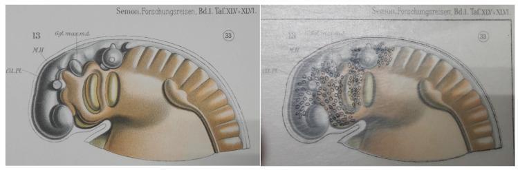
Embryological anatomy of the pharyngeal arches (Carnegie Stage 13 human embryo)
Sources:
- The Developing Human: Clinically Oriented Embryology (Moore & Persaud), Table 9.1, pp. 463-487
- Langman's Medical Embryology, pp. 306-309