
Time taken for the surgery of keeping titanium rod fixation for radius of left hand
radial shaft fracture AND intramedullary nailing AND operative time
how long does radius fracture surgery take plate or rod fixation operative time
| Type of fixation | Typical surgical time |
|---|---|
| Distal radius (wrist) plate and screws | 60 to 90 minutes, per a distal radius ORIF guide |
| Radial shaft plate fixation (open) | About 60 to 120 minutes. This is my estimate, not a figure from a source I found. |
| Intramedullary rod or nail (closed, through a small incision) | Often similar to plating, and shorter if the fracture reduces easily. |
- Plate or rod: Most "titanium fixation" of the radius uses a plate and screws, especially at the wrist. A true rod is an intramedullary nail, used mainly for shaft fractures. Campbell's says plating gives better length and rotational stability and restores the radial bow, but it needs a longer operative time and a larger incision than nailing (Campbell's Operative Orthopaedics 15th Ed 2026, Complications section, middle-third forearm fractures).
- Rod versus plate in studies: One comparison of both-bone forearm fractures found no difference in operative time between nailing and plating. Nailing caused almost no blood loss because the fracture was reduced closed, while open plating lost a mean of about 60 cc. Nailed fractures healed in about 10 weeks versus 14 weeks with plates (Rockwood and Green's Fractures in Adults 10th ed 2025, Outcomes section).
- Things that lengthen surgery: These include a comminuted or open fracture, a joint-surface (intra-articular) fracture, an old fracture, nailing that needs the fracture opened, an associated ulna fracture or wrist ligament repair, and the surgeon's experience.
- Anaesthesia and hospital stay: Regional block (arm numbing) or general anaesthesia is used. Most patients go home the same day or the next day.
- Recovery: Bone healing takes about 10 to 14 weeks. Gentle wrist movement often starts 1 to 2 weeks after plate fixation.
Shaft was broken and what does intramedullary means and nailing and plating
intramedullary nail forearm radius shaft fracture radiograph
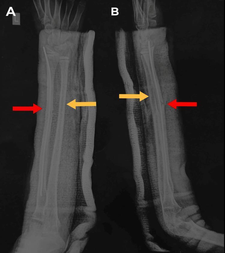
This diagnostic radiograph displays two views of a pediatric left forearm: an anterior-posterior (AP) view (A) and a lateral view (B). The image shows the radius and ulna post-surgical intervention for mid-shaft fractures. In both views, a titanium elastic nail system (TENS) nail is clearly visible as a radiopaque intramedullary rod within the radial shaft (indicated by red arrows), providing internal fixation and stabilization of the radius fracture. The ulna (indicated by yellow arrows) demonstrates residual bowing characteristic of plastic deformation, which has undergone corrective reduction. The bones are currently immobilized within an external radiopaque cast or splint, visible as the textured layer surrounding the soft tissue. This imaging illustrates standard pediatric orthopedic management of forearm fractures using flexible intramedullary nailing to maintain alignment while allowing for biological healing. Key educational concepts include intramedullary fixation techniques, management of pediatric plastic deformation of the ulna, and post-operative radiographic assessment of forearm alignment.
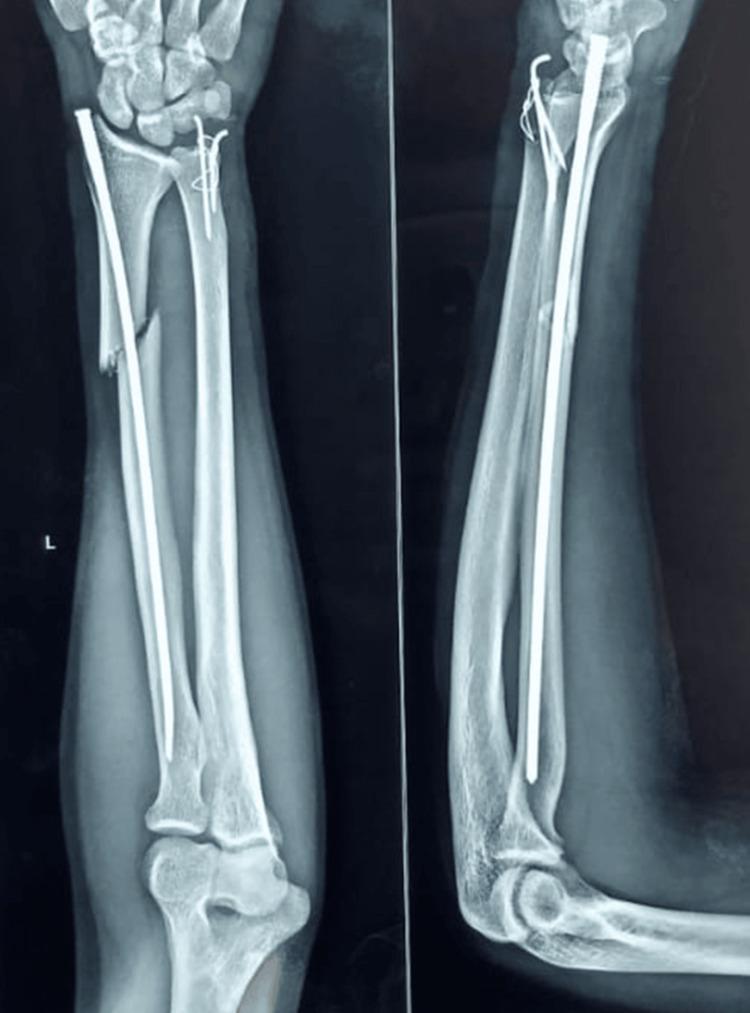
Anteroposterior (AP) and lateral radiographic views of a left adult forearm showing immediate post-operative orthopedic fixation of radial and ulnar fractures. The radiograph visualizes the radius, ulna, distal humerus, and carpal bones. A mid-to-distal radial shaft fracture, appearing transverse to short oblique with mild cortical comminution, is stabilized by a long metallic intramedullary (IM) screw nail extending from the distal radius to the proximal shaft. The distal ulna exhibits a fracture of the ulnar styloid process, which has been reduced and fixated using a tension band wiring (TBW) construct consisting of two thin Kirschner wires (K-wires) and a figure-of-eight tension wire loop. The hardware provides internal splinting for the radial diaphysis and compression for the ulnar styloid fragment. Alignment of the forearm bones is maintained, and the elbow and wrist joints are included in the field of view. This image serves as a clinical example of intramedullary osteosynthesis and tension band techniques in forearm trauma management.
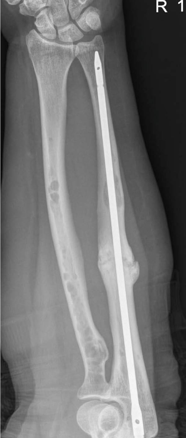
Anteroposterior (AP) radiograph of the right forearm demonstrating the radius and ulna with surgical hardware and underlying pathology. An intramedullary (IM) nail is present within the ulnar canal, extending from the proximal to the distal shaft to provide internal fixation. The mid-shaft of the ulna shows evidence of a healed pathological fracture with exuberant, consolidated callus formation consistent with radiographic union. Throughout both the radius and ulna, multiple discrete, well-defined radiolucent 'punched-out' lesions are visible, characteristic of multiple myeloma involvement. The distal and proximal radioulnar joints and the visible carpals appear stable. This diagnostic image illustrates the orthopedic management of a pathological fracture secondary to plasma cell dyscrasia, highlighting the use of IM nailing for stabilization and the characteristic bony manifestations of myelomatous disease in the long bones.
plate and screw fixation radius shaft fracture radiograph
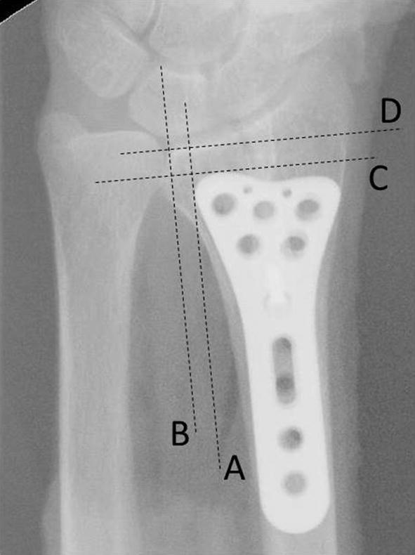
This posteroanterior (PA) radiograph of the distal radius and ulna demonstrates a volar locking plate in situ, used for internal fixation of a distal radius fracture. The image illustrates radiographic assessment parameters for plate positioning. Two vertical reference lines (A and B) are drawn parallel to the radial shaft to measure the plate-to-radius ulnar border (PRU) distance; Line A extends from the ulnar edge of the plate, and Line B follows the ulnar border of the radius. Two horizontal reference lines (C and D) are drawn perpendicular to the radial shaft to measure the plate-to-distal radius (PDR) distance; Line C marks the distal edge of the plate, while Line D marks the distal articular surface of the radius. The anatomically precontoured volar plate features a proximal shaft with elongated compression slots and a distal head with multiple circular locking screw holes. This imaging is used in orthopedic surgery to ensure the implant is positioned proximally to the 'watershed line' to prevent flexor tendon irritation or rupture.
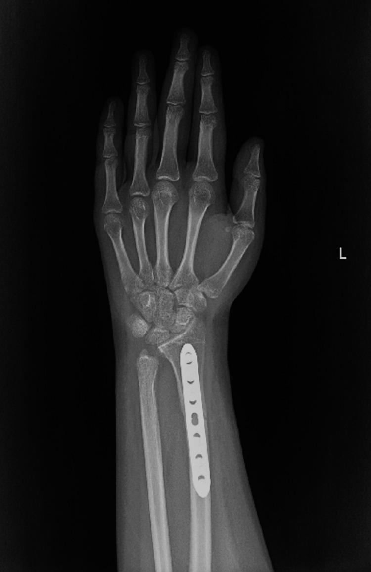
This posterior-anterior (PA) radiograph of the left wrist and hand displays the distal forearm, carpal bones, metacarpals, and phalanges. A prominent metallic internal fixation device, specifically a locking compression plate (LCP), is positioned along the distal radius. The plate features seven screw holes; however, the lack of visible radiopaque screws indicates they have been removed, or the imaging depicts the state after hardware extraction while the plate remains. The radius and ulna exhibit stable anatomical alignment with evidence of bone union following the surgical reduction of a distal third radius shaft fracture. The distal ulna, previously treated for a Salter-Harris type II fracture, shows the absence of Kirschner wires (K-wires), consistent with post-operative hardware removal. No acute secondary fractures, joint space narrowing, or carpal dislocations are visible. This diagnostic image illustrates successful orthopedic management of pediatric forearm fractures involving both shaft and growth plate components.
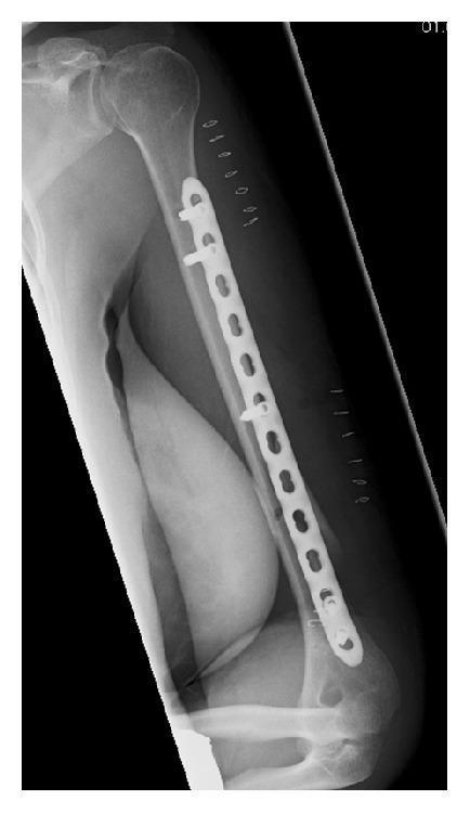
This postoperative AP (anteroposterior) radiograph displays the left humerus following surgical stabilization of a fracture. A long, radiopaque internal fixation plate is positioned along the lateral aspect of the humeral shaft, extending from the proximal humerus to the distal metaphysis near the elbow joint. The device is a 12-hole plate secured with five bicortical screws: two placed in the proximal cluster, one in the mid-shaft, and two in the distal cluster. The bone shows satisfactory alignment post-reduction. Superficially, multiple small, U-shaped radiopaque skin staples are visible along the lateral soft tissue envelope, indicating the site of the surgical incision. This imaging demonstrates the principles of minimally invasive plate osteosynthesis (MIPO) in orthopedic trauma surgery, where long plates are used to bridge fracture sites with sparse screw fixation to maintain biological environment and mechanical stability. The anatomy includes the humeral head, glenohumeral joint, humeral shaft, and the proximal radius and ulna at the elbow.
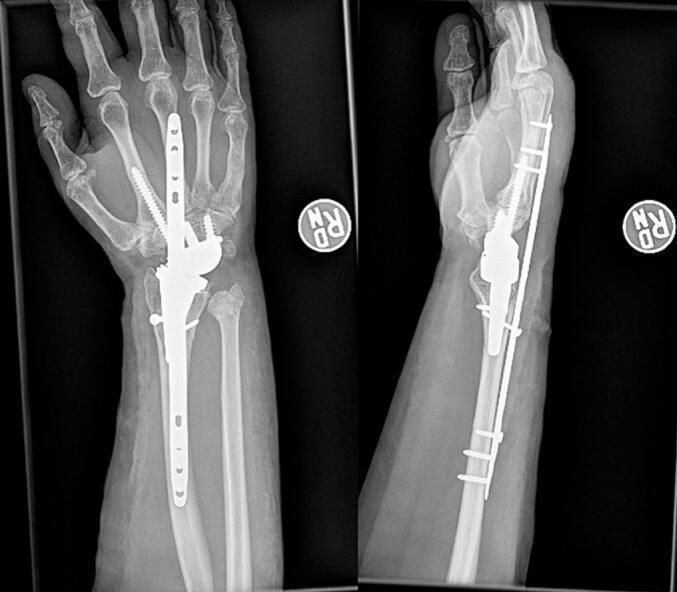
Anteroposterior (AP) and lateral radiograph views of a right wrist demonstrating post-operative management of a periprosthetic distal radius fracture. The images show a Total Wrist Arthroplasty (TWA) in situ, with the radial component seated in the distal radius and the carpal component fixated to the metacarpals. A dorsal spanning plate (DSP) is utilized for internal fixation, bridging the fracture site by extending from the radial diaphysis to the third metacarpal. The plate is secured with multiple cortical screws proximally in the radial shaft and distally in the metacarpal. A discrete lag screw is visible at the radial fracture site to provide interfragmentary compression. There is also evidence of a distal ulna fracture. The imaging highlights complex orthopedic salvage techniques for peri-implant fractures where primary bone stock may be compromised, emphasizing the use of a bridging construct to bypass the prosthetic joint and provide mechanical stability during healing.
Key terms
- Shaft (diaphysis): The long middle section of a bone, between the two ends (the elbow end and the wrist end).
- Intramedullary: "Inside the marrow canal." Long bones are hollow tubes with a central canal that holds bone marrow, so intramedullary means placed inside that canal.
- Nailing (intramedullary nailing): The surgeon passes a thin metal rod (the "nail") into the canal. It runs through the break and holds the two bone ends in line from the inside. This is the "rod" you described.
- Plating (plate and screws): The surgeon opens the skin over the fracture and lays a flat metal plate on the outside of the bone. Screws through the plate hold the bone pieces together. The plate acts as an external splint attached to the bone.
How the two compare
| Intramedullary nail | Plate and screws | |
|---|---|---|
| Position | Inside the bone canal | On the surface of the bone |
| Incision | Small, often at the wrist | Longer, directly over the fracture |
| Fracture reduction | Often done closed (without opening the fracture site) | Done open, with the fracture exposed |
| Blood loss | Minimal | Greater |
| Stability | Less rotational control, so a splint or cast is usually needed afterwards | Better length and rotational stability, and restores the natural curve of the radius |
| Healing time in one study | About 10 weeks | About 14 weeks |
What it looks like on X-ray
Surgery time for a shaft fracture
Ulna is not broken but just a dislocation at the metacarpal and ulna joint And radius broken at one third part from the metacarpal joint that distl one third
Galeazzi fracture AND surgical treatment
Galeazzi fracture radiograph distal radius shaft fracture with distal radioulnar joint dislocation plate fixation
What this means for your surgery
- The surgeon opens the forearm, usually from the palm side (volar approach) because the fracture is in the distal half. A back-of-forearm approach is sometimes used.
- The radius is lined up exactly and fixed with a plate and screws. An exact reduction of the radius usually brings the DRUJ back into place on its own.
- The surgeon then tests whether the DRUJ is stable.
- Stable: nothing more is needed.
- Unstable: the joint is pinned with Kirschner wires, with or without repair of the triangular fibrocartilage complex (TFCC). The TFCC is the ligament and cartilage complex that stabilizes the joint. A broken ulnar styloid base is fixed as well.
- Will not reduce: the DRUJ is opened through a small cut on the back of the wrist, because soft tissue such as a tendon may be blocking it.
Likely operating time
After surgery
- Stable DRUJ: A long-arm splint or cast is used for 3 to 6 weeks. In selected stable cases, elbow movement starts right away and forearm rotation starts about 2 weeks after surgery.
- Unstable or repaired DRUJ: A long-arm cast is used for 3 to 6 weeks. If the DRUJ dislocated toward the back, the forearm is usually held with the palm facing up (supination). If it dislocated toward the palm, it is held with the palm down (pronation). The position is chosen during surgery based on where the joint is most stable.
- Pins: If wires were placed across the joint, they are removed later. Do not rotate the forearm until you are cleared, because the pins can break.
- Outcomes: With a well-reduced radius and DRUJ, 80 to 92% of patients have satisfactory results. A poor reduction can leave the joint out of line and cause pain or reduced forearm rotation (Rockwood and Green's).
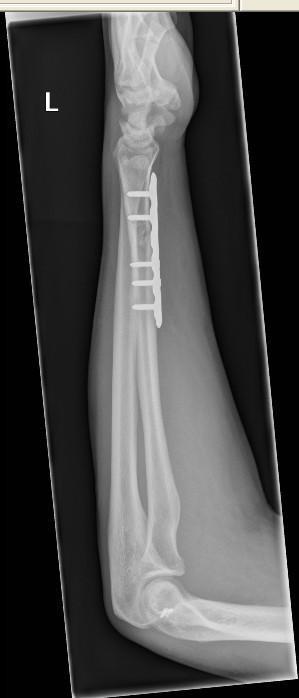
- Is the DRUJ stable after the plate is placed?
- Might wires or a ligament repair be needed?
- How long will the cast or splint stay on?
- Left image (front view): The radius has a fracture in its lower half, toward the wrist. The ulna looks intact along its length.
- Middle image (side view): The radius fragments look displaced and overlapping. That means shortening and angulation, not a clean crack. The wrist bones near the top look out of their normal position relative to the forearm.
- Right image: This is a view that includes the elbow. It is probably there to check the elbow and radial head, which is routine when a forearm injury is suspected.
- Ulna: I see no obvious break in the shaft, which fits what you told me.
- A displaced distal radial shaft fracture with an unbroken ulna is the typical pattern.
- Overlap and shortening of the radius usually mean the distal radioulnar joint (DRUJ) is disrupted too. On a film like this, a widened gap between the radius and ulna at the wrist is a clue. I can't judge that gap reliably from a glared photo.
- A fracture this displaced won't hold with a cast, so it needs surgery. That agrees with plating, as I described earlier.
- Is the DRUJ dislocated, and in which direction?
- Is there any ulnar styloid fracture?
- Will wires or a ligament repair be needed after the plate is placed?
- Is there any nerve or circulation concern, such as numb fingers or swelling?
Durj means
- Distal means the end farthest from the body, which here is the wrist end.
- Radio-ulnar means between the radius (thumb side) and the ulna (little-finger side).
- Joint means the point where the two bones meet.
But there dislocation at the ulnar and metacarpal joint region but how it will be durj
distal radioulnar joint anatomy diagram ulnar head sigmoid notch TFCC
Why the ulnar side of the wrist is the DRUJ
- Radius and ulna: These are the two forearm bones. At the wrist end, the end of the ulna (the ulnar head) lies right beside the end of the radius.
- Carpal bones: These are eight small wrist bones.
- Metacarpals: These are the long hand bones that lead to the fingers.
Other possibilities
| Possible injury | What it is | How it relates to the radius fracture |
|---|---|---|
| DRUJ dislocation | The ulnar head comes out of its notch on the radius | This is the classic partner of a distal radial shaft fracture (Galeazzi) |
| Carpal (wrist bone) dislocation | Wrist bones come out of position, for example around the lunate | Less common, but it can happen with high-energy injuries |
| Carpometacarpal dislocation | The hand bones come out of position at the base of the hand | Rare, and it would be a separate injury |
How to confirm
- Ask the surgeon: "Is the dislocation at the DRUJ (the joint between the radius and ulna), or in the wrist bones (carpals)?"
- Check the X-ray report: Look for the words "distal radioulnar joint," "DRUJ," "perilunate" or "carpal."
- CT scan: Surgeons sometimes order one to see the DRUJ clearly, because ordinary X-rays can miss it.
Sorry I'm taking about carpels from the beginning not the metacarpal
perilunate dislocation lateral wrist radiograph lunate carpal dislocation
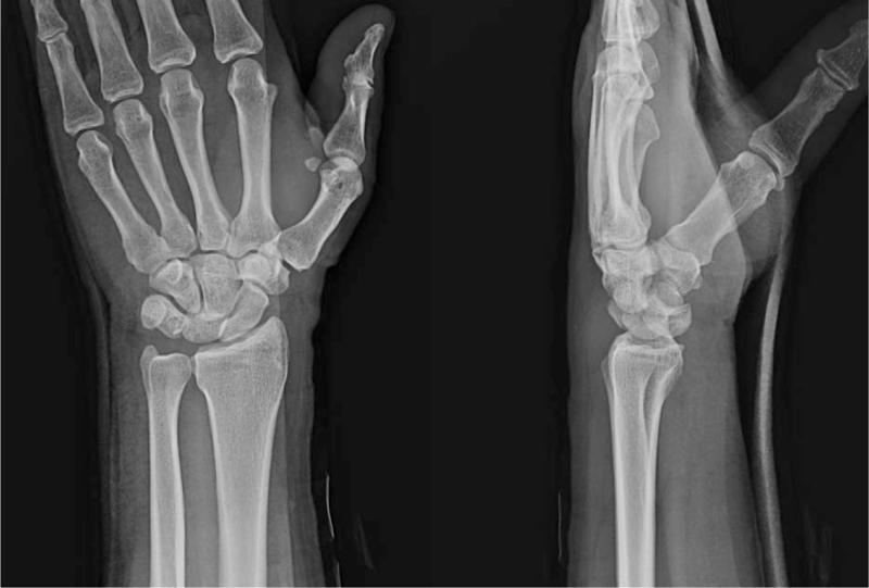
This diagnostic image consists of two X-ray radiographs of a human wrist: a posteroanterior (PA) view and a lateral view. In the PA radiograph, there is a significant disruption of the normal carpal arcs, with the carpal bones appearing disorganized and crowded. The scaphoid is shortened, suggesting flexion, and the joint spaces between the proximal and distal rows are irregular. In the lateral radiograph, the diagnostic hallmark of a dorsal perilunate dislocation (PLD) is visible: the lunate remains in its normal anatomical articulation within the lunate fossa of the distal radius, but the capitate and the remainder of the carpus are displaced dorsally (posteriorly) relative to the lunate. This 'spilled teacup' appearance is absent here, as that would signify a lunate dislocation; instead, the persistent radio-lunate alignment with dorsal capitate displacement confirms a perilunate dislocation. These images illustrate high-energy carpal instability often associated with ligamentous ruptures, specifically of the scapholunate and lunotriquetral interosseous ligaments.
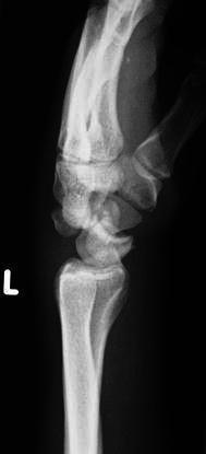
This diagnostic image is a lateral plain radiograph of the left wrist demonstrating a volar perilunate dislocation. The imaging highlights a significant disruption of the normal carpal alignment, specifically regarding the relationship between the distal radius, lunate, and capitate bones. In a normal lateral view, these structures should form a linear, collinear arrangement (the 'apple in a cup' configuration). Here, while the lunate maintains its articulation with the distal radius (remaining in the 'cup'), the capitate and the rest of the distal carpal row are markedly displaced volarly (towards the palmar aspect) relative to the lunate. This malalignment signifies a high-energy ligamentous injury and carpal instability. The radiograph serves as a critical educational tool for identifying carpal dislocations, emphasizing the loss of Gilula's arcs and the disruption of the central column alignment of the wrist, which are essential diagnostic criteria in orthopedic and emergency medicine.
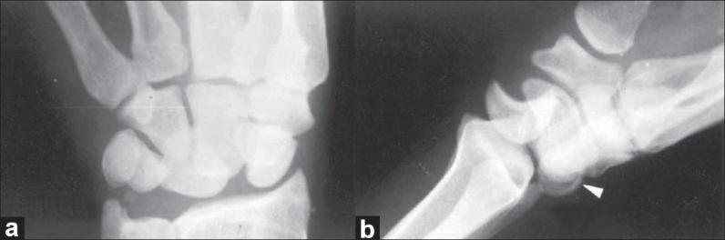
This diagnostic imaging set consists of two X-ray radiographs of the wrist demonstrating a dorsal (posterior) perilunate dislocation. View (a) is an anteroposterior (AP) skiagram showing a clear disruption of the normal carpal arches (Gilula's lines) and a transverse avulsion fracture at the tip of the ulnar styloid process. View (b) is a lateral radiograph illustrating the characteristic dorsal displacement of the capitate and remainder of the carpus relative to the lunate, which remains aligned with the distal radius. A distinct avulsion fracture of the posterior (dorsal) horn of the lunate bone is visible, indicated by a white arrowhead. These findings are clinically significant for diagnosing complex carpal instability and associated ligamentous or bony injuries following high-energy trauma. The images serve as an educational example of perilunate injury patterns, emphasizing the importance of evaluating both AP and lateral views to identify carpal bone malalignment and subtle avulsion fragments.
What the combined injury probably is
What the surgery involves
- Radius shaft: Plate and screws, as described before.
- Carpal dislocation:
- The first step is a closed reduction, putting the bones back in place without cutting. If that works, the wrist is splinted. If it fails, or if the median nerve is squeezed (acute carpal tunnel syndrome), open surgery is needed, and it is urgent in that case.
- Open surgery uses a cut on the back of the wrist, sometimes with a second cut on the palm side to release the carpal tunnel.
- The bones are aligned using the lunate as the reference. They are then held with Kirschner wires. The torn ligaments are repaired with small suture anchors.
- The wires are usually cut under the skin and removed after 8 to 12 weeks (Rockwood and Green's, Authors' Preferred Treatment for Perilunate Dislocation).
Time estimate
Points to watch
- Delay worsens the outcome. About 20% of perilunate injuries are missed at first. Late treatment can lead to pain, weakness, stiffness, carpal tunnel syndrome and early arthritis (Rockwood and Green's).
- Warning signs: Numbness or tingling in the thumb, index and middle fingers, severe swelling, a cold or pale hand, or uncontrolled pain should be reported right away.
- Recovery is longer than for the radius alone. Expect a cast or splint, wire removal in 2 to 3 months, and then hand therapy for stiffness. Doctors warn that the outlook for these injuries is guarded, but early and accurate fixation gives the best chance of a good result.
Questions for the surgeon
- What exactly is dislocated? Is it a perilunate or lunate dislocation, or a radiocarpal dislocation?
- Is there any carpal bone fracture, for example of the scaphoid?
- Will the carpal bones be pinned or have ligament repair?
- Is there any sign of median nerve compression?
- When will the pins come out?