
Make me revise the bronchial asthma in pharmacology,I have my viva in 20minutes
Bronchial Asthma - Pharmacology Viva Rapid Revision
1. Definition (Quick Opener)
2. Pathophysiology (Know for Mechanism Questions)
- Early phase (0-30 min): Allergen activates mast cells via IgE → releases histamine, leukotrienes (LTC4, LTD4, LTE4), and prostaglandins → immediate bronchoconstriction
- Late phase (4-8 hrs): Eosinophil and T-lymphocyte infiltration → sustained inflammation, mucus, airway remodeling
- Key mediators: Histamine, LTs, PGD2, PAF, TNF-α, IL-4, IL-5, IL-13
- cAMP ↑ → bronchodilation; cGMP ↑ → bronchoconstriction
3. Drug Classification
| Category | Drugs | Role |
|---|---|---|
| β2-agonists (SABA) | Salbutamol (albuterol), terbutaline | Reliever - acute bronchospasm |
| β2-agonists (LABA) | Salmeterol, formoterol | Controller - long-term prevention |
| Inhaled corticosteroids (ICS) | Beclomethasone, budesonide, fluticasone | Controller - first-line anti-inflammatory |
| Anticholinergics | Ipratropium (SAMA), tiotropium (LAMA) | Bronchodilator - adjunct |
| Methylxanthines | Theophylline, aminophylline | Bronchodilator - adjunct/oral |
| Leukotriene modifiers | Montelukast, zafirlukast (blockers); zileuton (synthesis inhibitor) | Controller - mild persistent asthma |
| Mast cell stabilizers | Cromolyn sodium, nedocromil | Prophylactic - exercise-induced |
| Biologics/Anti-IgE | Omalizumab | Severe allergic asthma |
| Anti-IL-5 | Mepolizumab, reslizumab, benralizumab | Severe eosinophilic asthma |
4. Key Drug Mechanisms (Most Asked in Viva)
Beta-2 Agonists
- MOA: Bind β2 receptors on airway smooth muscle → activate adenylyl cyclase → ↑cAMP → bronchodilation. Also inhibit mast cell mediator release.
- SABA onset: 5 min; duration: 4-6 hrs
- SABA (Salbutamol): Drug of choice for acute attack; metered dose inhaler (MDI) preferred
- LABA (Salmeterol): Onset 15-30 min; duration 12 hrs. Long lipophilic side chain anchors it in membrane.
- Formoterol: LABA with fast onset (like SABA) - can be used for both relief and maintenance (SMART regimen with budesonide)
- SE: Tachycardia (β1), tremor, hypokalemia (K+ shift into cells), tolerance with overuse
Corticosteroids (ICS)
- MOA: Bind glucocorticoid receptor → nuclear translocation → inhibit transcription of inflammatory cytokines (IL-4, IL-5, IL-13, TNF-α). Reduce eosinophil and mast cell infiltration. Reduce bronchial hyperreactivity. Do NOT directly relax smooth muscle.
- ICS drugs: Beclomethasone, budesonide, fluticasone, mometasone, ciclesonide
- SE of ICS: Oral candidiasis, dysphonia (hoarseness) - prevented by rinsing mouth after use
- Systemic steroids (IV hydrocortisone, oral prednisolone): Used in severe acute attacks and status asthmaticus
Theophylline
- MOA: PDE inhibitor → prevents cAMP breakdown → bronchodilation. Also adenosine receptor antagonist (blocks adenosine-induced bronchoconstriction).
- Narrow therapeutic index: 10-20 mcg/mL (therapeutic); >20 mcg/mL toxic
- SE: Nausea/vomiting (early), arrhythmias, seizures (toxic levels)
- Drug interactions: Cimetidine, erythromycin, ciprofloxacin INCREASE levels (inhibit CYP1A2). Smoking, rifampicin DECREASE levels.
- Aminophylline: Water-soluble theophylline salt; given IV in acute severe asthma
Anticholinergics
- MOA: Block M3 muscarinic receptors on bronchial smooth muscle → inhibit acetylcholine-induced bronchoconstriction
- Ipratropium bromide (SAMA): Quaternary ammonium - poor systemic absorption, no CNS effects. Used as add-on in acute severe asthma (combined with salbutamol nebulization)
- Tiotropium (LAMA): 24-hr duration; now approved for asthma maintenance (add-on to ICS). Dissociates rapidly from M2 receptors (preserving ACh feedback inhibition).
- SE: Dry mouth, urinary retention (rare), worsening glaucoma if sprayed in eye
Leukotriene Modifiers
- Montelukast, zafirlukast: CysLT1 receptor blockers → block LTC4, LTD4, LTE4 effects
- Zileuton: 5-lipoxygenase inhibitor → blocks ALL leukotriene synthesis (including LTB4)
- Uses: Mild persistent asthma, aspirin-sensitive asthma (ASA exacerbates via 5-LOX shunting), exercise-induced asthma, allergic rhinitis with asthma
- Montelukast SE: Neuropsychiatric effects (nightmares, depression, suicidal ideation - black box warning)
Cromolyn Sodium / Nedocromil
- MOA: Stabilize mast cell membranes → prevent degranulation → no mediator release. Block Cl- channels. Also inhibit sensory nerve activation.
- Only prophylactic - no bronchodilator effect, useless in acute attack
- Best indication: Exercise-induced asthma (inhaled before exercise), allergen-induced asthma
- SE: Very safe; minor throat irritation
Omalizumab (Anti-IgE)
- MOA: Monoclonal antibody that binds free serum IgE → prevents IgE binding to mast cell FcεRI receptors → blocks allergic cascade
- Use: Moderate-to-severe allergic asthma not controlled by ICS+LABA
- Given: SC injection every 2-4 weeks
- SE: Injection site reactions; rare anaphylaxis (observe for 30-60 min after each dose)
5. Stepwise Management (GINA-based)
| Step | Treatment |
|---|---|
| Step 1 (mild intermittent) | SABA as needed (salbutamol PRN) |
| Step 2 (mild persistent) | Low-dose ICS + SABA PRN |
| Step 3 (moderate persistent) | Low-dose ICS + LABA OR medium ICS |
| Step 4 (severe persistent) | Medium/high ICS + LABA |
| Step 5 (very severe) | Add tiotropium / omalizumab / anti-IL-5 biologic |
| Acute severe attack | Nebulized salbutamol + ipratropium, IV hydrocortisone, O2, IV aminophylline if needed |
6. Status Asthmaticus Management
- Oxygen - maintain SpO2 >94%
- Nebulized salbutamol (2.5-5 mg) + ipratropium (0.5 mg) - repeat every 20 min x3
- IV/oral corticosteroids - hydrocortisone 100-200 mg IV or prednisolone 40-60 mg oral
- IV aminophylline - if no response (load 5 mg/kg over 20 min, then infusion)
- IV magnesium sulfate (2 g IV) - bronchodilation via Ca2+ channel blockade
- Heliox, intubation - in life-threatening cases
7. High-Yield Viva Points
| Question | Answer |
|---|---|
| Drug of choice - acute attack | Salbutamol (inhaled SABA) |
| Drug of choice - chronic prevention | Inhaled corticosteroid (ICS) |
| Drug not used in acute attack | Cromolyn sodium, salmeterol |
| Theophylline therapeutic range | 10-20 mcg/mL |
| SABA + LABA with fast onset (SMART regimen) | Formoterol + budesonide |
| Aspirin-exacerbated asthma treatment | Montelukast (zileuton) |
| MOA of ipratropium vs salbutamol | M3 block vs β2 agonist |
| Why ICS preferred over systemic steroids? | Topical effect, minimal systemic SE |
| Biologic for allergic asthma | Omalizumab (anti-IgE) |
| Biologic for eosinophilic asthma | Mepolizumab (anti-IL-5) |
| SE of ICS to warn patient | Oral candidiasis - rinse mouth after use |
| What increases theophylline toxicity? | Cimetidine, erythromycin, ciprofloxacin |
| Formoterol vs salmeterol | Formoterol: fast + long; Salmeterol: slow + long |
8. Quick Mechanism Diagram (Memorize)
Allergen → IgE on mast cells → Mast cell activation
       ↓
Histamine, LTs, PGD2 → Bronchoconstriction + inflammation
       ↓
β2 agonists → ↑cAMP → Smooth muscle relaxation (RELIEF)
ICS → ↓Cytokines, ↓eosinophils (CONTROL)
Anticholinergics → Block M3 → ↓ACh bronchoconstriction
Montelukast → Block CysLT1 → ↓LT-mediated bronchoconstriction
Theophylline → ↓PDE → ↑cAMP → Bronchodilation
Explain theopyline part more clearly
Theophylline - Complete Explanation
What is it?
Mechanism of Action (3 Proposed Mechanisms)
1. PDE (Phosphodiesterase) Inhibition - Primary MOA
- Normally, cAMP is broken down by the enzyme phosphodiesterase (PDE)
- Theophylline inhibits PDE → cAMP is NOT broken down → intracellular cAMP accumulates
- High cAMP → smooth muscle relaxation → bronchodilation
- Specifically: PDE3 inhibition relaxes airway smooth muscle; PDE4 inhibition reduces cytokine release (anti-inflammatory effect at lower doses)
Theophylline → inhibits PDE → ↓breakdown of cAMP → ↑cAMP → Bronchodilation
2. Adenosine Receptor Antagonism - Secondary MOA
- Adenosine (a local mediator) normally acts on airway smooth muscle → causes bronchoconstriction + histamine release from mast cells
- Theophylline blocks adenosine receptors → prevents adenosine-induced bronchoconstriction
- Note: Some xanthines without adenosine antagonism (e.g., enprofylline) still cause bronchodilation, so this is NOT the sole mechanism
3. Histone Deacetylase (HDAC) Enhancement - Newer MOA
- Inflammatory gene transcription requires acetylation of histones
- Low-dose theophylline enhances the action of HDAC → reduces inflammatory gene transcription
- This is the same pathway by which corticosteroids work - so low-dose theophylline actually potentiates ICS effects
- This explains why theophylline has anti-inflammatory activity at sub-bronchodilator doses
Pharmacokinetics (Very Important for Viva)
| Parameter | Detail |
|---|---|
| Oral bioavailability | Nearly 100% |
| Protein binding | ~40% |
| Metabolism | Hepatic - CYP1A2 (mainly) |
| Half-life | ~8 hrs in adults (shorter in smokers, longer in neonates/elderly/liver disease) |
| Excretion | Urine (as demethylated xanthines - NOT uric acid) |
| Volume of distribution | ~0.5 L/kg |
Therapeutic Drug Monitoring (Narrow Therapeutic Index)
| Level (mcg/mL) | Effect |
|---|---|
| < 5 | Sub-therapeutic, no bronchodilation |
| 10-20 | Therapeutic range |
| 20-30 | Nausea, vomiting, headache, insomnia, tremor |
| > 30 | Cardiac arrhythmias (atrial tachycardia, PVCs, ventricular tachycardia) |
| > 40-100 | Seizures, hypotension, death |
Drug Interactions (Very Commonly Asked)
Drugs that INCREASE theophylline levels (toxic):
| Drug | Mnemonic |
|---|---|
| Cimetidine | CERE - these raise theophylline |
| Erythromycin | |
| Ciprofloxacin | |
| Allopurinol | |
| Oral contraceptives | |
| Fluvoxamine |
Drugs that DECREASE theophylline levels (sub-therapeutic):
| Drug |
|---|
| Rifampicin |
| Phenytoin |
| Carbamazepine |
| Phenobarbitone |
| Smoking (polycyclic aromatic hydrocarbons induce CYP1A2) |
Viva tip: Smokers need higher doses of theophylline. When they quit smoking, levels rise - monitor carefully.
Pharmacological Effects Across Organ Systems
| System | Effect |
|---|---|
| Lungs | Bronchodilation (main therapeutic use); also reverses diaphragm fatigue in COPD |
| CNS | Mild arousal at low doses; nervousness, insomnia; seizures in toxicity |
| CVS | Tachycardia, positive inotrope, vasodilation (except cerebral vessels - causes vasoconstriction); arrhythmias in toxicity |
| Kidney | Weak diuresis (↑GFR + ↓tubular Na+ reabsorption) |
| GIT | ↑gastric acid secretion → avoid in peptic ulcer disease |
| Skeletal muscle | Improves contractility; reverses diaphragmatic fatigue |
Clinical Uses
- Acute severe asthma / Status asthmaticus: IV aminophylline (loading dose 5-6 mg/kg over 20-30 min, then maintenance infusion of 0.5-0.9 mg/kg/hr). Use only if patient is not already on oral theophylline.
- Chronic asthma: Oral sustained-release theophylline as add-on to ICS (Step 3-4). Less popular now due to narrow TI.
- COPD: Add-on for patients with persistent symptoms.
- Apnea of prematurity: Methylxanthines (theophylline/caffeine) stimulate respiratory center - caffeine preferred now.
Toxicity and Management
- Early: Nausea, vomiting (most common first symptom), headache, tremor, insomnia
- Intermediate: Tachycardia, hypokalemia (due to β2-adrenergic activation), hyperglycemia
- Severe: Ventricular arrhythmias, seizures (resistant to regular anticonvulsants), hypotension
- Activated charcoal - repeated doses (theophylline undergoes enterohepatic circulation, so repeat doses are effective)
- Whole bowel irrigation - especially for sustained-release formulations (toxicity can be delayed for hours)
- Propranolol / esmolol (β-blockers) - specific antidote for β2-mediated tachycardia and hypotension
- Benzodiazepines - for seizures
- Hemodialysis - if serum level >100 mcg/mL or intractable seizures
Roflumilast - Selective PDE4 Inhibitor (Bonus)
- Selective PDE4 inhibitor developed to reduce theophylline's toxicity
- FDA approved for COPD (reduces exacerbations), NOT asthma
- SE: Nausea, headache, diarrhea (limits use)
Quick Recall Table
| Feature | Detail |
|---|---|
| Class | Methylxanthine (1,3-dimethylxanthine) |
| MOA | PDE inhibition (↑cAMP) + adenosine receptor block + HDAC enhancement |
| Therapeutic range | 10-20 mcg/mL |
| IV form | Aminophylline |
| Toxicity sign | Vomiting (early), arrhythmias, seizures |
| Antidote for tachycardia in OD | Propranolol |
| Levels increased by | Cimetidine, erythromycin, ciprofloxacin |
| Levels decreased by | Rifampicin, phenytoin, smoking |
| Unique SE vs β2-agonists | Seizures, arrhythmias, GI stimulation |
Histone deactylase deeply
histone acetylation deacetylation inflammatory gene transcription mechanism
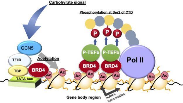
A pathophysiology diagram illustrating the molecular mechanism of transcription elongation via gene body epigenetics in response to carbohydrate signaling. The process begins with a 'Carbohydrate signal' activating GCN5, which facilitates histone acetylation (marked as 'Ac') near the TATA box, TBP, and TFIID complex. BRD4 (bromodomain-containing protein 4) is then recruited to these acetylated histone marks within the gene body region. Once bound, BRD4 interacts with P-TEFb (positive transcription elongation factor b) to catalyze the phosphorylation of RNA polymerase II (Pol II) at the Serine 2 residue of its C-terminal domain (CTD), indicated by 'P' labels. This coordinated epigenetic and enzymatic sequence promotes active mRNA transcription. The diagram highlights the relationship between dietary signals, histone modification, and the transcriptional regulation of metabolic genes, which is clinically relevant to the study of metabolic syndromes and type 2 diabetes.
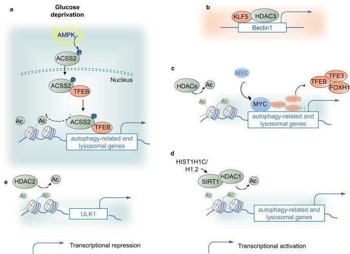
A multi-panel pathophysiology diagram illustrating the epigenetic regulation of autophagy via histone acetylation and deacetylation. (a) Under glucose deprivation, AMPK phosphorylates ACSS2, promoting its nuclear translocation; ACSS2 binds TFEB to facilitate histone H3 acetylation (Ac) at autophagy and lysosomal gene promoters, leading to transcriptional activation. (b) KLF5 and HDAC3 bind the Beclin1 promoter, resulting in transcriptional repression. (c) MYC and Class I HDACs cooperatively repress autophagy-related and lysosomal genes by removing acetylation marks and displacing transcription factors like TFEB, TFE3, and FOXH1. (d) Linker histone HIST1H1C/H1.2 upregulates SIRT1 and HDAC1 to maintain H4K16 deacetylation, which paradoxically leads to ATG protein expression and autophagy promotion. (e) HDAC2 activity on the ULK1 promoter maintains deacetylation; reduction in HDAC2 leads to increased H3K27 acetylation but correlates with reduced ULK1 expression in specific clinical contexts like diabetic renal fibrosis. The diagram uses blue arrows to denote transcriptional activation and right-angled arrows to denote repression, highlighting the diverse roles of histone-modifying enzymes in cellular homeostasis.
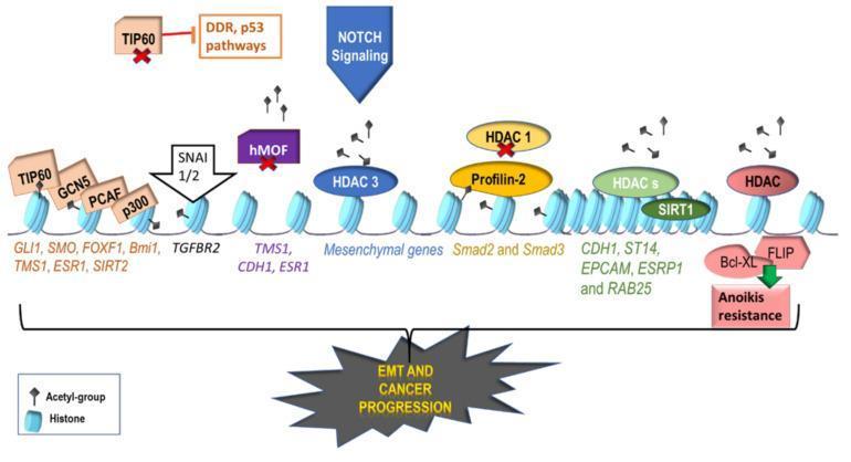
This pathophysiology diagram illustrates the role of histone acetylation and deacetylation in regulating genes associated with Epithelial-Mesenchymal Transition (EMT) and cancer progression. The central structure depicts a linear chromatin strand with blue, bead-like nucleosomes (histone complexes). Small black flag icons represent acetyl groups. On the left, Histone Acetyltransferases (HATs) such as TIP60, GCN5, PCAF, and p300 are shown adding acetyl groups to promote the expression of genes like GLI1, SMO, and FOXF1. The transcription factors SNAI1/2 are depicted activating TGFBR2 expression. Moving right, the diagram shows the repressive actions of Histone Deacetylases (HDACs) and SIRT1, which remove acetyl groups to downregulate tumor suppressor genes like CDH1 (E-cadherin). Notable regulatory interactions include Notch signaling activating HDAC3, and Profilin-2 inhibiting HDAC1 to enhance Smad2/3 activation. The far right illustrates how FLIP and Bcl-XL overexpression contributes to anoikis resistance. Red 'X' marks indicate common downregulations (e.g., TIP60, hMOF) that favor malignancy. The collective epigenetic modifications are summarized as drivers of EMT and metastatic cancer progression.
Histone Deacetylase (HDAC) in Asthma - Deep Explanation
Step 1: Start with the Basics - What is a Histone?
Step 2: Acetylation vs Deacetylation
| Enzyme | Action | Effect on Gene |
|---|---|---|
| HAT - Histone AcetylTransferase | Adds acetyl groups (Ac) to histones | Histones loosen → DNA unwinds → gene ON (transcription happens) |
| HDAC - Histone DeACetylase | Removes acetyl groups from histones | Histones tighten → DNA coils up → gene OFF (transcription stops) |
- Acetylation = gene ON (chromatin open, euchromatin)
- Deacetylation = gene OFF (chromatin closed, heterochromatin)
Step 3: What Happens in Asthma (The Inflammatory Process)
Allergen/IL-1β/TNF-α
        ↓
Activates IKKβ (IκB Kinase beta)
        ↓
Activates NF-κB (Nuclear Factor kappa B) - the "master inflammatory switch"
        ↓
NF-κB moves into nucleus
        ↓
Recruits coactivators (like CBP - CREB Binding Protein)
CBP has HAT (Histone AcetylTransferase) activity
        ↓
HAT adds acetyl groups to histones
        ↓
Chromatin OPENS → Inflammatory genes transcribed
        ↓
IL-4, IL-5, IL-13, TNF-α, GM-CSF produced
        ↓
Eosinophil infiltration, bronchoconstriction, mucus, airway remodeling
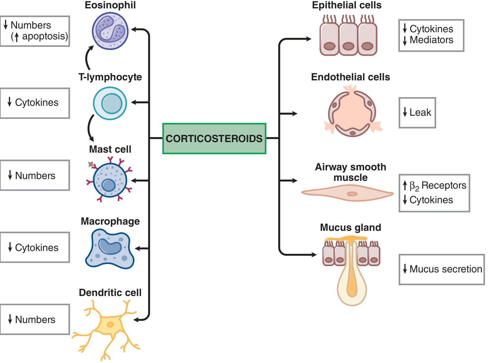
Step 4: How Corticosteroids Switch Off Inflammation (via HDAC2)
Corticosteroid enters cell
        ↓
Binds Glucocorticoid Receptor (GR) in cytoplasm
        ↓
GR-steroid complex moves into nucleus
        ↓
Does TWO things simultaneously:

1. Directly inhibits HAT activity (prevents further acetylation)
2. Recruits HDAC2 to the site of already-acetylated inflammatory genes
        ↓
HDAC2 removes acetyl groups from histones
        ↓
Chromatin CLOSES → Inflammatory genes switched OFF
        ↓
↓ IL-4, IL-5, IL-13, TNF-α, GM-CSF etc.
        ↓
Anti-inflammatory effect in asthma
Step 5: Where Theophylline Fits In - The Key Pharmacology Point
Low-dose theophylline
        ↓
Directly activates HDAC2 (nuclear enzyme)
        ↓
HDAC2 deacetylates histones at inflammatory gene sites
        ↓
Inflammatory genes switched OFF
        ↓
Anti-inflammatory effect (independent of PDE inhibition)
Why this makes theophylline + ICS a powerful combination:
| Drug | Mechanism | Role |
|---|---|---|
| ICS (corticosteroid) | Recruits HDAC2 to inflammatory gene sites | Brings HDAC2 to the right location |
| Low-dose theophylline | Activates/enhances HDAC2 enzyme activity | Makes HDAC2 work better once it's there |
Step 6: The COPD/Steroid Resistance Connection
- Corticosteroids cannot recruit a functional HDAC2
- Inflammatory genes stay ON
- Patient becomes steroid-resistant
Step 7: Clinical Implication Summary
| Scenario | HDAC2 Status | Effect |
|---|---|---|
| Normal airway on ICS | HDAC2 functional; steroid recruits it | Good anti-inflammatory response |
| COPD / Steroid resistance | HDAC2 reduced/dysfunctional | Steroids ineffective |
| COPD + Low-dose theophylline | HDAC2 restored | Steroid sensitivity restored |
| Mild/moderate asthma + Low-dose theophylline + ICS | HDAC2 enhanced | Synergistic anti-inflammatory effect |
The Full Pathway in One Picture
INFLAMMATION PATH (Bad):
NF-κB → HAT → Histone ACETYLATION → Genes ON → Cytokines → Asthma

CORTICOSTEROID PATH (Good):
ICS → Glucocorticoid Receptor → Recruits HDAC2 → Histone DEACETYLATION → Genes OFF

THEOPHYLLINE ENHANCEMENT:
Low-dose theophylline → Activates HDAC2 directly → Potentiates ICS effect
One-Line Summary for Viva
"Inflammatory stimuli (via NF-κB) activate HAT, which acetylates histones and switches ON inflammatory genes. Corticosteroids recruit HDAC2 to deacetylate these histones and switch the genes OFF. Low-dose theophylline directly activates HDAC2, enhancing the steroid effect - this explains its anti-inflammatory action at sub-bronchodilator plasma levels."